CLINICAL SCIENCE
Color Doppler imaging of the superior ophthalmic
vein in patients with Graves’ orbitopathy before and
after treatment of congestive disease
Ma´rio L. R. Monteiro,I Rodrigo B. S. Moritz,IHe´lio Angotti-Neto,IJoseph E. BenabouII
IDivision of Ophthalmology, Hospital das Clı´nicas of the University of Sa˜o Paulo Medical School, Sa˜o Paulo, Brazil.IIDepartment of Radiology, Hospital das Clı´nicas of the University of Sa˜o Paulo Medical School, Sa˜o Paulo, Brazil.
OBJECTIVE:To compare superior ophthalmic vein blood flow parameters measured with color Doppler imaging in patients with congestive Graves’ orbitopathy before and after treatment and in normal controls.
METHODS:Twenty-two orbits from 12 patients with Graves’ orbitopathy in the congestive stage and 32 orbits from 16 normal controls underwent color Doppler imaging studies. Color Doppler imaging was repeated after treatment in the group of patients with Graves’ orbitopathy, which included orbital decompression in 16 orbits and corticosteroids in six orbits. The findings for each group were compared.
RESULTS: In the group of orbits with congestive disease, superior ophthalmic vein flow was detected in 17 orbits (anteroposteriorally in 13 and in the opposite direction in four) and was undetectable in five. After treatment, superior ophthalmic vein flow was detected and anteroposterior in 21 and undetected in one orbit. In normals, superior ophthalmic vein flow was detected anteroposterior in 29 orbits and undetectable in three orbits, indicating a significant difference between groups. There was also a significant difference between controls and congestive Graves’ orbits and between congestive orbits before and after treatment, but not between controls and patients after treatment. A comparison of superior ophthalmic vein flow parameters revealed a significant difference between the groups. The superior ophthalmic vein flow was significantly reduced in the congestive stage compared with the flow parameters following treatment and in the untreated controls.
CONCLUSIONS: Superior ophthalmic vein flow was significantly reduced in the orbits affected with congestive Graves’ orbitopathy and returned to normal following treatment. Congestion appears to be a contributing pathogenic factor in the active inflammatory stage of Graves’ orbitopathy.
KEYWORDS: Color Doppler imaging; Graves’ orbitopathy; Superior ophthalmic vein; Congestive orbitopathy; Endocrine exophthalmos.
Monteiro MLR, Moritz RBS, Angotti-Neto H, Benabou JE. Color Doppler imaging of the superior ophthalmic vein in patients with Graves’ orbitopathy before and after treatment of congestive disease. Clinics. 2011;66(8):1329-1333.
Received for publication onFebruary 3, 2011;First review completed onMarch 15, 2011;Accepted for publication onApril 20, 2011 E-mail: [email protected]
Tel.: 55 11 3661-7582
INTRODUCTION
Graves’ orbitopathy (GO) is an autoimmune inflamma-tory process that affects the periorbital and orbital tissues, mainly the extraocular muscles. The enlargement of these muscles is responsible for most of the manifestations of the disease, including lid retraction, proptosis, extraocular muscle restriction, dysthyroid optic neuropathy, and con-gestive signs such as conjunctival hyperemia, chemosis, and lid swelling.1,2GO occurs before, during, or after the onset
of hyperthyroidism and, less frequently, in euthyroid or hypothyroid patients.
GO is divided into a congestive stage and a fibrotic stage. In the congestive or inflammatory stage, autoimmunity is believed to play a key role in the process leading to inflammatory cellular infiltration of the muscles and transformation of fibroblasts into adipose tissue. The fibrotic stage is characterized by fibrosis in the orbital tissues with residual manifestations of the disease such as proptosis and strabismus.3,4
The pathogenesis of the acute inflammatory stage has been attributed to autoimmunity, and treatment of patients in this stage has traditionally been based on corticosteroids and radiotherapy. Superior orbital vein (SOV) congestion also plays an important role in the inflammatory stage, as demonstrated by computed tomography5-7 and color Doppler imaging (CDI) studies.8-11 In a recent study using
CDI, we demonstrated that SOV flow was significantly reduced in orbits with congestive GO and with myogenic fibrotic GO, but not in orbits with fibrotic lipogenic orbitopathy.11 This finding was consistent with previous studies using CDI in patients with GO at different stages.8-10,12Our previous study lends support to the notion that venous congestion plays a significant role in the pathogenesis of the active stage of the orbitopathy and suggests that patients can benefit from relief of SOV congestion by clinical or surgical treatment.11 To our knowledge, however, no previous study has evaluated CDI flow parameters in patients with congestive orbito-pathy before and after clinical and/or surgical treatment of the disease. The purpose of this study was to evaluate SOV blood flow parameters using CDI in patients with active GO before and after treatment of the disease. We investigated the effect of treatments such as orbital decompression and/ or corticosteroids in CDI-measured SOV flow parameters.
METHODS AND MATERIALS
This interventional, prospective, and cross-sectional study was conducted between October 2005 and September 2010. The study followed the principles of the Declaration of Helsinki. Approval from the Institutional Review Board Ethics Committee was obtained, and all of the participants gave their informed consent.
A total of 12 patients (seven men, five women) with congestive GO were examined. The mean age of the patients was 53.2 (SD 9.5) years. The diagnosis of GO was established according to previously published criteria.1All of the patients had Graves’ disease but became euthyroid under treatment. Sixteen normal subjects (four men, twelve women), with a mean age of 48.1 (SD 10.9) years, were included in the study as a control group. The normal subjects were defined as healthy euthyroid volunteers without ocular diseases.
The patients underwent a complete ophthalmic examina-tion that included best corrected visual acuity, applanaexamina-tion tonometry, pupillary reactions, extraocular motility evalua-tion, slit lamp examinaevalua-tion, evaluation of eyelid and soft tissue inflammation, measurement of the lid fissure, Hertel exophthalmometry, fundoscopy, and visual field evaluation with standard automated perimetry using the 24-2 SITA-Standard strategy (Humphrey Field Analyzer, Carl-Zeiss Meditec, Dublin, CA). Goldmann manual perimetry was also used when necessary to confirm optic nerve involve-ment. Both orbits of the patients were scanned with a 16-slice multidetector computed tomography scanner (Brilliance 16; Philips Medical Systems, Nederland B.V., the Netherlands).
GO was classified as active (congestive) or fibrotic according to a 10-item clinical activity score based on four of five well-known classical signs of inflammation (pain, redness, swelling, and impaired function). The clinical activity score included orbital pain, redness of the eyelid or conjunctiva, swelling of the conjunctiva (chemosis), caruncle, or eyelid and impaired function represented by decreased visual function or decreased eye movement.3One point was given per eye for each item present. The evaluated items corresponded to symptoms (orbital ache and gaze-evoked pain), signs (conjunctival redness, eyelid erythema, eyelid edema, chemosis, and swelling of the plica or caruncle) and indications of progressive disease in at
least two examinations.3,13 Patients with active congestive disease had a clinical activity score of four or more points, whereas patients classified with fibrotic disease, not included in this study, had a clinical activity score equal to or lower than two. To avoid any possible doubt regarding disease activity in this study, patients with a score of three were also excluded. Eye movement restrictions in the field of action of the superior rectus muscle (elevation) due to inferior rectus restriction and of the lateral rectus muscle (abduction) due to medial rectus restriction were graded from 0 (no limitation) to 4 (absence of eye movement from primary position in the muscle’s field of action). Grade 1, 2 and 3 restrictions indicated 75%, 50%, and 25% excursion, respectively, from the primary position, either by elevation (restriction caused by the inferior rectus) or by abduction (restriction of the medial rectus). A combined restriction index ranging from 0 to 8 was calculated by adding the two scores. Orbits were also classified according to the presence or absence of dysthyroid optic neuropathy. The diagnostic criteria for dysthyroid optic neuropathy were diminished best corrected visual acuity not caused by changes in transparency, presence of a relative afferent pupillary defect and presence of visual field defect on automated or Goldmann perimetry. To qualify as an abnormal visual field on standard automated perimetry, at least three adjacent abnormal points at thep,0.05 level or two adjacent points with one abnormal at thep,0.01 level on the pattern deviation plot were required.14On the Goldmann perime-try, visual field was considered abnormal in the presence of central or paracentral scotoma, localized depression of the isopters greater than 10 degrees or generalized constriction of the isopters.15,16 When visual acuity was normal and visual field abnormalities were present on standard auto-mated perimetry, a repeat examination using autoauto-mated or the Goldmann manual perimetry was performed in order to rule out false-positive results. All clinical assessments were performed within one week of the CDI study.
After the ophthalmic examination, the patients were transferred to the Radiology Department for CDI with a 6-12 megahertz linear-array transducer coupled to a Logiq 9 Doppler scanner from General Electric (Santa Clara, California, USA). Maximum and minimum blood flow in the SOV was determined in both eyes with the patient resting on a bed with 30 degrees head elevation. During the examination, the patients were asked to remain still with both eyes closed and to fixate straight ahead. The transducer was gently placed over the closed eyes (right eye first), and care was taken to avoid applying pressure to the eye. Blood flow velocity was measured in the superior nasal part of the SOV, anterior to the point where it crosses the optic nerve. Velocity was measured in each vessel several times until obtaining at least two good readings, maintaining the angle between the sound beam and the blood flow direction less than 30 degrees. Only the highest values were used for statistical analysis. All CDI measurements were performed by the same experienced professional (JEB) who was blinded to the clinical status of the patients with GO. Maximum and minimum SOV blood flow velocities were registered, and the mean values and standard deviations were calculated.
of congestive disease. After treatment, GO was inactive in all orbits according to the clinical activity score classification (a score of two or less).
Statistical analysis
The descriptive statistics included mean values¡SD for
normally distributed variables after the analysis of histo-grams and the Shapiro-Wilk test confirmed the normality assumption. We used an unpaired t-test for comparisons of the three groups (normal controls, active GO before treatment and GO after treatment) and a paired t-test for comparison of active GO before and after treatment.
Each group of orbits was initially classified according to whether the flow in the SOV was anteroposterior, absent (not detected), or posteroanterior (reverse). The observed proportions were compared using Fisher’s exact test. For further statistical analyses, the anteroposterior flow was expressed in positive numbers, undetected flow was assigned a value of zero, and the posteroanterior flow was expressed in negative numbers. Apvalue of less than .05
was considered statistically significant.
RESULTS
A total of 76 CDI studies were performed: 22 orbits from 12 patients (seven men and five women, mean age¡ SD: 53.1¡9.5 years) with active GO (clinical activity score index of four or higher), the same 22 orbits after treatment (activity score index of two or less), and 32 orbits from 16 control subjects (twelve women and four men; mean age ¡ SD:
48.3¡10.9). There was no significant age difference between
the patients and controls (p= 0.231). Before treatment, the
exophthalmometry measurements ranged from 22 to 28 mm (mean ¡ SD: 25.52¡1.96 mm) in GO patients, and the restriction index ranged from 0 to 8 (mean¡SD: 5.0¡1.93). Before treatment, 21 orbits of patients with GO were categorized as having predominantly myogenic disease (with a restriction grade of 3-8). One orbit was categorized as having predominantly lipogenic disease (with muscle restriction graded as 0). In addition, 15 orbits presented with dysthyroid optic neuropathy before treatment.
In the normal controls, SOV was detected in 29 orbits and was undetectable in three. Reverse blood flow was not observed. In orbits with active congestive disease, SOV flow was absent in five, present in 13 and reversed in four orbits before treatment. After treatment, SOV flow in the same 22 orbits was anteroposterior (n = 21) or undetectable (n = 1) (Table 1). A significant difference was found between GO orbits before treatment and the control orbits (p =0.007,
Fisher’s exact test). No significant difference was found
between the GO orbits after treatment and the control orbits (p= 0.638).
Table 2 compares SOV flow values for the two GO groups and for normal controls. The results of the unpaired t-test revealed a significant difference between GO patients before treatment and controls with regard to the maximum (p= 0.009), minimum (p= 0.013), and differential (p= 0.005)
SOV flow. There was no significant difference between the GO patients after treatment and the controls with regard to the maximum, minimum, and differential flow (p= 0.703,
0.599 and 0.969, respectively). The paired t-test revealed a significant difference in the maximum (p= 0.007), minimum
(p= 0.012) and differential (p= 0.006) SOV flow in GO
patients before and after treatment (Table 2).
DISCUSSION
Autoimmunity is likely the primary mechanism involved in the pathogenesis of Graves’ orbitopathy. It is, however, suspected that orbital venous blood flow congestion contributes to the development of clinical signs and symptoms (e.g., proptosis, muscle restriction, periorbital swelling, and chemosis) during the active stage of the disease,17 as suggested by a number of clinical and experimental findings. Previous studies have reported prompt improvement in congestive signs after orbital decompression, suggesting that venous congestion is an important factor in the pathogenesis of the disease.18 In another study, venous drainage impairment and subsequent resolution was visible on fluorescein angiographs of the optic discs from patients with dysthyroid optic neuropa-thy.19 Hudson et al.6 suggested that the orbital SOV enlargement observed on computed tomography scans of patients with dysthyroid optic neuropathy may be a contributing factor in the development of optic nerve impairment. A previous study indicated that experimentally induced orbital venous stasis can closely mimic many of the clinical changes that occur in GO.17
CDI allows simultaneous imaging of the anatomic structures by B-mode ultrasonography with superimposed color-coded vascular flow. The current study confirmed a significant difference in CDI parameters between patients with GO in the active stage and controls or orbits with GO following treatment. Reverse and absent blood flow were observed in four and five out of 22 orbits with active GO, respectively. Flow may be undetectable in eyes of normal controls for several reasons, including insufficient sensitiv-ity of the device for measuring very small venous flow rates, or anatomic flow variations. In abnormal orbits, absent flow is most often related to the impairment of venous return at the orbital apex, leading to a venous flow of zero or close to
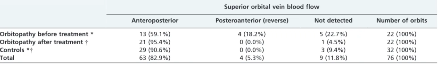
Table 1 -Detection and direction of blood flow in the superior ophthalmic vein using color Doppler imaging in patients with Graves’ orbitopathy and control subjects.
Superior orbital vein blood flow
Anteroposterior Posteroanterior (reverse) Not detected Number of orbits Orbitopathy before treatment * 13 (59.1%) 4 (18.2%) 5 (22.7%) 22 (100%)
Orbitopathy after treatment{ 21 (95.4%) 0 (0.0%) 1 (4.5%) 22 (100%)
Controls *{ 29 (90.6%) 0 (0.0%) 3 (9.4%) 32 (100%)
Total 63 (82.9%) 4 (5.3%) 9 (11.8%) 76 (100%) *Fisher’s exact test (
p= 0.007, controls compared with orbitopathy before treatment).
{Fisher’s exact test (
zero. Although flow may occasionally be absent in normal subjects, we believe the absence of flow is most likely an indication of venous congestion. In our study, a direct comparison revealed the absence of flow in five of 22 congestive GO orbits but only in three out of 32 normal controls and one of 22 GO orbits after treatment. Conversely, reverse blood flow has not been reported in normal subjects20and may indicate more severely impaired orbital venous drainage. While reverse blood flow occurred in four of 22 active GO orbits, it did not occur in any of the 32 control orbits or in the 22 GO orbits following treatment. A comparative analysis of our groups also indicated that SOV was significantly reduced in orbits with congestive GO compared with control orbits or the GO orbits following treatment, suggesting that venous congestion is most likely a contributing factor to the pathogenesis of this condition.
Our findings of reduced SOV flow in patients with active GO are consistent with results from several previous studies. Nakase et al.8found reversed SOV flow in 15% of 39 orbits with GO, but not in controls (n = 22). Reversed SOV flow was present in 36% of 14 orbits with GO and apical crowding. Nakase et al. suggested that these findings were a strong indication of severe venous stasis in the orbits, possibly related to the development of dysthyroid optic neuropathy. No data were provided, however, regarding the disease activity status of each orbit. Benning et al.12 found a significantly smaller maximum SOV blood flow velocity in patients with GO than in normal subjects and a slight correlation between reduced venous outflow in the SOV and disease severity. Somer et al.10 used CDI in 48 orbits of 24 patients with GO and in 20 orbits of 10 healthy volunteers. The authors found the mean venous blood flow velocity to be significantly decreased in patients with GO. No SOV flow, however, was detected in 18 orbits with GO. Alp et al.9 performed CDI on 111 patients with Graves’ disease and 46 healthy controls. Patients with Graves’ disease were divided into two groups: 69 with GO and 42 with no orbitopathy. Reverse blood flow in the SOV was observed in 13% of patients with GO. The authors found blood flow velocities in the ophthalmic artery and the central retinal artery to be significantly higher in patients with GO than in orbitopathy-free patients with Graves’ disease or healthy controls. Blood flow was significantly reduced in the SOV. The study included only patients who were not receiving local or systemic treatment for GO, and no disease activity criteria were provided.
Despite reporting significant changes in orbital blood flow in patients with GO, none of the aforementioned studies were designed to directly compare findings for
different forms of GO. Only a few studies have attempted to correlate disease activity, based on the clinical activity score,13 with CDI blood flow parameters. Yanik et al.21 studied 118 patients with Graves’ disease, 25 without orbitopathy and 93 with activity-scored GO.13The authors found a negative correlation between orbital blood flow parameters and their clinical activity scores.21 They excluded patients requiring urgent orbital decompression or receiving systemic medication, thereby likely excluding subjects with more severe forms of orbitopathy. The study did not separate patients in predominantly myogenic or lipogenic forms of the disease. In a recent study, we evaluated patients with GO after clearly distinguishing between active and inactive as well as between myogenic (restrictive) and lipogenic (non-restrictive) GO.14Our find-ings indicated that most patients with congestive GO had reduced or reversed maximum SOV flow. Patients with fibrotic GO in the myogenic form also displayed a significant reduction in SOV flow compared with normal controls. Patients with fibrotic lipogenic disease did not differ significantly from the controls.
In the current study, we compared the same group of active GO patients before and after treatment. The results indicated dramatic differences in the SOV flow parameters between the active stage of the disease and post-treatment GO or normal controls, indicating that venous congestion is an important element in the active stage of the disease.
The CDI data provided by this study and by previous studies may be useful for the management of GO. Treatment of GO in the acute stage is traditionally immunosuppressive (using corticosteroids or immunosuppressants), but con-gestive signs sometimes remain despite adequate treatment and may require orbital decompression. Persistently reduced or reversed SOV blood flow despite adequate treatment may be an indication for decompressive surgery in select patients. The current data appear to confirm this assumption, as most of our patients were in fact treated with orbital decompression.
In conclusion, the fact that SOV flow was significantly reduced in the congestive stage of GO and normalized after treatment of active orbitopathy suggests that venous congestion plays a significant role in the pathogenesis of the disease. Our data lends support to the notion that clinical improvement with treatment is due to the control of not only the autoimmune process but also orbital venous congestion. On the basis of these findings, we believe that if diminished SOV flow persists despite adequate clinical treatment, orbital decompression should be considered to improve congestive signs. Further studies with more
Table 2 -Comparison of blood flow parameters in the superior ophthalmic vein (SOV) in patients with Graves’ orbitopathy and controls.
Mean (range) velocity (cm/s)
SOV maximum velocity * SOV minimum velocity * Differential flow velocity * Congestive GO before
treatment (22 orbits)
3.07*{ (-13.16 – 9.55)
2.11*{ (-10.0 – 7.26)
0.96*{ (-4.22 – 6.66)
GO after treatment (22 orbits)
7.80 * (0 – 11.96)
5.31 * (0 – 8.75)
2.48 * (0 – 4.43)
Normals (32 orbits)
7.49{ (0 – 11.68)
5.02{ (0 – 8.91)
2.47{ (0 – 5.48) *
p,0.05, paired t-test, comparing GO orbits before and after treatment.
{
patients are necessary confirm these findings and to better evaluate potential differences in congestion relief between clinical and surgical treatment of the congestive stage of GO.
ACKNOWLEDGMENTS
This work was supported by a grant from Conselho Nacional de Desenvolvimento Cientı´fico e Tecnolo´gico, CNPq (No 309709/2007-5), Brası´lia, Brazil.
Study registered on ClinicalTrial.gov number: NCT00697528
REFERENCES
1. Bartley GB, Gorman CA. Diagnostic criteria for Graves’ ophthalmopathy. Am J Ophthalmol. 1995;119:792-5.
2. Bartley GB, Fatourechi V, Kadrmas EF, Jacobsen SJ, Ilstrup DM, Garrity JA, et al. Clinical features of Graves’ ophthalmopathy in an incidence cohort. Am J Ophthalmol. 1996;121:284-90.
3. Mourits MP, Koornneef L, Wiersinga WM, Prummel MF, Berghout A, van der Gaag R. Clinical criteria for the assessment of disease activity in Graves’ ophthalmopathy: a novel approach. Br J Ophthalmol. 1989;73:639-44, doi: 10.1136/bjo.73.8.639.
4. Bartalena L, Pinchera A, Marcocci C. Management of Graves’ ophthal-mopathy: reality and perspectives. Endocr Rev. 2000;21:168-99, doi: 10. 1210/er.21.2.168.
5. Monteiro ML, Goncalves AC, Silva CT, Moura JP, Ribeiro CS, Gebrim EM. Diagnostic ability of Barrett’s index to detect dysthyroid optic neuropathy using multidetector computed tomography. Clinics. 2008;63:301-6, doi: 10.1590/S1807-59322008000300003.
6. Hudson HL, Levin L, Feldon SE. Graves exophthalmos unrelated to extraocular muscle enlargement. Superior rectus muscle inflammation may induce venous obstruction. Ophthalmology. 1991;98:1495-9. 7. Nugent RA, Belkin RI, Neigel JM, Rootman J, Robertson WD, Spinelli J,
et al. Graves orbitopathy: correlation of CT and clinical findings. Radiology. 1990;177:675-82.
8. Nakase Y, Osanai T, Yoshikawa K, Inoue Y. Color Doppler imaging of orbital venous flow in dysthyroid optic neuropathy. Jpn J Ophthalmol. 1994;38:80-6.
9. Alp MN, Ozgen A, Can I, Cakar P, Gunalp I. Colour Doppler imaging of the orbital vasculature in Graves’ disease with computed tomographic correlation. Br J Ophthalmol. 2000;84:1027-30, doi: 10.1136/bjo.84.9.1027. 10. Somer D, Ozkan SB, Ozdemir H, Atilla S, Soylev MF, Duman S. Colour Doppler imaging of superior ophthalmic vein in thyroid-associated eye disease. Jpn J Ophthalmol. 2002;46:341-5, doi: 10.1016/S0021-5155(02)00485-9.
11. Monteiro ML, Angotti-Neto H, Benabou JE, Betinjane AJ. Color Doppler imaging of the superior ophthalmic vein in different clinical forms of Graves’ orbitopathy. Jpn J Ophthalmol. 2008;52:483-8, doi: 10.1007/ s10384-008-0594-y.
12. Benning H, Lieb W, Kahaly G, Grehn F. [Color duplex ultrasound findings in patients with endocrine orbitopathy]. Ophthalmologe. 1994;91:20-5.
13. Mourits MP, Prummel MF, Wiersinga WM, Koornneef L. Clinical activity score as a guide in the management of patients with Graves’ ophthalmopathy. Clin Endocrinol (Oxf). 1997;47:9-14, doi: 10.1046/j. 1365-2265.1997.2331047.x.
14. Monteiro ML, Portes AL, Moura FC, Regensteiner DB. Using frequency-doubling perimetry to detect optic neuropathy in patients with Graves’ orbitopathy. Jpn J Ophthalmol. 2008;52:475-82, doi: 10.1007/s10384-008-0579-x. 15. Neigel JM, Rootman J, Belkin RI, Nugent RA, Drance SM, Beattie CW, et al. Dysthyroid optic neuropathy. The crowded orbital apex syndrome. Ophthalmology. 1988;95:1515-21.
16. Panzo GJ, Tomsak RL. A retrospective review of 26 cases of dysthyroid optic neuropathy. Am J Ophthalmol. 1983;96:190-4.
17. Saber E, McDonnell J, Zimmermann KM, Yugar JE, Feldon SE. Extraocular muscle changes in experimental orbital venous stasis: some similarities to Graves’ orbitopathy. Graefes Arch Clin Exp Ophthalmol. 1996;234:331-6, doi: 10.1007/BF00220709.
18. Trobe JD, Glaser JS, Laflamme P. Dysthyroid optic neuropathy. Clinical profile and rationale for management. Arch Ophthalmol. 1978;96:1199-209.
19. Yoshikawa K, Higashide T, Inoue T, Inoue Y. Fluorescein angiographic findings in optic discs with dysthyroid optic neuropathy. Orbit. 1991;10:89-96, doi: 10.3109/01676839109023088.
20. Erickson SJ, Hendrix LE, Massaro BM, Harris GJ, Lewandowski MF, Foley WD, et al. Color Doppler flow imaging of the normal and abnormal orbit. Radiology. 1989;173:511-6.