Effects of Epinephrine in Local Dental Anesthesia in Patients with
Coronary Artery Disease
Ricardo Simões Neves, Itamara Lucia Itagiba Neves, Dante Marcelo Artigas Giorgi, Cesar José Grupi, Luís Antonio
Machado César, Whady Hueb, Max Grinberg
Instituto do Coração do Hospital das Clínicas da Faculdade de Medicina da Universidade de Sâo Paulo – FMUSP – São Paulo, SP - Brazil
Summary
Background: The use of vasoconstrictors for local anesthesia in patients with coronary heart disease is controversial in the literature, and there is concern regarding risk of cardiac decompensation.
Objective: To evaluate electrocardiographic and blood pressure parameters during restorative dental procedure under local anesthesia with and without a vasoconstrictor in patients with coronary artery disease.
Methods:6L[W\WZRSDWLHQWVZHUHLQFOXGHGLQWKHVWXG\DJHVUDQJLQJIURPWRPHDQ
RIZKRPZHUHPDOH7KLUW\SDWLHQWVZHUHUDQGRPO\DVVLJQHGWRUHFHLYHOLGRFDLQHZLWKHSLQHSKULQHHSLQHSKULQH JURXSDQGWKHUHPDLQLQJSDWLHQWVOLGRFDLQHZLWKRXWHSLQHSKULQHQRQHSLQHSKULQHJURXSIRUORFDODQHVWKHVLD$OO SDWLHQWVXQGHUZHQWKRXUDPEXODWRU\EORRGSUHVVXUHPRQLWRULQJDQGG\QDPLFHOHFWURFDUGLRJUDSK\7KUHHSHULRGV ZHUH FRQVLGHUHG LQ WKH VWXG\ EDVHOLQH ² UHFRUGLQJV REWDLQHG GXULQJ WKH PLQXWHV SULRU WR WKH SURFHGXUH SURFHGXUH²UHFRUGLQJVREWDLQHGIURPWKHEHJLQQLQJRIDQHVWKHVLDWRWKHHQGRIWKHSURFHGXUHDQGKRXUV
Results: There was an increase in blood pressure in both groups during the procedure, compared with baseline values; EXWZKHQWKHWZRJURXSVZHUHFRPSDUHGQRVLJQLILFDQWGLIIHUHQFHZDVGHWHFWHGEHWZHHQWKHP+HDUWUDWHUHPDLQHG XQFKDQJHGLQERWKJURXSV1R67VHJPHQWGHSUHVVLRQ!PPRFFXUUHGHLWKHUDWEDVHOLQHRUGXULQJWKHSURFHGXUH 6HYHQSDWLHQWVH[SHULHQFHGPRUHWKDQWHQDUUK\WKPLDHSLVRGHVSHUKRXUGXULQJWKHSURFHGXUHIRXULQ WKHQRQHSLQHSKULQHJURXSDQGWKUHHLQWKHHSLQHSKULQHJURXS
Conclusion:1RGLIIHUHQFHZDVREVHUYHGLQEORRGSUHVVXUHKHDUWUDWHRUHYLGHQFHRILVFKHPLDDQGDUUK\WKPLDVLQHLWKHU
group. The use of vasoconstrictor has proved to be safe within the range of the present study.
Key words: Epinephrine/adverse effects; anesthesia, local; dental restoration, permanent; coronary arteriosclerosis.
0DLOLQJDGGUHVV5LFDUGR6LP}HV1HYHV
Rua Alves Guimarães 689/11 - 05410-001 – São Paulo, SP - Brazil E-mail: [email protected]
Manuscript received October 4, 2006; revised manuscript received December 28, 2006; accepted February 13, 2007.
Introduction
The literature is controversial regarding the use of vasoconstrictors in local dental anesthesia in patients with heart disease. Most studies were conducted either with healthy patients1-10 or those with heart disease of various etiologies11,12. In addition, many authors13-15 failed to include a control group comparing the effects of the anesthetic with and without a vasoconstrictor, making it difficult to extrapolate these results to CAD patients. We investigated the behavior of systemic blood pressure and 24-hour electrocardiogram (Holter monitoring) in patients with clinical symptoms of stable angina and on drug therapy, positive exercise testing, and angiographically proven coronary stenosis > 70% in at least one major artery and who underwent restorative dental treatment under anesthesia with and without a vasoconstrictor.
Methods
Sixty-two patients were followed up on at the outpatient clinic of the Coronary Care Unit of Incor (Hospital das Clínicas Heart Institute of the University of SãoPaulo Medical School), 51 (82.3%) of whom were male. Ages ranged from 39 to 80 (mean 58.7 ± 8.8), and body mass index (BMI), from 18.8 to 39.4 (mean 27.4).
Twenty-four patients (38.7%) were diagnosed with systemic hypertension and 24, with diabetes (38.7%) All patients continued their medication for coronary artery disease and possible comorbidities, especially beta-blockers (87.1%), lipid-lowering agents (87.1%), antiplatelets (83.9%), and nitrates (54.8%).
Patients were enrolled after Institutional Ethics Committee approval, and all of them read and signed an Informed Consent after receiving thorough verbal explanation of the study and its attendant risks.
Inclusion criteria were the following:
- Personal: patients of both genders from 30 to 80 years of age;
group and 32 in the non-epinephrine group) underwent a single restorative procedure under inferior alveolar nerve block anesthesia.
Statistical analysis - For quantitative variables, minimum and maximum values were observed and means, medians, and standard deviations were calculated. For qualitative variables, the absolute and relative frequencies were calculated.
The following tests were used: Student’s t test to compare means between two groups; one-way analysis of variance (ANOVA)19, using Bonferroni’s multiple comparison test18, to compare four groups; Fisher’s exact test for proportions;
repeated-measures ANOVA19 to assess groups behavior
taking into account the medical conditions studied; in addition to Friedman’s nonparametric test and the Mann-Whitney nonparametric18 test when the assumption of data normality was rejected. The significance level was set at 5% (p = 0.05).
Results
Duration of procedures: length range was wider in the epinephrine group (12 to 94 minutes, mean 35 ± 15) than in the non-epinephrine group (22 to 69 minutes, mean 40 ± 2). However, means between both groups did not differ (p = 0,200). No clinical events were observed.
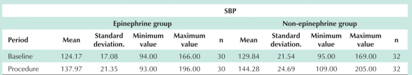
Blood pressure behavior - There was a rise in systolic and diastolic blood pressure from baseline to the procedure in both groups (14 mm Hg and 5 to 7 mmHg, respectively), when they were evaluated separately. However, when the epinephrine and non-epinephrine groups were compared, no significant difference was found (Tables 1 and 2). Moreover, no differece was found when mean systolic (p = 0.2076) and diastolic (p = 0.5936) blood pressure were compared between the baseline, procedure, sleep and wake periods (Figure 1).
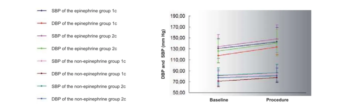
The number of anesthetic cartridges used, one (1 c) or two (2 c) with or without epinephrine, did not differ between the epinephrine and non-epinephrine groups regarding mean systolic (p = 0.2083) and diastolic (p = 0.1183) blood pressure (Figure 2), and there was no influence by the use or not of beta-blockers.
24-hour ambulatory electrocardiography - Fifty-six patients underwent 24-hour electrocardiography. For technical reasons (motion artifacts and noise > 2%), six patients were excluded from the study: three from the non-epinephrine group and lumen stenosis) and exercise stress test positive for myocardial
ischemia performed within less than three months in the absence of recent acute myocardial infarction;
- Dental: fully or partially dentate requiring lower molar, premolar, or canine restoration.
Exclusion criteria were the following:
- Clinical: neoplasias, septicemia, pregnancy, unstable angina, and malignant hypertension.
Coronary angiography revealed single-vessel disease in seven (11.3%) patients, two-vessel disease in 18 (29%) patients, and three-vessel disease in 37 (59.7%) patients.
Thirty patients were randomly assigned to receive 2% lidocaine and 1:100,000 epinephrine (epinephrine group), and 32 to receive 2% lidocaine without epinephrine (non-epinephrine group) for local anesthesia. In the (non-epinephrine group, 15 patients were given one anesthetic cartridge (1.8 mL) of 2% lidocaine plus 1:100,000 epinephrine and 15 patients, two anesthetic cartridges (3.6mL). In the non-epinephrine group, 15 patients were given one cartridge of lidocaine 2% without vasoconstrictor and 17 patients, two cartridges.
All patients underwent 24-hour electrocardiography (Holter) and ambulatory blood pressure monitoring (ABPM). They were instructed to report any chest pain or discomfort, noting the time of the event in the diary provided by the Holter Laboratory. Three recording periods were considered: 1) baseline – one hour before anesthesia; 2) procedure – from the beginning of anesthesia until the patient left the dental chair; post-procedure – until the 24 hours were completed.
To assess systolic blood pressure (SBP), diastolic blood pressure (DBP), and heart rate (HR), the mean values recorded during these three periods were considered.
An ischemic episode was defined as either ST-segment HOHYDWLRQPPRUKRUL]RQWDORUGRZQVORSLQJ67VHJPHQW GHSUHVVLRQ PP IURP EDVHOLQH ODVWLQJ IRU DW OHDVW RQH minute and reverting to baseline levels for at least one minute during the study periods (baseline, procedure and post-procedure).
The presence of cardiac arrhythmia was considered when more than ten ventricular extrasystoles (VES) and supraventricular extrasystoles (SVES)16 occurred per hour during the study periods.
Dental procedure - Each patient (30 in the epinephrine
Table 1 - Distribution of means, standard deviation, minimum, and maximum SBP at baseline and during the procedure according to the presence or not of epinephrine in the anesthetic solution
SBP
Epinephrine group 1RQHSLQHSKULQHJURXS
Period Mean Standard deviation.
Minimum value
Maximum
value n Mean
Standard deviation.
Minimum value
Maximum value n
Baseline 124.17 17.08 94.00 166.00 30 129.84 21.54 95.00 169.00 32 Procedure 137.97 21.35 93.00 196.00 30 144.28 24.69 109.00 205.00 32
SBP - systolic blood pressure; Epinephrine group - lidocaine plus epinephrine; Non-epinephrine group - lidocaine without epinephrine. Comparing groups
three from the epinephrine group.
Mean heart rate did not differ between the epinephrine and non-epinephrine groups at baseline, during the procedure (p = 0.1967), and over the 24 hours (p = 0.8417) (Tables 3 and 4), as well as in the assessment based on the number of anesthetic cartridges used, that is, one or two with or without epinephrine.
ST-segment depression - Ten patients (17.9%) had
ST-segment depression greater than 1 mm from baseline, six (20.7%) in the non-epinephrine group (14.8%) and four
(14.8%) in the epinephrine group, but no significant difference was found between both groups (p = 0.731). No events were recorded at baseline or during the procedure, and all episodes occurred at least two hours after the dental procedure had been completed.
Cardiac arrhythmias - Sinus rhythm prevailed throughout
the 24-hour monitoring period. Over the 24 hours, 17 patients (30.4%), ten (34.5%) belonging to the non-epinephrine group and seven (25.9%) belonging to the epinephrine group, experienced supraventricular extrasystoles (SVES) and/or ventricular extrasystoles (VES). During the procedure, however, only seven (12.5%) patients had arrhythmias, four (13.8%) in the non-epinephrine group and three (11.1%) in the epinephrine group, but no significant difference was found between the two groups (p = 1.00).
Discussion
7KLVVWXG\LQFOXGHG&$'SDWLHQWVZLWKRIOXPHQ stenosis; most of them (59.7%) had triple-vessel disease. In keeping with the literature20,21, the study sample was predominantly male (82.3%) .
The presence of classical risk factors for coronary artery disease was common, particularly metabolic disorders, such as diabetes mellitus (38.7% vs7.5% of the population mean), and circulatory conditions, such as systemic hypertension (38.7%vs20% of the population mean)22.
Table 2 - Distribution of means, standard deviation, minimum, and maximum DBP at baseline and during the procedure according to the presence or not of epinephrine in the anesthetic solution
DBP
Epinephrine group 1RQHSLQHSKULQHJURXS Period Mean Standard
deviation.
Minimum value
Maximum
value n Mean
Standard deviation.
Minimum value
Maximum value n
Baseline 75.70 10.34 51.00 96.00 30 79.28 13.76 56.00 106.00 32 Procedure 81.33 10.83 59.00 109.00 30 83.75 14.22 66.00 117.00 32
DBP - diastolic blood pressure; Epinephrine group - lidocaine plus epinephrine; Non-epinephrine group - lidocaine without epinephrine. Interaction between
JURXSVDQGSHULRGVS &RPSDULVRQEHWZHHQSHULRGVS ,QGLYLGXDOJURXSV²S
Fig. 1 -Distribution of SBP and DBP means during the baseline, procedure, sleep, and wake periods according to the presence (epinephrine group) or not (non-epinephrine group) of epinephrine in the anesthetic solution. SBP - systolic blood pressure; DBP - diastolic blood pressure.
SBP of the epinephrine group SBP of the non-epinephrine group
DBP of the epinephrine group DBP of the non-epinephrine group
D
B
P
a
n
d
SB
P
(m
m
H
g
)
Periods
Bas elin
e
Slee p
Proc edur
e Wak
e
Fig. 2 -Distribution of SBP and DBP means at baseline and during the procedure according to the presence (epinephrine group) or not (non-epinephrine group) of
HSLQHSKULQHLQWKHDQHVWKHWLFVROXWLRQDQGWKHQXPEHURIFDUWULGJHVXVHGRQHFDUWULGJHFWZRFDUWULGJHVF6%3V\VWROLFEORRGSUHVVXUH'%3GLDVWROLF
blood pressure.
D
B
P
a
n
d
SB
P
(m
m
H
g
)
Baseline Procedure
SBP of the epinephrine group 1c
DBP of the epinephrine group 1c
SBP of the epinephrine group 2c
DBP of the epinephrine group 2c
SBP of the non-epinephrine group 1c
DBP of the non-epinephrine group 1c
SBP of the non-epinephrine group 2c
The prevailing use of beta-blockers, acetylsalicylic acid, lipid-lowering agents, and nitrates is also consistent with that found in a series of CAD patients20,23,24. The percentages of patients on beta-blockers and nitrates (87.1% and 54.8%, respectively) were similar to those reported by Leviner et al25 (60% and 85%, respectively), in 1992; nevertheless, these rates were higher than those reported by Cintron et al13 (27.5% and 32.5%) in a sample of 40 patients with recent acute myocardial infarction.
In order to make all study phases homogeneous, patients were enrolled based on their need of dental care. This approach enabled the length of the procedure and anesthetic dosages to be standardized, something that usually is not seen in endodontic and surgical procedures, thereby contributing to reduce the influence of stress, which has always been borne in mind.
Moreover, for practical purposes, caries treatment is the predominant procedure in dental office daily routine. In spite of this, few researchers, among them Leviner et al25, Cioffi et al26, Corah27, and Oliver et al28, have studied the effects of vasoconstrictors in patients undergoing dental anesthesia for restorative treatment.
In this study, there was a significant increase in SBP and DBP in both groups of patients from baseline to the procedure, when they were analyzed separately. However, when both groups were compared, no significant difference was found (baseline, procedure, sleep, and wake periods).
The same was true regarding the anesthetic solution dosage delivered: 1.8 ml (one cartridge) and 3.6 ml (two cartridges). Therefore, in our opinion, blood pressure increase was independent of the presence or absence of vasoconstrictor in
the anesthetic solution, and should be attributed to the dental procedure in general.
Our rates contrast with those reported by Cintron et al13, who did not detect changes in blood pressure before, during or after the procedure, using the same kind of anesthetic solution. These authors, however, used a lower amount of anesthetic agent and, thus, less vasoconstrictor (10 µg vs 18 or 36 µg in our study).
Vaderheyden et al15 and Findler et al29 reported lower rates than ours, but in smaller samples (20 and 26 patients, respectively); in addition, they failed to include a control group to compare the effects of anesthetics with and without vasoconstrictors.
Other authors11,12 have evaluated heterogeneous groups, that is, patients with heart diseases of various etiologies, thus making it difficult to compare their results with our findings.
It has been suggested that adverse drug interactions between nonspecific beta-blockers30, 31 may cause an excessive rise in blood pressure; however, this was not the case with our patients, most of whom (54.8%) were taking nonselective beta-blockers.
No change in HR was found from baseline to the procedure in both of the study groups, with either one or two cartridges of anesthetic solution; however, beta-blocker influence should be considered. Our rates are similar to those reported by other authors who studied CAD patients, such as Cintron et al13 and Findler29, but unlike those reported by Leviner et al25.
ST-segment assessment showed no evidence of myocardial ischemia either at the baseline period or during the procedure. Ten patients experienced ischemic episodes, and all of them
7DEOH'LVWULEXWLRQRIPHDQVVWDQGDUGGHYLDWLRQPLQLPXPDQGPD[LPXP+5DWEDVHOLQHDQGGXULQJWKHSURFHGXUH
according to the presence or not of epinephrine in the anesthetic solution
+5ESP
Epinephrine group 1RQHSLQHSKULQHJURXS Period Mean Standard
deviation.
Minimum value
Maximum
value n Mean
Standard deviation.
Minimum value
Maximum value n
Baseline 63 8.2 48 90 27 66 11.8 48 108 29
Procedure 63 8.4 48 95 27 66 10.8 48 103 29
+5KHDUWUDWH(SLQHSKULQHJURXSOLGRFDLQHSOXVHSLQHSKULQH1RQHSLQHSKULQHJURXSOLGRFDLQHZLWKRXWHSLQHSKULQH&RPSDULQJJURXSV S &RPSDULQJWKHVDPHSHULRGVLQERWKJURXSV²S &RPSDULQJSHULRGVLQDVLQJOHJURXS²S
7DEOH'LVWULEXWLRQRIPHDQVVWDQGDUGGHYLDWLRQPLQLPXPDQGPD[LPXP+5RYHUKRXUV
according to the presence or not of epinephrine in the anesthetic solution
+5ESP
Epinephrine group 1RQHSLQHSKULQHJURXS Period Mean Standard
deviation.
Minimum value
Maximum
value n Mean
Standard deviation.
Minimum value
Maximum value n
24 hours 68 7.8 45 86 27 69 8.8 53 86 29
References
1. Report of a Working Conference Jointly Sponsored by the American Dental Association and American Heart Association. Management of Dental Problems in Patients with Cardiovascular Disease. J Am Dent Assoc. 1964;68:333-42.
2. Gortzak RA, Oosting J, Abraham-Inpijn L. Blood pressure response to routine restorative dental treatment with and without local anesthesia: continuous noninvasive blood pressure registration with a finger manometer. Oral Surg Oral Med Oral Pathol. 1992;73(6):677-81.
3. Knoll-Kohler E, Frie A, Becker J, Ohlendorf D. Changes in plasma epinephrine concentration after dental infiltration anesthesia with different doses of epinephrine. J Dent Res. 1989;68(6):1098-101.
4. Troullos ES, Goldstein DS, Hargreaves KM, Dionne RA. Plasma epinephrine levels and cardiovascular response to high administered doses of epinephrine contained in local anesthesia. Anesth Prog. 1987;34(1):10-3.
5. Williams RM, Keyes M, Becker DJ, Williams RA, Wasserman F. Electrocardiographic changes during oral surgical procedures under local anesthesia. Oral Surg Oral Med Oral Pathol. 1963;16:1270-5.
6. Lamb DH, Plant R. Patient anxiety in the dentist’s office. J Dent Res. 1972;51(4):986-9.
7. Wong M, Lytle WR. A comparison of anxiety levels associated with root canal therapy and oral surgery treatment. J Endod. 1991;17(9):461-5.
8. Howitt JW, Stricker G. Sequential changes in response to dental procedures. J Dent Res. 1970;49(5):1074-7.
9. Major E, Winder M, Brook AH, Berman DS. An evaluation of nitrous oxide in the dental treatment of anxious children: a physiological and clinical study. Br Dent J. 1981;151(6):186-91.
10. Rapp GW. Some physiologic responses to high-speed handpiece noises. Dent Dig. 1971;77(3):136-40.
11. Niwa H, Sugimura M, Satoh Y, Tanimoto A. Cardiovascular response to epinephrine-containing local anesthesia in patients with cardiovascular disease.
Oral Surg Oral Med Oral Pathol Oral Radiol Endod. 2001;92(6):610-6.
12. Hasse AL, Heng MK, Garrett NR. Blood pressure and electrocardiographic response to dental treatment with use of local anesthesia. J Am Dent Assoc. 1986;113(4):639-42.
13. Cintron G, Medina R, Reyes AA, Lyman G. Cardiovascular effects and safety of dental anesthesia and dental interventions in patients with recent uncomplicated myocardial infarction. Arch Intern Med. 1986;146(11):2203-4.
14. Abraham-Inpijn L, Borgmeijer-Hoelen A, Gortzak RA. Changes in blood pressure, heart rate, and electrocardiogram during dental treatment with use of local anesthesia. J Am Dent Assoc. 1988;116(4):531-6.
15. Vanderheyden PJ, Williams RA, Sims TN. Assessment of ST segment depression in patients with cardiac disease after local anesthesia. J Am Dent Assoc. 1989;119(3):407-12.
16. Lorga AM, Lorga Filho AM. Arritmias ventriculares: tratamento e indicações de estudo eletrofisiológico. In: Timerman A, Cesar LAM, editors. Manual de cardiologia SOCESP. São Paulo: Atheneu; 2000. p. 472-6.
17. Malamed SF. Manual de anestesia local. 4ª ed. Rio de Janeiro: Editora Guanabara Koogan; 2001.
18. Rosner B. Fundamentals of biostatistics. 2nd ed. Boston: PWS Publishers; 1986.
19. Timm NH. Multivariate analysis with applications in education and psychology. Monterrey: CA Brooks/Cole; 1975.
20. Vale AAL, Martinez TRL. Fatores de risco coronário: quais os já consagrados e sua importância na gênese da doença coronária? In: Timerman A, César LAM, Bertolami MC, Ferreira JFM, eds. Manual de Cardiologia SOCESP. São Paulo: Atheneu; 2000. p. 99-102.
21. Lotufo PA. Epidemiologia das doenças isquêmicas do coração no Brasil. In: Lessa I, ed. O adulto brasileiro e as doenças da modernidade: São Paulo: HUCITEC/ABRASCO; 1998. p. 115-22.
22. Malerbi DA, Franco LJ. Multicenter study of the prevalence of diabetes occurred at least two hours after the procedure had been
completed. In our opinion, there were no grounds to associate these results to vasoconstrictor use, since as many as six patients who had ischemia belonged to the non-epinephrine group and, thus, were free of the vasoconstrictive effect of this drug.
Analyzing the data in the patients’ diaries, we could not correlate the time of ischemia with their reports. Our data strongly suggest that the recorded ischemic episodes were secondary to the heart disease itself. However, this might be explained by the fact that all patients were medicated, since other investigators32 have found that the use of medication may reduce both the incidence and duration of ischemia episodes during normal daily life activities.
Our data superimpose those reported by Vanderheyden et al (1989)15. In the study of Hasse et al12, however, the incidence of ST-segment depression was significantly higher in CAD patients than in non-CAD patients. These authors, though, established a criterion of 0.5 mm, rather than 1 mm, considered indicative of ST-segment depression or elevation by cardiologists33,34, thus producing false positives.
Several authors12,35 have detected arrhythmias in normal patients undergoing dental treatment, but these are particularly exacerbated in patients with cardiovascular disease. In our
study, 12.5% patients experienced more than ten cardiac arrhythmias per hour during the dental procedure16, a figure that rose proportionally over the 24 hours. However, no difference was found between the epinephrine and non-epinephrine groups.
The use of 0.018 mg or 0.036 mg of epinephrine in the local anesthetic solution was safe and well tolerated by all patients. Further studies with specific groups of cardiac patients are needed for the benefit of the dentist-physician-patient relationship.
Conclusion
We came to the conclusion that blood pressure did not behave differently with epinephrine-containing and epinephrine-free anesthetic solution. Heart rate did not change during the procedure, nor was there evidence of myocardial ischemia and cardiac arrhythmia, using anesthetic solution with and without vasoconstrictor, in patients taking drugs, the majority of whom were on beta-blockers.
Potential Conflict of Interest
mellitus and impaired glucose tolerance in the urban Brazilian population aged 30-69 yr. The Brazilian Cooperative Group on the Study of Diabetes Prevalence. Diabetes Care. 1992;15(11):1509-16.
23. Gibbons RJ, Chatterjee K, Daley J, Douglas JS, Fihn SD, Gardin JM, et al. ACC/AHA/ACP-ASIM guidelines for the management of patients with chronic stable angina: executive summary and recommendations. A Report of the American College of Cardiology/American Heart Association Task Force on Practice Guidelines (Committee on Management of Patients with Chronic Stable Angina). Circulation. 1999;99(21):2829-48.
24. Dargie HJ, Ford I, Fox KM. Total Ischaemic Burden European Trial (TIBET). Effects of ischaemia and treatment with atenolol, nifedipine SR and their combination on outcome in patients with chronic stable angina. The TIBET Study Group. Eur Heart J. 1996;17(1):104-12.
25. Leviner E, Tzukert AA, Mosseri M, Fisher D, Yossipovitch O, Pisanty S, et al. Perioperative hemodynamic changes in ischemic heart disease patients undergoing dental treatment. Spec Care Dentist. 1992;12(2):84-8.
26. Cioffi GA, Chernow B, Glahn RP, Terezhalmy GT, Lake CR. The hemodynamic and plasma catecholamine responses to routine restorative dental care. J Am Dent Assoc. 1985;111(1):67-70.
27. Corah NL. Psychologic stress in a video-simulated dental restoration. J Dent Res. 1969;48(3):444-7.
28. Oliver C, Hirschman R. Voluntary heart rate control and perceived affect. J
Dent Res. 1982;61(1):8-10.
29. Findler M, Galili D, Meidan Z, Yakirevitch V, Garfunkel AA. Dental treatment in very high risk patients with active ischemic heart disease. Oral Surg Oral Med Oral Pathol. 1993;76(3):298-300.
30. Mito RS, Yagiela JA. Hypertensive response to levonordefrin in a patient receiving propranolol: report of case. J Am Dent Assoc. 1988;116(1):55-7.
31. Niwa H, Shibutani T, Kim Y, Takagi J, Asahi Y, Sakiyama K, et al. Hemodynamic effects of acebutolol and propranolol during intraoral injection of epinephrine contained in local anesthetic solution. J Jap Dent Soc Anesthesiol. 1997;25:23-8.
32. Selwyn AP, Ganz P. Myocardial ischemia in coronary disease. N Engl J Med. 1988;318(16):1058-60.
33. Rozanski A, Bairey CN, Krantz DS, Friedman J, Resser KJ, Morell M, et al. Mental stress and the induction of silent myocardial ischemia in patients with coronary artery disease. N Engl J Med. 1988;318(16):1005-12.
34. Mason RE, Likar I, Biern RO, Ross RS. Multiple -lead exercise electrocardiography: experience in 107 normal subjects and 67 patients with angina pectoris, and comparison with coronary cinearteriography in 84 patients. Circulation. 1967;36(4):517-25.