Sabrina Kívia Correia Gama1, Fernando Antônio Lima Habib2,
Antônio Luiz Barbosa Pinheiro3, Telma Martins de Araújo4
Effectiveness of CO
2
laser in removal of papillary gingival
hyperplasia
Introduction: Laser applications have increased in a variety of dental procedures, especially in surgeries of soft
tissues. Radiation is not invasive and is very well tolerated by tissues. CO2 laser acts in small vessels promoting blood coagulation, making it possible to work in a controlled way. Patients undergoing fixed orthodontic therapy often present injuries of gingival hyperplasia, originating esthetical and functional problems.
Objective: This study aimed at evaluating the CO2 laser effectiveness in removal of hyperplasia lesions in gingival
papilla regions of patients with fixed orthodontic appliances. For this, ten patients were chosen and in these 75 teeth with gingival hyperplasia were identified. Measures from the papilla to incisal edge were performed with the use of a digital caliper. Besides that, the individuals were submitted to previous examinations to the surgical procedure with laser: Full blood count, blood coagulation profile and fasting blood glucose. After this, patients were submitted to the surgery for lesion removal, carried out at the Laser Center of FOUFBA, utilizing a CO2 laser machine (Sharplan 20C, Tel Aviv, Israel).
Results: It was showed that laser provided a significant increase (p<0,001) in the distance from the papilla to the
incisal edge of the teeth, with no tissue contraction, aspects which were maintained for over two months.
Conclusion: It can be concluded that CO2 laser has proved to be effective in removal of papillary gingival
hyper-plasia lesions.
Keywords: Laser. Gingival hyperplasia. Orthodontics.
How to cite this article: Gama SKC, Habib FAL, Pinheiro ALB, Araújo TM. Effec-tiveness of CO2 laser in removal of papillary gingival hyperplasia. Dental Press J Or-thod. 2012 Mar-Apr;17(2):33.e1-6.
Submitted: August 27, 2008 - Revised and accepted: November 26, 2008
» The authors report no commercial, proprietary, or financial interest in the products or companies described in this article.
Contact address: Sabrina Kívia Correia Gama
Av. Araújo Pinho, 62 – 7º andar – Canela – CEP: 40110-912 – Salvador / BA – Brazil E-mail: [email protected]
1 PhD in Laser, Dental School, Bahia Federal University (FOUFBA).
2 Associate Professor, Department of Orthodontics, FOUFBA.
3 Head Professor, Department of Surgery and Maxillo-Facial Traumatology,
FOUFBA.
4 Head Professor of Orthodontics, FOUFBA. Coordinator of Specialization Course of
INTRODUCTION
There are many reasons for the development of gingival hyperplasia. Most of them develop due to lack of buccal hygiene. However, in some situations, this can be induced by medications or by genetic
disor-ders.2 Patients undergoing fixed orthodontic therapy
often show gingival hyperplasia,18 which usually is
at-tributed to inflammation.5
The quest for esthetic is one of the main reasons leading patients to orthodontic therapy. Considering the smile, it can be noticed the growing appeal from modern society in search of beautiful and healthy
smiles.1 The desirable characteristics of shape and
proportion of teeth, gingival esthetic and relation be-tween teeth and gingiva are well defined in the
litera-ture.13 The ideal gingival contour is characterized by
the shape of the interdental papilla and gingival edges
equally aligned in the cervical region of teeth.14
In order to achieve success in the treatment of orthodontic patients suffering with hyperplasia, the integration between orthodontist, periodontist
and maxillofacial surgeon is important.2 Gingival
hyperplasia can be treated initially just with oral hygiene control. When the persistence of this hyper-plasic tissue occurs, the treatment of choice is gin-givectomy, consisting of the surgical removal of the hyperplasic tissue, along with mechanical removal
of plaque and an effective control of oral hygiene.18
The advantages of conventional gingivectomy, per-formed with a scalpel, are the low costs and dura-bility of instruments; however, they need constant and proper sterilization and the sharp edges must be
effective to avoid further tissue damage.12 The
elec-trosurgery is other genre of treatment for oral sur-geries. Nevertheless, its use results in a substantial thermal damage to adjacent tissues, which is imme-diately recognized by a strong smell and by the accu-mulation of tissues remains at the application site. Its use is restricted to patients with pacemakers and
patients that already underwent radiotherapy.11
Another kind of treatment for gingival
hyperpla-sia is the use of laser.8 “Laser” is a shortening for Light
Amplification by Stimulated Emission of Radiation.15 Its use in Dentistry, by means of many therapeutical properties, has called the attention of professionals and researchers, since development of Rubi laser in
1960, by Theodore Maiman.11
Equipment can conduct a large amount of ener-gy to tissues, with amazing accuracy. The action of this technology in different tissues results, accord-ing to the type of laser, in thermic, photochemical
and non-linear effects.10
It is the special characteristics of this type of light
that gives it important therapeutical properties.16
Radiation is non invasive and is well tolerated by
tis-sues.10 Laser can release energy in a continuous or
pulsating way. In the continuous mode, tissues tend to absorb more energy, resulting in higher warm-ing. Now, with pulsating mode, the cooling between energy pulses is possible. The amount of energy re-leased during the process leads to collateral effects
and postoperative discomfort.14
There are three main types of lasers being used as instruments for surgical therapy in the oral cavity: The Neodymium lasers — YAG (Nd:YAG), of Argon
(Ar) and Carbon dioxide (CO2).19 The wavelengths of
lasers are not equal and determines their visibility
and biological effects.6 In CO
2 laser the long
wave-length has the advantage of being highly absorbed by tissues with large quantity of water, presenting easi-er evaporation and providing the removal of lesions
without causing any deep burn.15
CO2 laser acts in small vessels promoting blood
coagulation, being possible to work in a controlled
way.15 For this reason, it is widely used in surgery of
vascular lesions. Its use transforms a contaminated or infected wound in a sterile wound. It is of utmost importance in patients where the infection control is critical, e.g., patients with immunological lems, bacterial endocarditis, among others prob-lems. The possibility of diffusion of abnormal cells due to the sealing of lymphatic vessels, low
contrac-tion and low scarring9 and also the decrease of
post-operative pain may occur.9,15,17
When cutting, evaporating or coagulating the tissue, the pain remains just for a few seconds after surgery, and it is due to the formation of thermal neuromas. The final esthetic result is much more
acceptable than in conventional gingivectomy.15
CO2 laser is a fast and effective method for treating
the use of sutures and surgical cement is unneces-sary because the wound is left to heal by secondary
intention,9 being recovered by a biological ‘cement’
resulting from the superficial coagulation of
pro-teins,12 which makes this technique to be more used
for the treatment of gingival hyperplasia in patients with mental retardation, for one can avoid the
non-intentional remotion of surgical cement.4
Another advantages of this technique is the re-duction in the volume of contraction of the surgical wound due to a lower quantity of myofibroblasts when
compared to conventional surgical procedures3,20
re-ducing scar size and improving their quality.11
In this way, this paper aimed at evaluating the
ef-fectiveness of CO2 laser use for removal of
hyperpla-sia lesions in areas of the gingival papilla of patients with fixed orthodontic appliances.
MATERIAL AND METHODS
The experimental approach employed was ap-proved by the Ethics Committee in Research of Maternidade Climério de Oliveira of Bahia Federal University under the protocol number 111/2005. To carry out this research 10 patients undergoing orth-odontic treatment were chosen at the Center of Or-thodontics and Facial Orthopedics Prof. José Édimo Soares Martins - FOUFBA, with Edgewise Standard appliance, patients with gingival hyperplasia in up-per and/or lower anterior region. All patients partic-ipating of this research signed the Term of Free and Informed Consent. In these patients 75 teeth were identified with gingival hyperplasia in the anterior region. Patients making use of any anticonvulsant medication or presenting motor deficiency were ex-cluded from this research.
Initially individuals underwent previous ex-aminations to perform the laser surgical procedure, which were complete blood count, blood coagulation profile and fasting blood glucose.
Prior to surgical intervention patients underwent the following steps: Prophylaxis and distance mea-suring from the papilla to the incisal edge.
- Prophylaxis: Orthodontic archwires were re-moved and prophylaxis with bicarbonate jet (Profi-dent - Dabi Atlante®) was performed taking care to position the instrument tip at a cervical-incisal
di-rection, 450 of inclination in relation to the tooth.
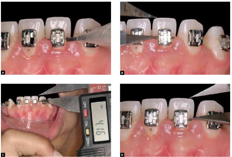
- Measuring the distance from the papilla to the incisal edge: With the use of a graphite pencil, corre-sponding marks were made at the level of the inter-dental papilla on the right (RGP) and left side (LGP) on the buccal surface of the tooth (Fig 1A). Using a digital caliper graduated from 0 to 150 mm, the mea-surement of the distance from the incisal edge to the marking previously made was performed (Fig 1 B, C and D). All data were recorded on a clinical form.
- Laser surgery: Surgical procedures were
per-formed with CO2 laser (Sharplan 20C, Tel Aviv, Israel),
wavelength of λ 10,600 nm, 5W average power, focus
of 2 mm and the beam was focused/unfocused, direct current. The surgeries were performed at FOUFBA Laser Center, by a single professional, with academic degree in Periodontics and Laser.
After having received intraoral anesthetic with 2% lidocaine containing Epinephrine 1: 100,000, the patients had the hyperplastic lesions removed with
incision and/or vaporization with the CO2 laser. The
tissue removal was followed by surgical gingival con-tour. No sutures or surgical cement were used. After surgery, in case of pain, an analgesic that the patient already had the habit of taking was prescribed and they should follow a hygiene protocol for 15 days. This protocol consisted on brushing the region with a Colgate Professional Ultra Soft gum® brush, along with the Colgate Total® toothpaste, three times a day and flossing, twice a day. As for food, patients were instructed to make use of cold food or ice cold only during the day of the surgery. The measurement of the distance of the papilla to the incisal edge oc-curred four times: Prior to surgery – Time 0, immedi-ately after surgery – Time 1, after one month – Time 2 (Fig 2) and two months after the surgical procedure – Time 3. Immediately after measuring, the
orth-odontic archwires in use werereseated.
All data were tabulated and subjected to statisti-cal analysis. Student t tests, ANOVA and Tukey were used to assess the difference between the groups, with a significance level of 5%.
RESULTS
A
C
B
D
where normal distribution was identified for all variables and therefore the mean and standard de-viation were the estimates used in testing statistics. In Figure 3, the variation in the distance from the papilla to the incisal edge of the right side is seen in relation to time.
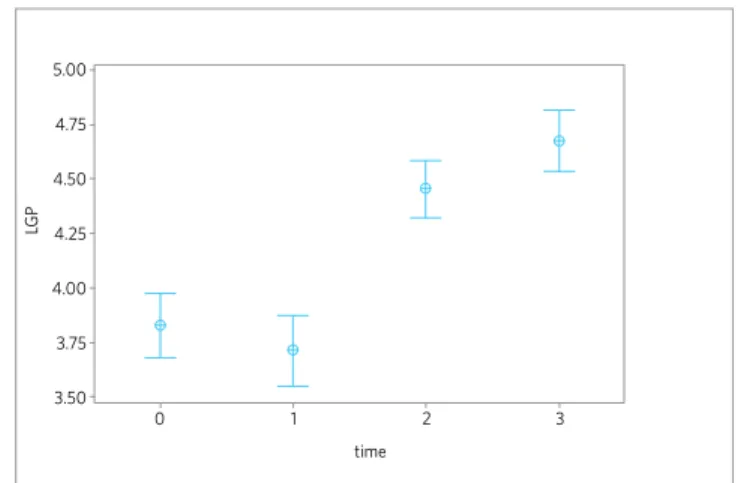
The distance from the papilla to the incisal edge of the left side in relation to time is shown in Figure 4.
DISCUSSION
The laser treatment provides the lengthening of
the clinical crowns of teeth.14 This work confirms
this statement, as it can be observed that in Figures 3 and 4, from Time 0 (pre-surgical) to Time 2 (1 month later), there was a statistically significant increase (p<0.001) in the distance from the papilla to the in-cisal edge on both sides, right and left. This result remained throughout Times 2 and 3, presenting no
Figure 1 - A) Marking the height of the papilla before surgery. B, C and D) Measuring the distance from the papilla to the incisal edge.
Figure 2 - One month after the surgical procedure.
5.00 5.00
RG
P
LG
P
time time
4.75 4.75
4.50 4.50
3.50 3.50
0 1 2 3 0 1 2 3
4.25 4.25
4.00 4.00
3.75 3.75
Figure 3 - Mean and confidence interval at 95% of the distance from the papilla to the incisal edge of the right side (RGP) in relation to time.
Figure 4 - Mean and confidence interval at 95% of the distance from the papilla to the incisal edge of the left (LGP) in relation to time.
statistically significant differences (p<0.001) be-tween them. With this finding, it can be inferred that there was no tissue contraction after the laser
gingivectomy, agreeing with the literature.6,3 The
measures did not show statistically significant dif-ferences between Time 0 and Time 1, on both sides. This probably occurred due to the difficulty on deter-mining the apex of the papilla right after the surgical procedure. There was no need of using the sutures or surgical cement, agreeing with the literature that as-serts that the surgical wound is left to heal by second intention, without the use of sutures or surgical
ce-ment.9,12 After the surgical procedure with the CO2
laser, there is pain reduction.7,9,15 In this research,
despite the recommendation made to patients for, in the case of pain, making use of analgesic medication, all of them said that they did not use this medicine and showed that the procedure is not uncomfortable.
As for food, they were instructed to make use of cold food or ice cold, only on the day of surgery. Pa-tients only complained of the odor exhaled during the surgical act, but all of them said that would ac-cept the procedure again, if necessary. Initially, the indication of the use of chlorhexidine gluconate
0.12% for 30 seconds once a day was suggested, as an adjunct of the mechanical cleaning, but this step was suspended, because immediately after surgery, pa-tients had very sensitive gums and two days after, the region had already healed. The patients responded well to the guideline regarding hygiene.
It was possible to work in a more controlled way because of the non-occurrence of bleeding during the surgical act, agreeing with the literature that
asserts that the CO2 laseracts on small vessels
pro-moting coagulation.15 The laser turns an infected
wound in a sterile wound.9 In this work, no cases of
infection were observed.
CONCLUSION
The CO2 laser presented itself effective for the
1. Araújo TM, Machado AW, Nascimento MH, Machado JW. Ortodontia e Dentística na recuperação da estética do sorriso: relato de um caso clínico. Rev Clín Ortod Dental Press. 2005;4(5):64-71.
2. Clocheret K, Dekeyser C, Carels C, Willems G. Idiopathic gingival hyperplasia and orthodontic treatment: a case report. J Orthod. 2003;30(1):13-9.
3. Freitas AC, Pinheiro ALB, Oliveira MG, Ramalho LMP. Assessment of the behavior of myofibroblasts on scalpel and CO2 laser wounds: an immunohistochemical study in rats. J Clin Laser Med Surg. 2002;20(4):221-5.
4. Hoed-Petersen B. The potential use of CO2 laser gingivectomy for phenytoin-induced gingival hyperplasia in mentally retarded patients. J Clin Periodontol. 1993;20(10):729-31.
5. Jarjoura K. Soft tissue lasers. Am J Orthod Dentofacial Orthop. 2005;127(5):527-8. 6. Luomanem M. Processo de cicatrização nas cirurgias com laser. In: Brugnera A Jr,
Pinheiro ALB. Lasers na Odontologia moderna. 1a ed. São Paulo: Pancast; 1998. p. 222-31.
7. Mason C, Hopper C. The use of CO2 laser in the treatment of fibromatosis: a case report. Int J Paediatr Dent. 1994;4(2):105-9.
8. Neves LS,Silva CMS, Henriques JFC, Cançado RH, Henriques RP, Janson G. Utilização do laser na Odontologia. Rev Dental Press Ortod Ortop Facial. 2005;10(5):149-56.
9. Pinheiro ALB. Evolução histórica e classificação dos lasers. In: Brugnera A Jr, Pinheiro ALB. Lasers na Odontologia moderna. 1a ed. São Paulo: Pancast; 1998. p. 17-26.
10. Pinheiro ALB. Física dos lasers. In: Brugnera A Jr, Pinheiro ALB. Lasers na Odontologia Moderna. 1a ed. São Paulo: Pancast; 1998. p. 28-44.
11. Pinheiro ALB, Frame JW. Tratamento cirúrgico de lesões pré- malignas e malignas da cavidade oral. In: Brugnera A Jr, Pinheiro AL. Lasers na Odontologia moderna. 1a ed. São Paulo: Pancast; 1998. p. 196-206.
REFERENCES
12. Pinheiro ALB, Frame JW. Tratamento cirúrgico de patologias de tecidos moles do complexo maxilofacial. In: Brugnera A Jr, Pinheiro ALB. Lasers na Odontologia Moderna. 1a ed. São Paulo: Pancast; 1998. p. 177-92.
13. Sarver D. Principles of cosmetic dentistry in orthodontics: part 1. Shape and proporcionality of anterior teeth. Am J Orthod Dentofacial Orthop. 2004;126(6):749-53.
14. Sarver D, Yanosky M. Principles of cosmetic dentistry in orthodontics: part 2. Soft tissue laser technology and cosmetic gingival contouring. Am J Orthod Dentofacial Orthop. 2005;127(1):85-90.
15. Túner J, Hode L. Laser therapy: clinical practice and scientific background. Sweden: Prima Books; 2002. 571p.
16. Walsh LJ. The current status of laser applications in dentistry. Aust Dent J. 2003;48(3):146-55.
17. Wang X, Ishizaki NT, Matsumoto K. Healing process of skin after CO2 laser ablation at low irradiance: a comparison of continuous-wave and pulsed mode. Photomed Laser Surg. 2005;23(1):20-6.
18. Zachrisson BU. Ortodontia e Periodontia. In: Lindhe J. Tratado de Periodontia clínica e Implantodontia oral. 3ª ed. Rio de Janeiro: Guanabara Koogan; 1999. p. 537-80.
19. Zaffe D, Vitale MC, Martignone A, Scarpelli F, Botticelli AR. Morphological, histochemical and immunocytochemical study of CO2 and Er:YAG laser effect on oral soft tissues. Photomed Laser Surg. 2004;22(3):185-9.