R E S E A R C H
Open Access
Vasoconstrictor effect of Africanized honeybee
(Apis mellifera L.) venom on rat aorta
Paulo César P Sousa
1, Teresinha S Brito
1, Daniel S Freire
1, Rafael M Ximenes
1,5, Pedro Jorge C Magalhães
1,
Helena SA Monteiro
1, Renata S Alves
2, Alice Maria C Martins
2, Daniela O Toyama
3and Marcos H Toyama
4*Abstract
Background:Apis melliferastings are a problem for public health worldwide, particularly in Latin America due to the aggressiveness of its Africanized honeybees. Massive poisoning byA. melliferavenom (AmV) affects mainly the cardiovascular system, and several works have described its actions on heart muscle. Nevertheless, no work on the pharmacological action mechanisms of the AmV in isolated aorta has been reported. Thus, the present work aimed to investigate the actions of AmV and its main fractions, phospholipase A2(PLA2) and melittin, on isolated aorta rings and a probable action mechanism.
Results:AmV and the complex PLA2+ melittin (0.1-50μg/mL) caused contraction in endothelium-containing aorta rings, but neither isolated PLA2nor melittin were able to reproduce the effect. Endothelium removal did not change the maximum vasoconstrictor effect elicited by AmV. Ca2+-free medium, as well as treatment with phentolamine (5μM), verapamil (10μM), losartan (100μM), and U-73122 (10μM, a phospholipase C inhibitor), eliminated the AmV-induced contractile effects.
Conclusions:In conclusion, AmV caused contractile effect in aorta rings probably through the involvement of voltage-operated calcium channels, AT1 andα-adrenergic receptors via the downstream activation of
phospholipase C. The protein complex, PLA2+ melittin, was also able to induce vasoconstriction, whereas the isolated proteins were not.
Keywords:Apis mellifera, Venom, Mellitin, Phospholipase A2, Aorta
Background
Africanized honeybees are a poly-hybrid species found in South America, mainly in Brazil, known for their aggressiveness and involvement in mass attacks [1,2]. The clinical manifestations of bee envenomation are characterized by local and systemic toxicity. Among the systemic manifestations observed after bee stings are cardiovascular changes and renal damage. Despite the significant advances made in understanding the pathogenesis of nephrotoxic and ischemic acute renal failure caused by such envenomation, there have been few changes in terms of mortality [3].
A. mellifera venom (AmV) is a complex mixture of enzymes, peptides and amines. Among the components of the venom are biogenic amines, hyaluronidases,
phospholipases A2(PLA2) and melittin, which is its main
constituent, approximately 50% of the dry weight [4,5]. This active 26-amino-acid peptide is released from its precursor, promelittin, during biosynthesis and presents hemolytic activity. It also is the primary allergen found in bee venom [6]. It acts synergistically with phospholipase A2(PLA2) to enhance its toxic actions [7].
Despite published in vivocharacterization of the cardio-vascular effects of both melittin and PLA2, fewin vitro
ex-periments that clarify the action mode of these toxins are available in the scientific literature [8]. Some papers report the role of endothelium, lipoxygenase products, and nitric oxide (NO) in the vascular effects of melittin [9-11]. The present study aimed to investigate the vasoconstrictor effect of crudeA. melliferavenom (AmV) and its fractions PLA2,
melittin, and the native linked fraction (PLA2+ melittin),
on isolated aortic rings of rats.
* Correspondence:[email protected]
4Paulista Coast Experimental Campus, São Paulo State University
(UNESP–Univ Estadual Paulista), São Vicente, São Paulo State, Brazil
Full list of author information is available at the end of the article
Methods
Venom and reagents
A. mellifera venom was purchased from Bio-Agents Serpentarium (Brazil). All chemicals and solvents were of analytical grade and purchased from certified suppliers (Bayer, Sigma-Aldrich, Labtest Diagnostic and Applied Biosystems).
Animals
Male Wistar rats (250–300 g) were maintained under standard conditions of temperature and humidity in 12-hour light–dark cycles. The animals had fasted for 24 hours before any experimental procedure, and water was providedad libitum. All experiments were in accord-ance with the guidelines for the ethical use of experimental animals published by the Brazilian College on Experimental Animal Care (COBEA), with a project license approved by the Animal Ethics Committee of Federal University of Ceará (protocol number 01/2012).
Purification of PLA2and melittin fromApis melliferavenom The bee venom PLA2 was purified in two
chromato-graphic steps. Briefly, AmV was dissolved (10 mg/mL) in NaCl 0.9% and kept at 14°C under constant stirring. The protein content of the venom was precipitated with pure acetone for 24 hours, centrifuged (4500 xg) and lyophilized to obtain the protein fraction, which was used for the frac-tionation of PLA2. In the first step, the protein fraction was
subjected to a molecular exclusion chromatography, using a size exclusion column (1 x 60 cm) previously packed with Superdex®75 (GE Healthcare Life Sciences, USA). The column was attached to semi-preparative HPLC and equilibrated with a 0.2M ammonium bicarbonate buffer (pH 7.8). The protein fraction (15 mg) was dissolved in 250μL of the buffer, homogenized, and centrifuged at
4500 x g for five minutes. The supernatant was filtered through a 0.45μm filter and then injected into the column.
Fractionation of the venom was performed at a constant flow rate of 0.3 mL/minute and monitored at 280 nm. Each fraction was collected for three minutes (0.9 mL) and tested for phospholipase A2enzymatic activity.
After confirming the PLA2 enzymatic activity, the
fraction was subjected to a new chromatographic step on reversed-phase HPLC using a C18 semi-analytical column, which was equilibrated with buffer A (0.1% TFA), to isolate the PLA2and melittin. Samples of the fraction with PLA2
activity (3 mg) were dissolved in 250 mL of buffer A, clari-fied and injected into the chromatographic column. The elution was performed with a continuous linear gradient of buffer B (66% acetonitrile in 0.1% TFA) and the monitoring was done at 280nm. The samples from the first chromato-graphic step were analyzed by SDS-PAGE, using a running gel of 12% acrylamide and a stacking gel of 5% acrylamide, with bisacrylamide (0.32%) as cross-linker.
Mass spectrometry
The molecular masses of the PLA2and melittin were
de-termined by matrix-assisted laser desorption/ionization time-of-flight (MALDI-TOF) mass spectrometry using a Voyager-DE PROMALDI-TOF mass spectrometer (Applied Biosystems®, Life Technologies™, USA). One microliter of the sample dissolved in 0.1% TFA was mixed with 2μL of the matrix (α-cyano-4-hydroxycinnamic acid).
The matrix was prepared with 30% acetonitrile and 0.1% TFA (v/v). Ion masses were determined at an acceleration voltage of 25 kV, the laser operating at 2890μJ/com2, with
a 300 ns delay and a linear analysis mode.
Aortic ring assay
The rats were sacrificed by stunning followed by cervical dislocation. The thoracic aorta were removed and immersed in a perfusion medium (Krebs-Henseleit solution) at room temperature. After removing adhering fat and connective tissue, each aorta was cut transversally into cylindrical ring-like segments (1 x 5 mm) attached to steel wire triangular pieces (0.3 mm diameter), which were suspended in a 5 mL organ bath containing Krebs-Henseleit solution (pH 7.4) with the following composition (mM): NaCl 118.0, KCl 4.7, MgSO4 1.2, CaCl2 2.5, KH2PO4 1.2, NaHCO3 25.0 and glucose 11.1, continuously aerated at 37°C with 95% O2and 5% CO2.
In some aortic tissues, endothelium was removed im-mediately after dissection by gentle rubbing of the aortic lumen with a stainless steel wire. Endothelium-containing or denuded strips were stretched with a passive tension of 1 g while the tension was recorded using an isometric force transducer (ML870B60/C-V, AD Instruments, Australia) connected to a digital data acquisition system (PowerLab™ 8/30, AD Instruments). After an equilibration period of at least 60 minutes, control contractions were induced by adding a submaximal concentration (60 mM) of potassium chloride (KCl) to the bath. When two successive control contractions showed similar amplitudes, preparations were considered equilibrated. Contraction data were expressed as percentages of the potassium-induced contraction. Endothelium-containing or -denuded preparations were contracted at the beginning of the experiment with potas-sium (30 mM) and after establishment of a contractile plateau, they were challenged with 1μM of acetylcholine.
The lack of acetylcholine-induced vasorelaxant effects was interpreted as evidence that the preparation was effectively denuded of endothelium [3].
Effects of theA. melliferavenom (AmV) in isolated rat aorta
Concentration-effect curves of AmV were obtained by exposing a preparation in basal tonus to cumulatively increasing concentrations of AmV (0.1 to 50μg/mL) added
determined in preparations without functional endothelium. In order to investigate the vasoconstrictor effect of AmV, concentration-effect curves were constructed by exposing the preparations to cumulatively increasing concentrations of AmV in basal tonus in preparations maintained in a Ca2+-containing medium, in a Ca2+-free medium (containing 2 x 10⁻⁵M EGTA), and in the pres-ence of verapamil (10μM), a well-known L-type
voltage-operated Ca2+ channel (VOCC) blocker, phentolamine (5μM), anα-adrenergic blocker, losartan (100μM), an
angiotensin II AT1 receptor antagonist, and U73122 (10μM), a phospholipase C inhibitor.
Effects of the AmV fractions, PLA2and melittin,
on isolated rat aorta
In order to investigate whether the fractions PLA2 and
melittin were involved in the vasoconstrictor effects of the AmV, concentration-effect curves (0.1 to 50μg/mL) were
constructed by exposing the preparations to cumulatively increasing concentrations of the fractions, PLA2, melittin
and the complex PLA2+ melittin (1:1, w:w).
Statistical analysis
Data were reported as means ± SEM. The means were evaluated by ANOVA, followed by the Holm-Sidakpost hoctest. Emax values were compared using the unpaired Student’s t test. The acceptance level for statistically significant differences was set at 5% (p < 0.05).
Results
Fractionation ofA. melliferacrude venom
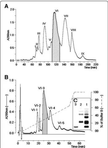
A. mellifera crude venom fractionation through size exclusion chromatography showed the presence of nine fractions, designated sequentially I to IX, as shown in Figure 1A. Only the fraction VI showed phospholipasic A2enzymatic activity. The SDS-PAGE analysis of fraction
VI revealed the presence of two major fractions as well as a minor electrophoretic band (results not shown). This fraction was subjected to a reverse phase HPLC. The chromatographic profile of the purification of the fraction VI showed five peaks, which were denominated se-quentially VI-1 to VI-5, where the most important fractions in terms of chromatogram area were VI-3 and VI-4 (Figure 1B). The analysis on SDS-PAGE of these fractions showed that VI-3 had a molecular mass of approximately 15 kDa (Figure 1C). Fraction VI-4 was not detected using a 10% acrylamide gel. This result suggested that fraction VI-4 must have a molecular mass lower than 10 kDa. PLA2enzymatic activity was found only in fraction
VI-3, whereas fraction VI-4 reduced the PLA2of fraction
VI-3. Finally, a MALDI-TOF analysis, to confirm the molecular homogeneity of PLA2and melittin, revealed
respective molecular masses of 15,343.31 Da and 3,101.03 Da for the PLA2and melittin.
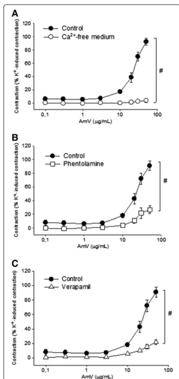
Effect ofA. melliferavenom and its fractions in isolated aorta
In endothelium-containing aorta preparations, AmV (0.1-50 μg/mL) induced contractile responses in a
concentration-dependent manner. The magnitude of the contraction induced by 50μg/mL of AmV corresponded
to 92.6 ± 5.5% (n = 5 rings) of a reference contraction induced by 60 mM K+. In the same preparation, the isolated PLA2or melittin (0.1-50μg/mL) did not induce
significant contractile responses. However, the complex PLA2+ melittin (0.1-50μg/mL) induced a vasoconstrictor
effect in a concentration-dependent manner, (n = 4 rings; p < 0.05; Figure 2A). The magnitude of the contractile effect at 50μg/mL was 66.1 ± 11.9% of the contraction
induced by K+ (60 mM). Endothelium removal did not change the maximum vasoconstrictor effect elicited by AmV (Figure 2B).
Figure 1Fractionation of AmV. (A)Chromatography of whole protein extract from honeybee venom using a molecular exclusion column packed with Superdex®75. The chromatographic run was carried out at a flow rate of 0.6 mL/h and monitored at 280 nm. (B) Reverse phase HPLC run yielded five main fractions designated from VI-1 to VI-5. Fraction VI-3 was confirmed as PLA2 by specific phospholipase
A2 assay and fraction VI-4 as melittin after MALDI-TOFF
Elucidation of the mode of action ofA. melliferavenom in isolated aorta
In aortic rings maintained in the Ca2+-free medium, the contractile effects induced by AmV were abolished and the magnitude of the maximal contraction was 3.7 ± 3.0% of the K+ (60 mM) value (n = 5; p < 0.05; Figure 3A), a value significantly lower than that observed in Ca2+-containing medium. Pretreatment of aortic rings with phentolamine (5 μM) also significantly reduced
AmV -induced contraction (50μg/mL) to 26.8 ± 5.6% of
the K+(60 mM) value (n = 5 rings; p < 0.05; Figure 3B). In order to evaluate the effect of a voltage-gated calcium channel blocker on AmV-induced contraction, prepara-tions were treated with verapamil (10μM) after which the
contractions elicited by AmV (50μg/mL) significantly
diminished to 21.7 ± 3.3% of the K+(60 mM) value (n = 4 rings; p < 0.05; Figure 3C).
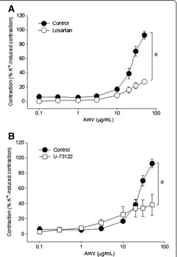
Aiming to verify whether angiotensin was involved in the contractions induced by AmV, an AT1 receptor antagonist Figure 2Characterization of the vasoconstrictor effect of AmV.
(A)Vasoconstrictor effect of AmV (0.1-50μg/mL) (●, n = 5) and the fractions: melittin (■, n = 5); PLA2(Ο, n = 5); and the complex
PLA2+ melittin (□, n = 5).(B)Effect of endothelium removal on
AmV vasoconstrictor effect. The concentration response curve of AmV (0.1-50μg/mL) on basal tone in endothelium-containing aorta preparations (●, n = 5), and in endothelium-denuded aorta preparations (Ο, n = 4). Vasoconstrictor effects are expressed as a percentage of the contractile response to K+(60 mM). Data are expressed as mean ± SEM and analyzed by ANOVA followed by the Holm-Sidakpost hoctest.
was employed. The preparations were treated with losartan (100 μM), and the contractions elicited by AmV were
decreased to 27.3 ± 2.4% of the K+ (60 mM) value (n = 4 rings; p < 0.05; Figure 4A). Moreover, pretreatment of aortic rings with U-73122 (10μM), a phospholipase C
inhibitor, significantly reduced AmV-induced contraction (50μg/mL) to 38.4 ± 5.6% of the K+(60mM) value,
re-spectively (n = 5 rings; p < 0.05; Figure 4B).
Discussion
The present work describes the isolation of three fractions from the venom ofA. mellifera, a mixed fraction of PLA2
and melittin, and both in pure protein form PLA2 and
melittin. These proteins together represent approximately 60% of the venom dry weight [12,13]. A vasoconstrictor effect of AmV and the complex PLA2 + melittin was
confirmed. However, the isolated PLA2and melittin had
no contractile effect on the aortic rings. Some authors reported that melittin facilitates the interaction of PLA2
with the cell membrane, exposing the membrane to the catalytic site of the enzyme due to its amphipathic characteristics [3]. In order to investigate the action mode ofA. mellifera-induced vasoconstriction, the involve-ments of extracellular Ca2+, voltage-gated calcium channels,
α2-adrenergic receptors, AT1 receptors and phospholipase
C were assayed.
In Ca2+-free medium, an inhibition of the contractile response of AmV was observed. Likewise, employing verapamil, a calcium channel blocker, produced an absence of the AmV-induced contractile response. We suggested that the vasoconstrictor properties are dependent on a trans-membrane Ca2+influx. Thus, the opening of voltage-operated calcium channels is involved in the mechanism proposed for the contractile effect of AmV.
In most cases, a smooth muscle contraction involves a combination of input and Ca2+release. The various types of smooth muscle may differ markedly in this regard: in the relative contribution of the two sources of Ca2+ for contraction (intracellular and extracellular) and in ion channels through which extracellular Ca2+can have access to the interior of the cell [14]. According to Jackson [15], the contraction of vascular smooth muscle occurs primarily by the influx of Ca2+ into the muscle cell. If the extracellular concentration of Ca2+ is reduced, the contraction is almost eliminated.
Adrenergic receptors consist of typical G-protein-coupled receptors. Each of these receptor classes, α1- and α2
-adrenergic receptors, is associated with a specific system of secondary messengers. The α1-adrenergic receptors
are coupled to phospholipase C and produce their effects mainly through the release of intracellular Ca2+stores, mediating vasoconstriction [16].
In an attempt to elucidate the pathway involved in the contraction of the aorta by AmV, anα-adrenergic
recep-tor blocker (phentolamine), an AT1 receprecep-tor antagonist (losartan), and a phospholipase C inhibitor (U-73122) were used. A significant reduction in the contractile effect of AmV on the aortic rings was observed, suggesting the involvement of these receptors in tissue contraction via phospholipase C.
This hypothesis is corroborated by Vinhoteet al. [17], who reported such contraction in aortic rings, induced byPolybia paulistawasp venom, showing that the effect was dependent on voltage-operated calcium channels, and thatα-adrenergic receptors were involved.
Conclusions
In conclusion, Apis mellifera venom causes a contractile effect on aorta rings probably through the involvement of voltage-operated calcium channels, AT1 and α-adrenergic
receptors via the downstream activation of phospholipase C. In contrast to the isolated proteins, the protein complex PLA2+ melittin was also able to induce vasoconstriction.
Figure 4Study on the probable action mode of AmV. Vasoconstrictor effects of AmV:(A)pretreatment with losartan (100μM;Ο, n = 5);(B)pretreatment with U-73122 (10μM;□, n = 5). Vasoconstrictor effects are expressed as a percentage of the contractile response to K+(60 mM). Data are expressed as mean ± SEM and
Ethics committee approval
The present study was approved by the Animal Ethics Committee of Federal University of Ceará (protocol number 01/2012). Moreover, all experiments were in accordance with the guidelines for the ethical use of experimental animals published by the Brazilian College on Experimental Animal Care (COBEA).
Competing interests
The authors declare that there are no competing interests.
Authors’contributions
PCPS, TSB, DSF, RSA, DOT and performed the biochemical and
pharmacological experiments. PJCM, HSAM, AMCM, MHT designed the study and discussed the results. RMX, AMCM and MHT drafted the manuscript. All authors read and approved the manuscript.
Acknowledgments
The authors would like to thank the National Council for Scientific and Technological Development (CNPq), the Coordination for the Improvement of Higher Education Personnel (CAPES) and the Cearense Foundation for Research Support (FUNCAP) for their financial support and Sidney Ann Pratt for language revision.
Author details
1Department of Physiology and Pharmacology, Federal University of Ceará,
Fortaleza, Ceará State, Brazil.2Department of Clinical and Toxicological Analyses, Federal University of Ceará, Fortaleza, Ceará State, Brazil.3Center of
Biological and Health Sciences, Mackenzie Presbyterian University, São Paulo, São Paulo State, Brazil.4Paulista Coast Experimental Campus, São Paulo State
University (UNESP–Univ Estadual Paulista), São Vicente, São Paulo State,
Brazil.5Department of Antibiotics, Federal University of Pernambuco, Recife,
Pernambuco State, Brazil.
Received: 6 April 2013 Accepted: 3 September 2013 Published: 25 September 2013
References
1. Kerr WE:The history of introduction of African bees to Brazil.South African Bee J1967,39(2):3–5.
2. Brizola-Bonacina AK, Alves VV, de Moraes M:Relation between the size of the acid gland and the quantity of venom produced in africanized bee,
Apis melliferaL. (Hymenoptera: Apidae), in the region of Dourados, MS, Brazil.Neotrop Entomol2006,35(2):210–214.
3. Furchgott RF, Zawadzki JV:The obligatory role of endothelial cells in the relaxation of arterial smooth muscle by acetylcholine.Nature1980,
288(5789):373–376.
4. De Melo MH, da Silva EA, Natal D:Africanized bees in a metropolitan area of Brazil: shelters and climatic influences.Rev Saúde Públ2003,37(2):237–241.
5. Ferreira Junior RS, Sciani JM, Marques-Porto R, Junior AL, Orsi R de O, Barraviera B, Pimenta DC:Africanized honey bee (Apis mellifera) venom profiling: Seasonal variation of melittin and phospholipase A(2) levels.
Toxicon2010,56(3):355–362.
6. de Graaf DC, Aerts M, Danneels E, Devreese B:Bee, wasp and ant venomics pave the way for a component-resolved diagnosis of sting allergy.J Proteomics2009,72(2):145–154.
7. Kreil G, Bachmayer H:Biosynthesis of melittin, a toxic peptide from bee venom. Detection of a possible precursor.Eur J Biochem1971,20(3):344–350.
8. Schumacher MJ, Egen NB:Significance of Africanized bees for public health. A review.Arch Intern Med1995,155(19):2038–2043.
9. Cerne K, Kristan KC, Budihna MV, Stanovnik L:Mechanisms of changes in coronary arterial tone induced by bee venom toxins.Toxicon2010,
56(3):305–312.
10. Forstermann U, Neufang B:Endothelium-dependent vasodilation by melittin: are lipoxygenase products involved?Am J Physiol1985,249(1 Pt 2):H14–19.
11. Rapoport RM, Ashraf M, Murad F:Effects of melittin on endothelium-dependent relaxation and cyclic GMP levels in rat aorta.Circ Res1989,64(3):463–473.
12. Chaud-Netto J, da Silva GP, Brochetto-Braga MR, Palma MS, Rodrigues A, Carmona EC:Influence of the collection methodology on theApis
melliferavenom composition: peptide analysis.Sociobiology2006,
47(3):761–770.
13. Schmidt JO:Toxinology of venoms from the honeybee genusApis.
Toxicon1995,33(7):917–927.
14. Yonamine CM, Costa H, Silva JAA, Muramoto E, Rogero JR, Troncone LRP, Camillo MAP:Biodistribution studies of bee venom and spider toxin using radiotracers.J Venom Anim Toxins incl Trop Dis2005,11(1):39–50.
15. Jackson WF:Ion channels and vascular tone.Hypertension2000,
35(1 Pt 2):173–178.
16. Webb RC:Smooth muscle contraction and relaxation.Adv Physiol Educ 2003,27(4):201–206.
17. Vinhote JFC, Torres AFC, Dantas RT, Praciano TP, Menezes RRPPB, Sousa DF, Brito TS, Lima FJB, Toyama MH, Magalhães PJ, Monteiro HSA, Martins-Nunes AMC:Renal and calcium-dependent vascular effects ofPolybia paulista
wasp venom.J Venom Anim Toxins incl Trop Dis2011,17(2):199–208.
doi:10.1186/1678-9199-19-24
Cite this article as:Sousaet al.:Vasoconstrictor effect of Africanized honeybee (Apis mellifera L.) venom on rat aorta.Journal of Venomous Animals and Toxins including Tropical Diseases201319:24.
Submit your next manuscript to BioMed Central and take full advantage of:
• Convenient online submission
• Thorough peer review
• No space constraints or color figure charges
• Immediate publication on acceptance
• Inclusion in PubMed, CAS, Scopus and Google Scholar
• Research which is freely available for redistribution