(
−
)-
α
-Bisabolol inhibits preferentially electromechanical coupling on
rat isolated arteries
R.J.B. de Siqueira
⁎
, H.V. Ribeiro-Filho, R.S. Freire, F. Cosker, W.B.S. Freire, A.A. Vasconcelos-Silva, M.A. Soares,
S. Lahlou, P.J.C. Magalhães
Department of Physiology and Pharmacology, School of Medicine, Federal University of Ceará, Fortaleza, CE, Brazil
a b s t r a c t
a r t i c l e
i n f o
Article history: Received 24 March 2014
Received in revised form 9 June 2014 Accepted 26 June 2014
Available online 14 August 2014
Keywords: (−)-α-Bisabolol
Docking analysis Electromechanical coupling Intracellular calcium Vascular smooth muscle
Previousfindings enable us to hypothesize that (−)-α-bisabolol acts as inhibitor of voltage-dependent Ca2+ channels in smooth muscle. The current study was aimed at consolidating such hypothesis through the recording of isometric tension, measurement of intracellular Ca2+as well as discovery of channel target using in silico analysis. In rat aortic rings, (−)-α-bisabolol (1–1000 µM) relaxed KCl- and phenylephrine-elicited contractions, but the IC50differed significantly (22.8 [17.6–27.7] and 200.7 [120.4–334.6] µM, respectively). The relaxation of phenylephrine contractions remained unaffected byL-NAME, indomethacin, 1H-[1,2,4]oxadiazolo[4,3-a] quinoxalin-1-one, tetraethylammonium, glibenclamide or KT-5720. Under Ca2+-free conditions, (−)-α-bisabolol did not alter the contractions evoked by phenylephrine or caffeine whereas it reduced those evoked by CaCl2 in KCl-, but not in PHE-stimulated preparations. Furthermore, it did not significantly alter the contrac-tions evoked by phorbol 12,13-dibutyrate or induced by the extracellular Ca2+restoration in cyclopiazonic acid-treated preparations. In mesenteric rings loaded with Fluo-4 AM, (−)-α-bisabolol blunted the tension and the cytosolic levels of Ca2+in response to K+but not to norepinephrine. Silico docking analysis of the Cavβ2a subunit of voltage-dependent Ca2+channel indicated putative docking sites for (−)-α-bisabolol. These
findings reinforce the ability of (−)-α-bisabolol to inhibit preferentially contractile responses evoked by Ca2+influx through voltage-dependent Ca2+channels.
© 2014 Elsevier Inc. All rights reserved.
1. Introduction
(−)-α-Bisabolol, also known as levomenol, is a sesquiterpenic monocyclic alcohol abundantly found as the major constituent of the es-sential oil of chamomile (Matricaria chamomillaL., Asteraceae). It has been attributed to this compound the healing attributes of chamomile, the feature that argues for the use of (−)-α-bisabolol in pharmaceutical formulations in virtue of its anti-inflammatory and antiallergic proper-ties[10]. In fact, it has been reported that (−)-α-bisabolol has protec-tive effects against gastric damage caused by acetylsalicylic acid[12, 26]. The antitumor activity of (−)-α-bisabolol, which is mediated by its proapoptotic effects, has also recently received special attention in the literature[2–4].
In the cardiovascular system, intravenous injections of increas-ing doses of (−)-α-bisabolol induced dose-dependent hypotension and bradycardia in awake rats[13]. However, the mechanism of ac-tion of these cardiovascular effects has not been addressed. The hy-potensive response to (−)-α-bisabolol could be mediated, at least partially, through an active vascular relaxation. We recently found that (−)-α-bisabolol induced endothelium-independent relaxation with a pharmacological potency that was higher in mesenteric than in aortic rings[5]. In only aortic rings, (−)-α-bisabolol relaxed KCl-induced contractions with pharmacological potency significantly higher than that observed in vessels contracted with phenyleph-rine. This data indicates that (−)-α-bisabolol counteracts contrac-tile responses that preferentially recruit voltage-gated Ca2 + channels, similar to what was reported for tracheal smooth muscle preparations[5].
Putative Ca2 +channel blocking activity as the underlying mech-anism for vasodilator action induced by (−)-α-bisabolol was previ-ously demonstrated by Vuorela et al.[28]. Such activity has been also reported for bisabolol oxide and (+)-T-cadinol, two analogues of (−)-α-bisabolol, in experiments with isolated papillary muscle from guinea-pig hearts [16] and binding assays with dihy-dropyridine on the voltage-operated Ca2 +channels[30]. However, direct demonstration that (−)-α-bisabolol is able to interfere with
–
⁎ Corresponding author at: Department of Physiology and Pharmacology, School of Medicine, Federal University of Ceará, R. Cel Nunes de Melo, 1127, Rodolfo Teófilo, 60430-270 Fortaleza, CE, Brazil. Tel.: +55 85 3366 8334; fax: +55 85 3366 8333.
E-mail address:[email protected](R.J.B. de Siqueira).
http://dx.doi.org/10.1016/j.vph.2014.06.006
1537-1891/© 2014 Elsevier Inc. All rights reserved.
Contents lists available atScienceDirect
Vascular Pharmacology
cytoplasmic Ca2 +dynamics was not demonstrated in smooth muscle cells hitherto.
Therefore, the present study was designed to test the hypothesis that pharmacological effects of (−)-α-bisabolol on vascular smooth muscle result from its ability to interfere with the intracellular levels of Ca2 +, especially on those events mediated electromechan-ically. Docking experiments and simultaneous measurement of me-chanical force and [Ca2 +]
iin Fluo-4 loaded mesenteric artery rings stimulated with either KCl or norepinephrine were performed to support the underlying mechanism involved in the vasorelaxant ef-fects of (−)-α-bisabolol.
2. Materials and methods
2.1. Animals
Male Wistar rats (250–300 g) were obtained from our local colonies (vivarium of the Department of Physiology and Pharmacology, Federal University of Ceará) and maintained under constant temperature (22 ± 2 °C) with a 12 h light/12 h dark cycle and free access to food and water. All animals were cared for in compliance with the Guide for the Care and Use of Laboratory Animals, published by the US Nation-al Institutes of HeNation-alth (NIH Publication 85-23, revised 1996). All proce-dures described herein were reviewed by and had prior approval from the local animal ethics committee (Protocol n° 48/09—CEPA).
2.2. Isolated ring-like aortic preparations
Rats were euthanized under sodium pentobarbital anesthesia and a segment of thoracic aorta was removed and immersed in perfusion me-dium at room temperature. After removing adhering fat and connective tissue, the aorta was cut transversally into cylindrical ring-like segments (1 × 5 mm) receiving careful transluminal insertion of steel wire trian-gular pieces (0.3 mm diameter) that allowed tissue suspension into 5-mL organ bath containing Krebs–Henseleit solution (continuously aer-ated at 37 °C with 5% CO2in O2). Endothelium-containing preparations were stretched with a passive tension of 1 g and tension was recorded using isometric force transducer (ML870B60/C-V, AD Instruments, Australia) connected to a data acquisition system (PowerLab™8/30, AD Instruments). After an equilibration period of 60 min, control con-tractions were induced by adding a submaximal concentration (60 mM) of KCl to the bath. When two successive contractions showed similar amplitude, preparations were considered in equilibrium. At the beginning of the experiment, each aortic or mesenteric ring was pre-contracted with phenylephrine (0.1μM) and thereafter challenged by acetylcholine (1μM) to evaluate the integrity of endothelium. Prepara-tions were considered to possess an intact endothelium when the vasorelaxant response to acetylcholine was 80% or greater. The concentration-effect curves were obtained by exposing the aortic prep-aration to cumulatively increasing concentrations of (−)-a-bisabolol (1–1000μmol/L), which was added to the bath and maintained at a given concentration for 10 min.
2.3. Simultaneous measurement of force and intracellular Ca2+in rings of
mesenteric artery
Vessel segments from the second branch of the superior mesenteric artery were dissected and maintained in oxygenated Krebs–Henseleit solution at room temperature. After removing adhering fat under a mi-croscope, cylindrical ring-like segments were obtained and carefully mounted in a confocal myograph chamber (DMT120CW Confocal Wire Myograph, Aarhus, Denmark) following the transluminal insertion of two tungsten wires (40μm diameter). Then, under constant temper-ature (37 °C), a resting tension of 11.8 kPa was applied to each isolated artery segment. The preparation was incubated by 50 min with thefl uo-rescent Ca2+indicator Fluo-4 AM (5
μM) supplemented with pluronic
acid (0.1% w/v) (Life Technologies, USA). After washing, the Ca2+ fl uo-rescence was registered with an inverted confocal microscope (Olym-pus, IX81) using a 20 × magnification and excitation/emission wavelengths of 488/505–515 nm. Sample rate was 1 frame/7 s and the intracellular Ca2+variations were expressed in relative variation of the initialfluorescence (F/F0). Simultaneously, the variations of the tension were recorded with an isometric force transducer connected to a data acquisition system (PowerLab™8/30, AD Instruments).
2.4. In silico docking analysis
We employed AutoDock 4.2[14]in docking experiments using three-dimensional molecular structure of the ligand (−)-α-bisabolol (constructed by the molecular editor Avogadro 1.1.0; [6]) in pharmacophoric alignments with macromolecules based on two crys-tallographic models of aβ-subunit isoform of voltage-gated L-type Ca2+channel, the conserved core Ca
vβ2aalone or in complex with the α-interaction domain (AID), namely AID-Cavβ2a[27]. The macromole-cules and ligand were prepared using the graphical interface AutoDockTools 1.5.6, which allowed the addition of polar hydrogens (Kollman) and partial charges (Gasteiger). For ligand, the number of ac-tive torsions was 5, and both macromolecules were considered rigid. Autogrid (part of the AutoDock package) allowed the construction of
af-finity gridfields, previously to the docking procedure. For all docking experiments, we used the genetic algorithm available in the software. In afirst step of docking experiments, we constructed a 0.59Å-spaced gridfield that covered the entire macromolecule of either Cavβ2aor AID-Cavβ2a. The numbers of energy evaluations and docking runs were 2,500,000 and 50, respectively, and other parameters were main-tained as default. According to the results of thefirst step, a second se-ries of simulations was performed by constructing two gridfields (with points spaced by 0.38Å), one centered on the Hook domain (res-idues 121–169) and the other centered near to the conserved hydro-phobic cleft namedα-binding pocket. Such region engages Cavβ2a with AID. The parameters were maintained as previously described. The cluster populations and binding energy as well as proximity of the residues were taken into account in the docking analysis.
2.5. Solutions and drugs
The perfusion medium used in isolated organ chamber was fresh modified Krebs–Henseleit solution (pH 7.4; in mM: 118.0 NaCl, 4.7 KCl, 1.18 KH2PO4, 1.18 MgSO4·7H2O, 2.50 CaCl2, 25.0 NaHCO3, and 11.1 glucose). Nominally Ca2 +-free solution was prepared by omit-ting CaCl2and adding ethylene glycol bis(2-aminoethyl ether)-N,N, N′N′-tetraacetic acid (EGTA). All drugs were of analytical grade puri-ty purchased from Sigma Co. (St. Louis, MO, USA). They were dis-solved directly in Krebs–Henseleit solution except nifedipine, cyclopiazonic acid, KT-5720 and 1H-[1,2,4]oxadiazolo[4,3-a] quinoxalin-1-one (ODQ) that were dissolved directly in DMSO (di-methyl sulfoxide). Solutions of (−)-α-bisabolol (bisabolol; Sigma-Aldrich) were prepared in Krebs–Henseleit solution containing Tween 80 (0.5%). Maximal percentage of Tween 80 in bath chamber was 0.06%. Substances were prepared as stock solutions and were brought to volume with Krebs–Henseleit solution in order to achieve a desired concentration in bath chamber.
2.6. Statistical analysis
Data are reported as mean ± SEM and n indicates the number of ex-periments. The IC50values (i.e., the (−)-α-bisabolol concentration in μM that relaxes a contraction by 50%) were calculated by interpolation from semi-logarithmic plots and are expressed as geometric mean [95% confidence interval]. The significance (pb0.05) of results was
U-test, and one- or two-way analysis of variance (ANOVA) followed by the Holm–Sidak post hoc test when appropriate.
3. Results
3.1. Vasodilator effects of (−)-α-bisabolol in aortic rings contracted with high KCl or phenylephrine
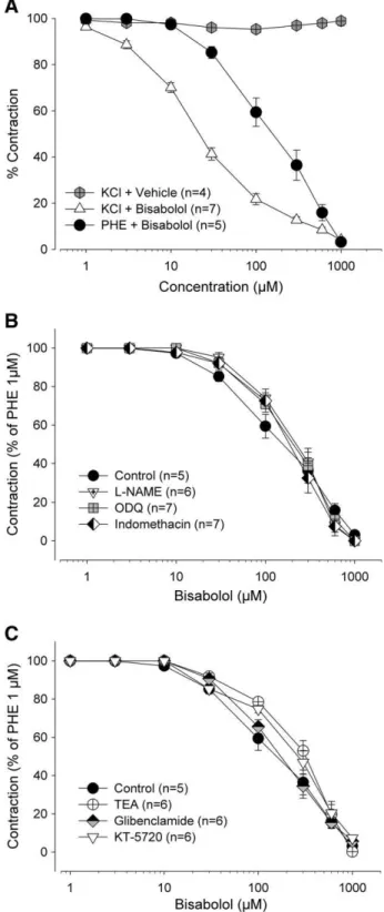
Aortic rings produced sustained contractions corresponding to 1.52 ± 0.15 (n = 6) and 1.30 ± 0.11 g (n = 7) in response to 1μM phenylephrine and 60 mM KCl (Fig. 1A). The cumulative addition of (−)-α-bisabolol (1–1000μM) fully relaxed the phenylephrine-and KCl-induced contractions with IC50values of 200.7 [120.4– 334.6] and 22.8 [17.6–27.7]μM, respectively. However, the potency of (−)-α-bisabolol in inducing relaxant effect was significantly (pb0.05, Mann–Whitney U-test) higher in preparations contracted by KCl. The cumulative addition of vehicle (Tween 80) does not exert significant effect on preparations precontracted with KCl 60 mM (Fig. 1A).
In a separate set of tissues contracted with phenylephrine (1μM), pretreatment withL-NAME (100μM), ODQ (10μM) or indomethacin (INDO, 10μM) (Fig. 1B) as well as with TEA (1 mM), glibenclamide (10μM) or KT-5720 (10μM) (Fig. 1C) did not change the vasodilator ef-fect of (−)-α-bisabolol as none of these pretreatments altered the IC50 value of (−)-α-bisabolol-induced vasorelaxation (Table 1; pN0.05, Mann–Whitney U-test).
3.2. Inhibitory effects of (−)-α-bisabolol on the contractions induced by
Ca2 + or Ba2 + in preparations maintained in the presence of KCl or
phenylephrine
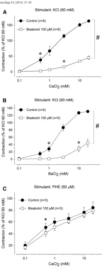
Considering that the phenylephrine-induced contraction was re-duced almost a half (59.4 ± 6.1%) by 100μM (−)-α-bisabolol, this concentration was used in all subsequent experiments. Under Ca2 +-free conditions (in the presence of 60 mM KCl and 10
μM EGTA), aortic rings contracted promptly in a concentration-dependent manner (pb0.001, ANOVA) when increasing
concentra-tions (0.1–20 mM) of either Ca2 +(Fig. 2A) or Ba2 +(Fig. 2B) were added in the extracellular medium (maximal effect of 163.2 ± 4.1 [n = 6] and 130.8 ± 3.4% [n = 5], respectively; values are % of the reference contraction induced by 60 mM KCl in Ca2 +-containing me-dium). The maximum contractile effect induced by Ca2 +or Ba2 +was significantly reduced (pb0.05, two-way ANOVA followed by Holm–
Sidak test) in the presence of (−)-α-bisabolol (100μM) to 45.2 ± 5.7 (Fig. 2A, n = 6) and 44.4 ± 10.3% (Fig. 2B, n = 5), respectively. Inter-estingly, when phenylephrine (60μM) was the agonist in Ca2+-free medium (containing 10μM EGTA and 1μM nifedipine), contractions due to Ca2 +(0.1
–20 mM) addition remained significantly (pN0.05,
two-way ANOVA) unaffected by 100μM (−)-α-bisabolol (Fig. 2C, n = 3).
3.3. (−)-α-Bisabolol does not interfere with the contractions mediated in-tracellularly by phenylephrine or caffeine in aortic rings under Ca2+-free
conditions
In aortic rings with intact endothelium under Ca2 +-free condi-tions (in the presence of 1 mM EGTA), phenylephrine (1μM) induced a transient (phasic) contraction that corresponded to 28.5 ± 3.4% (n = 8) of the K+-induced response under nominally normal Ca2 + (Fig. 3A). The previous addition of (−)-α-bisabolol (100μM, n = 6) did not change (pN0.05, paired Student's t-test) this phasic
con-traction (30.4 ± 4.6%). In other set of experiments with aortic prep-arations (with intact endothelium) maintained in Ca2 +-free medium at 25 °C, (−)-α-bisabolol (100μM) was also ineffective (pN0.05, paired Student's t-test) when caffeine (30 mM) was employed as the contractile agent. In fact, the transient contraction induced by
caffeine corresponded to 15.9 ± 2.0 and 23.4 ± 3.7% of the reference contraction induced by 60 mM KCl in the absence (n = 8) and in the presence (n = 6) of (−)-α-bisabolol, respectively (Fig. 3B).
Fig. 1.Effects of (−)-α-bisabolol on phenylephrine- or KCl-induced sustained
con-tractions in rat aortic rings. Concentration–response curves were constructed by the addition of bisabolol (1–1000μM) or his vehicle (Tween 80; 1–1000μM) on the steady state of induced contractions (panel A). Bisabolol was cumulatively added after phenylephrine (1μM)-induced contractions in the absence (control) or in the presence ofL-NAME (100μM), indomethacin (10μM), or ODQ (10μM) (panel B) or in the presence of TEA (1 mM), glibenclamide (10μM) or KT-5720 (10μM) (panel C). The results are mean ± S.E.M. and the number of experiments is indicated as n.
3.4. (−)-α-Bisabolol does not interfere with the contractions induced by in-ternal Ca2+store depletion or by phorbol 12,13-dibutyrate in aortic rings under Ca2+-free conditions
In order to assess the effects of (−)-α-bisabolol on contractions induced by Ca2+in
flux after internal Ca2+store depletion, aortic rings were exposed to successive stimuli (3–4 times) induced by phenylephrine (10μM) in Ca2+-free medium containing nifedipine (1μM) and cyclopiazonic acid (10μM) until testifying no measurable contraction in response to phenylephrine. After phenylephrine removal from the extracellular medium, the extracellular Ca2+was restored to 2 mM still in the presence of nifedipine and cyclopiazonic acid. This pro-cedure allowed aortic rings to produce sustained contractions corre-sponding to 54.7 ± 13.0% (n = 5) of the reference contraction induced by KCl (60 mM). Pretreatment with (−)-α-bisabolol (100μM) did not change significantly (pN0.05, paired Student's
t-test) this contraction induced by the Ca2+restoration (45.1 ± 8.5%, n = 5). In a separate set of experiments, the addition of the protein ki-nase C (PKC) activator phorbol 12,13-dibutyrate (1μM) to aortic rings with intact endothelium and maintained in Ca2+-free medium (with 1 mM EGTA) produces a sustained contraction corresponding to 171.1 ± 18.5% (n = 6) of the reference contraction. In the same aortic preparation, the addiction of (−)-α-bisabolol (100μM, n = 6) did not significantly changed (pN0.05, paired Student's t-test) the sustained
contraction (172.0 ± 19.6% of the reference contraction).
3.5. Inhibitory effects of (−)-α-bisabolol on K+-induced contractions occur
simultaneously to a decrease in cytoplasmic Ca2+levels in mesenteric rings
In Fluo-4 AM-loaded mesenteric vessels stimulated by KCl (mean amplitude of 3.50 ± 0.64 mN; n = 4), (−)-α-bisabolol (100μM, n = 4) significantly reduced thefluorescence ratio (F/F0) related to the cytoplasmic Ca2+to 50.7 ± 5.5% of the
fluorescence observed in control preparations (Fig. 4A, B, C), whereas it significantly reduced the contrac-tile effect of K+to 0.68 ± 0.10 mN (Fig. 4D) (p
b0.05, paired Student's
t-test). In contrast, such reduction induced by (−)-α-bisabolol was not seen when mesenteric vessels were stimulated by norepinephrine (60μM, n = 4). As one can see inFig. 4(panels E to H), neither the contractile force (Fig. 4H) nor the cytoplasmicfluorescence (Fig. 4G) was significantly changed by the pretreatment with 100μM (−)-α -bisabolol (pN0.05, paired Student's t-test, n = 4).
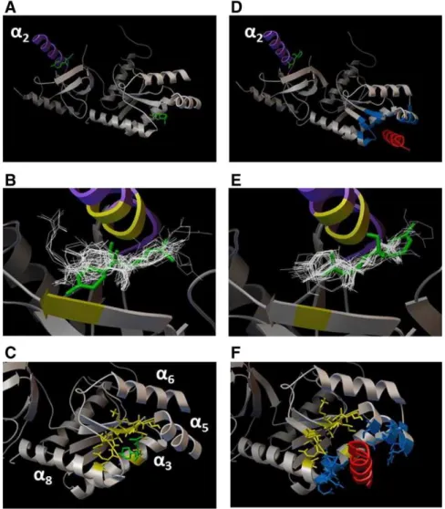
3.6. In silico analysis of the molecular interaction between (−)-α-bisabolol and Ca2+channels
Thefirst step of docking experiments indicated two preferential re-gions in the surface of Cavβ2asubunit (Fig. 5A–C) for (−)-α-bisabolol interactions. Thefirst one is located near theα2-helix in the HOOK
Table 1
IC50values for the relaxing effects induced by (−)-α-bisabolol in aortic rings (pretreated
or not withL-NAME, indomethacin, ODQ, TEA, glibenclamide or KT-5720) that were pre-contracted with 1μM phenylephrine or 60 mM KCl. Values are geometric mean [95% confidence interval].
Treatment IC50(μM)
Phenylephrine 200.7 [120.4–334.6]
+L-NAME (100μM) 213.5 [138.6–329.1] + Indomethacin (10μM) 190.1 [130.4–277.1]
+ ODQ (10μM) 216.9 [141.7–332.1]
+ TEA (1 mM) 318.3 [210.3–481.8]
+ Glibenclamide (10μM) 186.1 [114.5–302.4] + KT-5720 (10μM) 261.2 [179.3–380.5]
KCl 22.1 [17.6–27.7]a
ap
b0.05 by Mann–Whitney U-test vs. phenylephrine alone.
Fig. 2.Effects of (−)-α-bisabolol on contractions induced by cumulative addition of Ca2+
or Ba2+in aortic rings maintained in the presence of KCl or phenylephrine under Ca2+
-free conditions. The cumulative addition of Ca2+(0.1 to 20 mM; panels A and C) or
Ba2+(0.1 to 20 mM; panel B) contracted aortic rings stimulated with 60 mM KCl (panels
A and B) or 60μM phenylephrine (panel C) under Ca2+-free conditions in a
concentration-dependent manner (control; pb0.001, one-way ANOVA). Pretreatment with 100μM bisabolol significantly blunted the contractile response to exogenous Ca2+in KCl- but
not in phenylephrine-stimulated preparations. Results are mean ± S.E.M. and the number of experiments is indicated as n. *pb0.05,first significant effect by one-way ANOVA followed by Holm–Sidak test;#p
domain (Fig. 5A) as it revealed thefive biggest population clusters with binding energy lower than−5 kcal/mol. The other, located near theα -binding pocket, presented only two clusters, but with the lowest energy (−6.11 and 6.08 kcal/mol;Fig. 5A). When docking experiments were performed with the complex AID-Cavβ2a(Fig. 5D–F) the clusters were distributed only near the HOOK domain with binding energy of −6.17 kcal/mol (Fig. 5D).
Second step of analysis centering at the HOOK domain of the Cavβ2a revealed the occurrence of a hydrogen bond between docked (−)-α -bisabolol and LEU84 residue within a distance of 1.976Åin its best docked conformation (−6.34 kcal/mol). The closest residues with atoms closer than 4Åfrom docked (−)-α-bisabolol were VAL28, ARG29, TYR30, SER31, PRO81, LEU84, GLU85, MET87, ARG88, THR101, and PRO103 (Fig. 5B). When the analysis was centered at theα -binding pocket region, the lowest -binding energy was−6.88 kcal/mol to the best docked conformation and two hydrogen bonds (distance of 2.204 Å and 2.182 Å) were seen between (−)-α-bisabolol and ASN258, a residue of in the Cavβ2aα8-helix. The residues with atoms closer than 4Åfrom docked (−)-α-bisabolol were VAL215, LYS216, ILE217, SER218, SER219, MET129, LEU255, ASP256, GLU257, ASN258, and ALA263 (Fig. 5C). All these residues are part of the Cavβ2aα-binding pocket (residues highlighted in blue inFig. 5D). The results of the sec-ond step with AID-Cavβ2a subunit revealed a binding energy of
−6.49 kcal/mol in the best docked conformation of (−)-α-bisabolol near the HOOK domain (Fig. 5E), which established one hydrogen bond with ARG128 residue in the AID-Cavβ2aα2-helix (distance of 2.179 Å) (Fig. 5L) and VAL68, ARG69, TYR70, PRO121, LEU124, GLU125, MET127, THR217, and PRO219 and were the residues with atoms closer than 4Åfrom docked (−)-α-bisabolol (Fig. 5E).
4. Discussion
Previously we showed that in rat tracheal smooth muscle (−)-α -bisabolol induced its antispasmodic effects preferentially through inhi-bition of cellular mechanisms recruiting plasmalemmal voltage-dependent Ca2+in
flux[5]. The presentfindings using vascular smooth muscle rat preparations expand this previous work showing that such a mechanism is not peculiar to tracheal smooth muscle. This mode of ac-tion for the vasorelaxant effect of (−)-α-bisabolol was presently sup-ported by data obtained from simultaneous measurement of force and intracellular Ca2+in rings of mesenteric artery as well as from in silico docking analysis of the Cavβ2asubunit of voltage-dependent Ca2+ channel.
Putative participation of the vascular endothelium in modulating (−)-α-bisabolol-induced relaxation has been previously investigat-ed[5]. Our previous results show that the smooth muscle-relaxing activity of (−)-α-bisabolol in aortic preparations is independent of the integrity of endothelial layer as it remained unaltered by vascular endothelium removal[5]. The present study expands thisfinding showing that the vasorelaxant effects of (−)-α-bisabolol are resis-tant to pretreatment withL-NAME and indomethacin indicating
that neither oxide nitric nor prostaglandin release is involved. Fur-thermore, vasorelaxation of (−)-α-bisabolol could not be attributed to a putative ability of this substance in producing K+-channel open-ing as there was no effect of pharmacological blockade of potassium channels with either glibenclamide or TEA. Finally, neither soluble guanylate cyclase (sGC) nor adenylyl cyclase activation is involved in the mediation of vasorelaxant effects of (−)-α-bisabolol as they were unaffected following pre-incubation of aortic preparations with the sGC inhibitor ODQ and the cAMP-dependent protein kinase A inhibitor KT-5720, respectively. These data are consistent with a recent study showing vasodilatory effect of bioactive components fromMatricaria recutitaL., including the (−)-α-bisabolol, in porcine vessels[21].
High KCl induces membrane depolarization, which, in turn, opens the VOCCs, promotes Ca2 + in
flux, increases [Ca2+]
i, and elicits sustained contraction following myosin light chain phosphorylation
[25]. On the other hand, phenylephrine contracts vascular smooth mus-cle as result of theα1-adrenoceptor causing an initial phase of contrac-tion elicited by IP3-induced intracellular Ca2+release from sarcoplasmic reticulum, followed by a second sustained phase that develops slowly and depends on dihydropyridine-insensitive Ca2+ in
flux through receptor-operated channels (ROCCs)[29]. The present study showed that in aortic preparations with intact endothelium, (−)-α-bisabolol was able to inhibit KCl-induced contractions with pharmacological potency significantly higher (almost 10-fold increase) than that ob-served in the same preparations contracted with phenylephrine. This implies that in aortic preparations (−)-α-bisabolol also acts against contractile responses that recruit preferentially voltage-gated Ca2+ channels as was previously reported for the tracheal smooth muscle. Such mode of action of (−)-α-bisabolol is further supported by some experiments performed under Ca2+-free conditions. In aortic prepara-tions depolarized with high KCl maintained under such condiprepara-tions, the contractions induced by CaCl2, which are due to an increase in Ca2+ in-flux through VOCCs, were significantly reduced by (−)-α-bisabolol. Under the same conditions, (−)-α-bisabolol was equally effective against contractile responses to increasing concentrations of Ba2+, an ion that selectively passes through VOCCs but is poorly permeable on ROCCs[15,23]. However, in the presence of nifedipine to remove the
Fig. 3.(−)-α-Bisabolol does not interfere with the phasic contractions induced by
phen-ylephrine or caffeine in rat aortic rings under Ca2+-free conditions. Phasic (unsustained)
contractions were induced in aortic rings maintained in Ca2+-free medium after stimulus
with phenylephrine (1μM; panel A) or caffeine (30 mM; panel B) and their magnitude was expressed as % of the reference contraction induced by 60 mM KCl under Ca2+
-containing conditions. Pretreatment with bisabolol (100μM) did not change the magni-tude of these contractions (pN0.05, paired Student's t-test). Results are mean ± S.E.M. (n = 6–8 preparations by group).
indirect influence of VOCC-mediated Ca2+in
flux, (−)-α-bisabolol was ineffective against the CaCl2-induced contractions in aortic preparations pretreated with phenylephrine under Ca2+-free conditions. These
fi nd-ings do not only reinforce the hypothesis that (−)-α-bisabolol acts preferentially upon cellular mechanism that involves inhibition of plasmalemmal Ca2+in
flux by VOCCs (electromechanical coupling) in aortic rings but also argues against the idea of a myogenic action as the underlying mechanism by which (−)-α-bisabolol induces its vaso-dilator effect.
In this direction, putative effects of (−)-α-bisabolol on contractions induced by intracellular events were therefore investigated herein by studying the four following mechanisms under Ca2+-free conditions. Under such conditions, (−)-α-bisabolol did not significantly change (1) the development of transient contraction activated by a
phenylephrine-induced stimulus which is expected to result from Ca2+released from sarcoplasmic reticulum stores upon activation of IP3-sensitive Ca2+channels[7,20], (2) the caffeine-elicited transient contractions which are expected to result from Ca2+-induced Ca2+ re-lease from the sarcoplasmic reticulum via ryanodine receptors[11,22], (3) the sustained contraction in response to the PKC activator phorbol 12,13-dibutyrate[31,32]which results from cellular events that in-crease PKC-mediated sensitivity of contractile proteins to intracellular Ca2+[24], and
finally (4) the sustained contractions evoked by exoge-nous Ca2+ restoration after its internal store depletion with cyclopiazonic acid, an inhibitor of the sarco/endoplasmic reticulum Ca2+-ATPase (SERCA). This exogenous addition of Ca2+promotes sustained contractions by opening plasmalemmal store-operated Ca2+ channels (SOCCs)[17,19].
Fig. 5.In silico analysis of the molecular interaction between (−)-α-bisabolol and Ca2+channels. In silico experiments indicated two preferential sites for docking of bisabolol (green
mol-ecule) in the surface of Cavβ2asubunit alone (panels A–C) or in complex with AID (red domain in B), namely AID-Cavβ2a(D–F). Thefirst one is located near theα2-helix (purple helix) in
the HOOK domain, while the second one was seen near the sequence known asα-binding pocket (blue residues), but only in the Cavβ2amodel. Panels B and E show the 50 lower energy
docked conformations at HOOK domain and residues closer than 4Åare highlighted in yellow. Panels C and F show that bisabolol docked with its lowest energy conformation within a distance smaller than 4Åapart fromα-binding pocket residues, which are highlighted in yellow and compose the sequence of residues inα3,α5,α6, andα8helices. Note that when AID is
present (F), bisabolol shows no docked conformation inα-binding pocket.
Fig. 4.Inhibitory effects of (−)-α-bisabolol on force and cytoplasmicfluorescence measured simultaneously in rings of rat mesenteric artery. Rings from the second branch of the superior
mesenteric artery were stimulated with KCl or norepinephrine (NE; 60μM, in presence of verapamil) to produce typical increases in Ca2+-elicited cytoplasmicfluorescence (panels A–B
and E–F, respectively), at the same time that contractile responses were recorded (panels D and H, respectively). As one can see, the relativefluorescence (F/F0) and force were significantly
reduced by bisabolol (100μM) in KCl- (C and D, respectively) but not in norepinephrine-stimulated ones (panels G and H, respectively). All graphs were constructed with mean ± S.E.M. *pb0.05, paired Student's t-test (n = 4 preparations for each group).
It is then reasonable to conclude that the relaxant effects of (−)-α -bisabolol on vascular smooth muscle occur through its inhibitory prop-erties preferentially against contractions mediated by voltage-dependent Ca2+in
flux. This conclusion is strengthened by the current data obtained during simultaneous measurement of mechanical force and [Ca2+]
iin Fluo-4 loaded mesenteric artery rings stimulated with ei-ther KCl or norepinephrine. In fact, our results clearly show that (−)-α -bisabolol at 100μM decreases both the contractile force and the increase in the [Ca2+]
iin response to the KCl stimulus but not in response to nor-epinephrine. To the best for our knowledge, this is thefirst study dem-onstrating that (−)-α-bisabolol has the ability to blunt intracellular Ca2+after a contractile stimulus.
Crystal structures of Cavβ2awere employed in the docking experi-ments of the present study. Found in aortic tissues of rats[9], thisβ -subunit is an intracellular auxiliary protein that binds to the pore-formingα1 subunit via interaction with ADI, theα-interaction domain.
β-Subunit is involved with regulation of channel properties including its voltage-dependent activation[27]. In afirst analysis, two docking sites for (−)-α-bisabolol were revealed in Cavβ2a, one near the HOOK do-main, and the other near theα-binding pocket. The docking site for (−)-α-bisabolol in the HOOK region was located at theα2-helix and concentrated most docked clusters as in Cavβ2aas in AID-Cavβ2a. Al-though the HOOK region has been described to exert important regula-tory roles allowing interactions of Cavβwith other proteins such as calmodulin[18], the present in silico analysis could not definitely estab-lish whether the interaction of (−)-α-bisabolol with this subunit of voltage-gated Ca2+channels may result in inhibition of the contractile behavior in live cells.
In contrast, when we adopted the molecular model Cavβ2ain complex with theα-interaction domain (AID-Cavβ2a), only one docking site was seen probably in virtue of the spatial impairment imposed by AID in the present in silico approaching of (−)-α -bisabolol with the Cavβ2aα-binding pocket. Suchfinding appears of importance as this region is involved with the engagement between Cavβ2aand AID, a peptide consisting of 18 residues found in all Cav1 L-type pore-formingα1 subunit[1]. As previous studies established that the interaction betweenα and β subunits of the voltage-dependent Ca2 +channels is reversible even in intact cells[1,8], it is reasonable to postulate that (−)-α-bisabolol may produce its vaso-dilator effects through its putative ability to uncouple Cavβ2afrom AID. It is well-known that co-expression ofα1 andβsubunits may improve Ca2 +channel currents, although
βsubunits could not inter-fere directly with ion permeation through the channel[1]. Notwith-standing, considering that the direct blockade of the Ca2 +channel could not be discarded, if an inhibitory action can be confirmed as re-sult of such molecular interaction between (−)-α-bisabolol and Cavβ2a, then its vasodilator effects may likely be derived from an al-losteric influence of (−)-α-bisabolol on Cavβ2arather than from a di-rect blocking action on Ca2 +channel pore in
α1 subunit.
In conclusion, our findings indicate that (−)-α-bisabolol had concentration-dependent vasorelaxant effects which were independent of the involvement of nitric oxide and prostaglandin release as well as ATP-sensitive K+channels, sGC or adenylyl cyclase activation. Rather, the present confocal imaging and in silico analysis of data reinforce the hypothesis that (−)-α-bisabolol interfere preferentially with con-tractile responses evoked by Ca2+in
flux through voltage-dependent Ca2+channels.
Acknowledgments
We are indebted with Dr. Jairo Diniz Filho for his help with docking experiments. This work was supported by the“Conselho Nacional de Desenvolvimento Científico e Tecnológico (CNPq) (Edital INCT573928/ 2008-8)”through the“Instituto Nacional de Biomedicina do Semiárido Brasileiro (INCT-IBISAB)” and the “Programa Nacional de Pós-Doutorado”from the“Coordenação de Aperfeiçoamento de Pessoal de
Nível Superior (CAPES) (PNPD 2536/2011) and CNPq”. This manuscript is part of a PhD thesis developed by RJB de Siqueira.
References
[1]Buraei Z, Yang J. Theβsubunit of voltage-gated Ca2+channels. Physiol Rev 2010;90: 1461–506.
[2]Cavalieri E, Mariotto S, Fabrizi C, Carcereri A, Gottardo R, Leone S, Berra LV, Lauro GM, Ciampa AR, Suzuki H.α-Bisabolol, a nontoxic natural compound, strongly in-duces apoptosis in glioma cells. Biochem Biophys Res Commun 2004;315:589–94.
[3]Chen W, Hou J, Yin Y, Jang J, Zheng Z, Fan H, Zou G.α-Bisabolol induces dose- and time-dependent apoptosis in HepG2 cells via a Fas- and mitochondrial-related path-way, involves p53 and NFkB. Biochem Pharmacol 2010;80:247–54.
[4]Darra E, Abdel-Azeimb S, Manara A, Shoji K, Marechal JD, Mariotto S, Cavalieri E, Perbellini L, Pizza C, Perahia D, Crimi M, Suzuki H. Insight into the apoptosis-inducing action of alpha-bisabolol towards malignant tumor cells: involvement of lipid rafts and bid. Arch Biochem Biophys 2008;476:113–23.
[5]de Siqueira RJB, Freire WBS, Vasconcelos-Silva AA, Fonseca-Magalhães PA, Lima FJB, Brito TS, Mourão LTC, Ribeiro RA, Lahlou S, Magalhães PJC. In-vitro characterization of the pharmacological effects induced by (−)-α-bisabolol in rat smooth muscle
preparations. Can J Physiol Pharmacol 2012;90:23–35.
[6]Hanwell MD, Curtis DE, Lonie DC, Vandermeersch T, Zurek E, Hutchison GR. Avoga-dro: an advanced semantic chemical editor, visualization, and analysis platform. J Cheminform 2012;4:17.
[7]Hashimoto T, Hirata M, Itoh T, Kanmura Y, Kuriyama H. Inositol 1,4,5-trisphosphate activates pharmacomechanical coupling in smooth muscle of the rabbit mesenteric artery. J Physiol 1986;370:605–18.
[8]Hohaus A, Poteser M, Romanin C, Klugbauer N, Hofmann F, Morano I, Haase H, Groschner K. Modulation of the smooth-muscle L-type Ca2+channel alpha1 subunit (alpha1C-b) by the beta2a subunit: a peptide which inhibits binding of beta to the I– II linker of alpha1 induces functional uncoupling. Biochem J 2000;15:657–65.
[9]Hullin R, SingerLahat D, Freichel M, Biel M, Dascal N, Hofmann F, Flockerzi V. Calcium channelβsubunit heterogeneity: functional expression of cloned cDNA from heart, aorta and brain. EMBO J 1992;11:885–90.
[10]Kamatou GPP, Viljoen AM. A review of the application and pharmacological proper-ties ofα-bisabolol andα-bisabolol-rich oils. J Am Oil Chem Soc 2010;87:1–7.
[11]Karaki H, Weiss GB. Calcium release in smooth muscle. Life Sci 1988;44:111–22.
[12]Khayyal MT, el-Ghazaly MA, Kenawy SA, Seif-el-Nasr M, Mahran LG, KafafiYA, Okpanyi SN. Antiulcerogenic effect of some gastrointestinally acting plant extracts and their combination. Arzneimittelforschung 2001;51:545–53.
[13]Menezes IA, Barreto CM, Antoniolli AR, Santos MR, de Sousa DP. Hypotensive activity of terpenes found in essential oils. Z Naturforsch C 2010;65:562–6.
[14]Morris GM, Huey R, Lindstrom W, Sanner MF, Belew RK, Goodsell DS, Olson AJ. AutoDock4 and AutoDockTools4: automated docking with selective receptorfl exi-bility. J Comput Chem 2009;16:2785–91.
[15]Murray RK, Kotlikoff MI. Receptor-activated calcium influx in human airway smooth muscle cells. J Physiol 1991;435:123–44.
[16]Neuhaus-Carlisle K, Vierling W, Wagner H. Screening of plant extracts and plant constituents for calcium-channel blocking activity. Phytomedicine 1997;4:67–71.
[17]Noguera MA, D'Ocon MP. Evidence that depletion of internal calcium stores sensitive to noradrenaline elicits a contractile response dependent on extracellular calcium in rat aorta. 1993;110:861–7.
[18]Paarmann I, Spangenberg O, Lavie A, Konrad M. Formation of complexes between Ca2+-calmodulin and the synapse-associated protein SAP97 requires the SH3 domain-guanylate kinase domain-connecting HOOK region. J Biol Chem 2002;25: 40832–8.
[19]Putney Jr JW, McKay RR. Capacitative calcium entry channels. Bioessays 1999;21: 38–46.
[20]Rembold CM. Regulation of contraction and relaxation in arterial smooth muscle. Hypertension 1992;20:129–37.
[21]Roberts RE, Allen S, Chang AP, Henderson H, Hobson GC, Karania B, Morgan KN, Pek AS, Raghvani K, Shee CY, Shikotra J, Street E, Abbas Z, Ellis K, Heer JK, Alexander SP. Distinct mechanisms of relaxation to bioactive components from chamomile species in porcine isolated blood vessels. Toxicol Appl Pharmacol 2013;272:797–805.
[22]Sato K, Ozaki H, Karaki H. Multiple effects of caffeine on contraction and cytosolic free Ca2+levels in vascular smooth muscle of rat aorta. Naunyn Schmiedebergs Arch Pharmacol 1988;338:443–8.
[23]Saito K, Kitajima T, Uchida K, Kamikawa Y. Effects of Ba2+on norepinephrine-induced contraction of rat thoracic aorta in vitro. Pharmacology 2000;61:1–5.
[24]Somlyo AP, Somlyo AV. Ca2+sensitivity of smooth muscle and nonmuscle myosin II: modulated by G proteins, kinases, and myosin phosphatase. Physiol Rev 2003;83: 1258–325.
[25]Somlyo AV, Somlyo AP. Electromechanical and pharmacomechanical coupling in vascular smooth muscle. J Pharmacol Exp Ther 1968;159:129–45.
[26]Torrado S, Agis A, Jimenez ME, Cadorniga R. Effect of dissolution profile and (−
)-alpha-bisabolol on the gastrotoxicity of acetylsalicylic acid. Pharmazie 1995;50(2): 141–3.
[27]Van Petegem F, Clark KA, Chatelain FC, Minor Jr DL. Structure of a complex between a voltage-gated calcium channel beta-subunit and an alpha-subunit domain. Nature 2004;429:671–5.
[28]Vuorela H, Vuorela P, Tornquist K, Alaranta S. Calcium channel blocking activity: screening methods for plant derived compounds. Phytomedicine 1997;4:167–80.
[30]Zygmunt PM, Larsson B, Sterner O, Vinge E, Högestätt ED. Calcium antagonistic prop-erties of the sesquiterpene T-cadinol and related substances: structure–activity studies. Pharmacol Toxicol 1993;73:3–9.
[31]Yanagita T, Kobayashi H, Yamamoto R, Takami Y, Yokoo H, Yuhi T, Nakayama T, Wada A. Protein kinase C and the opposite regulation of sodium channel
alpha-and beta1-subunit mRNA levels in adrenal chromaffin cells. J Neurochem 1999;73: 1749–57.
[32]Walsh MP, Horowitz A, Clément-Chomienne O, Andrea JE, Allen BG, Morgan KG. Protein kinase C mediation of Ca(2+)-independent contractions of vascular smooth muscle. Biochem Cell Biol 1996;74:485–502.