Department of Neurology, Medical School, State University of Campinas (UNICAMP), Brazil:1Graduate Student,2Professor.
Prof. Dr. Benito P. Damasceno – Department of Neurology, Medical School, State University of Campinas / Box 6111 – 13083970 Campinas SP -Brazil. E-mail: [email protected].
Anosognosia in Alzheimer´s disease
A neuropsychological approach
Bárbara Bomfim Caiado de Castro Zilli
1, Benito Pereira Damasceno
2Abstract – Anosognosia is often found in Alzheimer´s disease (AD), but its relationship with
cognitive-behavioral changes is not well established.Objective: To verify if anosognosia is related to cognitive-behavioral disturbances, and to regional brain dysfunction as evaluated by neuroimaging.Methods: We included AD patients with Mini-Mental State Examination (MMSE) scores of 12 through 24, and Clinical Dementia Rating (CDR) scores of 1 or 2. Dementia diagnosis was based on DSM-IV and NINCDS-ADRDA criteria. We used Self-Consciousness Questionnaire (SCQ) and Denial of Illness Scale (DIS), and following neuropsychological counterproofs: WAIS-R digit span, Rey auditory verbal learning, verbal fluency test (category: animals), Cummings´ neuropsychiatric inventory (NPI) and Cornell scale for depression in dementia (CSDD).Results:
We studied 21 patients (12 men, 9 women) with AD (14 mild, 7 moderate), age 72.4±8.5 years, education 4.9± 4.2 years, and MMSE score 18.2±5. SCQ and DIS did not correlate to age, education, or regional cerebral per-fusion defects, but they tended to correlate to disease duration (and only SCQ also to MMSE). SCQ and DIS were correlated neither to CSDD, NPI, CDR, nor to any neuropsychological test. Significant correlations were found between SCQ and DIS, as well as between SCQ domain of “moral judgment” and MMSE.Conclusion:
SCQ and DIS were not correlated to age, education, disease duration, cognitive-behavioral measures, dementia severity, or regional cerebral perfusion defects, but were correlated to each other, suggesting SCQ and DIS eval-uate similar mental functions.
Key words:Alzheimers disease, dementia, anosognosia, agnosia, awareness.
Anosognosia na doença de Alzheimer: abordagem neuropsicológica
Resumo – Anosognosia é freqüentemente encontrada na doença de Alzheimer (DA), porém sua relação com
alterações cognitivo-comportamentais não está bem estabelecida.Objetivo: Verificar correlações entre anosog-nosia, alterações cognitivo-comportamentais e disfunções encontradas na neuroimagem (SPECT) da perfusão cerebral.Métodos: Incluimos pacientes com DA apresentando escores de 12 a 24 no Mini-Exame Mental (MEM) e 1 ou 2 na escala CDR (Clinical Dementia Rating). O diagnóstico de demência baseou-se nos critérios DSM-IV e NINCDS-ADRDA. Usamos o Questionário de Auto-Consciência (SCQ), a Escala de Negação de Doença (DIS); e contra-provas: span numérico do WAIS-R, aprendizado auditivo-verbal de Rey, fluência ver-bal (categoria: animais), inventário neuropsiquiátrico de Cummings (NPI) e a escala Cornell para depressão na demência (CSDD).Resultados: Estudamos 21 pacientes (12 homens, 9 mulheres) com DA (14 leve, 7 modera-da), idade de 72,4±8,5 anos, educação 4,9±4,2 anos e escore do MEM 18,2 ± 5. SCQ e DIS não relacionaram-se com idade, educação, defeito perfusional cerebral, CSDD, NPI, CDR ou testes neuropsicológicos, mas tender-am a relacionar-se com duração da doença (e apenas o SCQ ttender-ambém com o MEM). Correlações significativas foram encontradas entre o SCQ e a DIS, bem como entre o domínio de “julgamento moral” do SCQ e o MEM.
Conclusão: O SCQ e a DIS não se correlacionaram com idade, educação, duração da doença, alterações cogniti-vo-comportamentais, gravidade da demência ou defeito regional da perfusão cerebral, mas tenderam a correla-cionar-se entre si, sugerindo que SCQ e DIS avaliam funções mentais similares.
Anosognosia (Gk. “gnosis”, knowledge; “nosos”,
dis-ease) has been defined broadly as “apparent unawareness,
misinterpretation, or explicit denial of illness”
1, or as
im-paired insight for behavioral and cognitive problems.
Other authors2 have approached anosognosia as an
im-pairment of self-consciousness (SC). SC is then conceived
as the process by which the subject becomes the object of
their own awareness by realizing that they are perceiving
something (their own body or the outside world) or
reflecting on their own history (autobiography) or
proj-ects
2,3. SC is thus a complex mental function dependent
on memory and other cognitive functions, comprising
different aspects or degrees, which have to be taken into
account in evaluation
2.
Anosognosia has often been reported in Alzheimer´s
disease (AD), with a prevalence ranging between 15%
and 25%
4,5. AD involves pervasive changes of attention,
perception, memory, humor, and personality, and whose
relation with anosognosia is not well understood.
Even the relationship between anosognosia and
de-mentia severity has been inconsistent, partly because
other confounding variables (e.g., depression) have not
been taken into account. Smith et al.
6have found a
posi-tive correlation between dementia severity and degree of
anosognosia after controlling for depressive symptoms,
while other authors
7have found similar correlation even
without controlling for these symptoms.
As regards the role of depression, various authors
5,6,8-10have found it to be negatively correlated to anosognosia
in AD patients, while others have not
11. Migliorelli et al.
9found that patients with depressive symptoms
(dys-thymia) had lower anosognosia scores (i.e., greater
in-sight) than patients with AD who had either major
de-pression or no dede-pression. Therefore, it is important to
distinguish between major depression and depressive
symptoms when analyzing anosognosia
13.
The relationship between anosognosia and regional
brain dysfunction is not well established. Anosognosia
has been associated to right-hemisphere lesions involving
the parietal and temporal lobes, thalamus, and basal
gan-glia
13,14, as well as the frontal lobes
15-17. According to
Starkstein & Robinson
18in a study of stroke patients, the
presence of neglect and frontal-subcortical dysfunction
may constitute important predisposing factors.
Further-more, Reed et al.
4and Starkstein et al.
19have found
ano-sognosia associated with a deficiency in the perfusion of
the dorsolateral portion of the right frontal lobe. Thus,
Lopez et al.
15and Stuss & Benson
20suggest that the
frontal lobes play a relevant role in self-consciousness and
monitoring of cognitive tasks. As such, anosognosia
would result from a deficit in auto-monitoring due to
frontal lobe dysfunction. However, Starkstein et al.
12detected no significant difference between AD patients
with and without anosognosia, as regards their
perform-ance in tests of executive function.
The aim of this study was to verify relationships
be-tween anosognosia and cognitive deficits, depressive
symptoms, behavioral disturbances, and regional cerebral
blood flow in patients with mild to moderate AD. Our
hypotheses were that anosognosia would be more
fre-quent and severe (1) the longer the duration of the
dis-ease, (2) the more widespread the brain lesions, (3) the
more severe the cognitive deficits and dementia, and (4)
the lower the depressive scores.
Methods
We included patients with probable AD consecutively
attended at our university hospital, aged 45 to 95 years, and
presenting scores from 12 to 24 on Mini-Mental State
Ex-amination (MMSE)
21,22, and scores 1 or 2 on Clinical
De-mentia Rating (CDR)
23. Exclusion criteria were any
clini-cally significant cardiac, pulmonary, hepatic or renal
dis-ease, chronic exposition to neurotoxic compounds, or
pre-vious head trauma with loss of consciousness. Major
de-pressive disorder as a cause of dementia syndrome was
ex-cluded when not fulfilling DSM-IV criteria
24for
depres-sion or when cognitive complaints had not improved after
satisfactory treatment of depressive symptoms. All patients
gave their informed consent to participate, in accordance
with the rules of our Medical School Ethics Committee.
All patients provided a medical history, and
under-went physical, neurological, and neuropsychological
examination. Diagnosis of dementia was based on
DSM-IV criteria
24, as well as on NINCDS-ADRDA
25for AD.
Computed tomography (CT), magnetic resonance
imag-ing (MRI), cerebral blood flow imagimag-ing (SPECT
tomog-raphy using technetium-99m-HMPAO),
electroen-cephalography, cerebrospinal fluid analysis, and relevant
laboratory blood tests were performed to rule out other
causes of dementia. SPECT images were interpreted by
noting the location, extent and severity of the perfusion
defects, and subsequently classified into the following
perfusion patterns, according do Holman et al.
26: A,
Neuropsychological investigation comprised MMSE,
WAIS-R Digit Span for attention
27, Verbal Fluency test
(VF; category: animals´ names in one minute) for
execu-tive functions
28, Rey Auditory Verbal Learning test for
memory (RAVLT)
28,29, Neuropsychiatric Inventory (NPI)
30,
and Cornell Scale for Depression in Dementia (CSDD)
31.
CSDD was used to quantify depressive symptoms in our
dementia patients.
Assessment of anosognosia was carried out using the
Self-Consciousness Questionnaire
2and the Denial of
Illness Scale
18(see Appendix). The Self-Consciousness
Questionnaire (SCQ) has fourteen questions, four of
them concerning “identity” (Nos. 1, 5, 6, 7), three
“knowl-edge of cognitive disturbances” (metamemory or
meta-cognition: Nos. 2, 3, 4), one “self-evaluation of the
affec-tive state” (No. 8), two “knowledge about representation
of the body” (Nos. 9, 10), one on “anticipation”
(prospec-tive memory: No. 11), one “capacities for introspection”
(No. 12), and two “moral judgment” (Nos. 13, 14). The
answers were classified as relevant or correct (two points),
incorrect (no points), or partly correct (one point). The
higher the score, the greater the degree of
self-conscious-ness. The score obtained for each of these aspects of
con-sciousness was divided by the number of questions
corre-sponding to each aspect, giving a total maximum score of
14 points. The answers were checked with the patient´s
caregiver. The Denial of Illness Scale (DIS) consists of ten
items judged by the examiner and used to classify the
patients into those with mild, moderate, or severe
anosog-nosia. In DIS, the higher the score (0, 1 or 2) on each item,
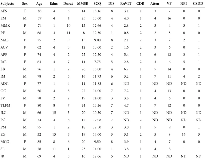
Table 1.Demographics and results of Mini-Mental State Examination, Self-Consciousness Questionnaire, Denial of Illness Scale, and
cognitive-behavioral evaluation.
Subjects Sex Age Educ Durat MMSE SCQ DIS RAVLT CDR Atten VF NPI CSDD
AFS F 83 4 5 14 13.16 8 3.1 1 3 7 0 0 EM M 77 4 4 25 13.00 4 4.0 1 4 16 0 0 MMK F 74 1 10 13 12.66 4 2.8 2 3 4 3 1 PF M 68 4 11 8 12.50 1 0.8 2 2 5 0 0 MAL F 75 2 9 15 9.00 8 2.1 2 3 7 2 1 ACV F 62 4 3 12 13.00 2 1.6 2 3 6 0 1 APP F 74 4 2 22 12.50 4 5.4 1 6 12 3 1 IAR F 63 4 7 14 7.75 5 2.8 2 3 6 5 1 LB M 76 1 2 26 13.00 4 6.2 1 5 14 0 0 IM M 78 2 5 16 11.73 6 3.2 1 7 11 4 2 ADC F 77 1 4 14 11.83 6 ND 1 ND ND ND ND OC M 56 4 8 27 14.00 7 7.2 1 4 13 0 0 FV M 78 2 2 19 14.00 3 3.8 1 4 6 0 0 TLFM F 80 8 7 24 13.26 7 4.7 1 7 12 0 0 JLC M 66 15 3 20 10.50 7 ND 1 ND ND ND ND PG M 74 4 8 17 12.08 7 ND 2 ND ND ND ND PM M 75 1 2 18 12.50 3 3.0 1 5 9 0 1 EG M 52 15 3 19 14.00 3 3.1 2 5 8 16 3 MCG F 85 8 6 20 9.50 8 3.9 1 4 7 0 0 SL M 78 11 1 23 14.00 1 3.8 1 4 8 1 1 JR M 69 4 5 16 12.66 5 ND 1 ND ND ND ND
patient refusal. SCQ scores were not related with age,
education, or regional perfusion defects found on SPECT
images, but tended to correlate with disease duration and
MMSE scores, though did not reach statistical
signifi-cance (r= –0.324 and r=0.317, respectively). In the
analy-sis of correlation between SCQ scores and SPECT
pat-terns (A, B, C, D, E, G; see Table 2), SCQ scores were
clas-sified into two subgroups: from 7 to 10.99, and from 11
to 14, since the minimum score was 7.75 and the
maxi-mum score, 14. Likewise, DIS scores did not show any
correlation to age, education, MMSE, or SPECT data, but
tended to correlate to disease duration (r=0.3208),
though did not reach statistic significance.
SCQ and DIS scores did not correlate with mood state
(CSDD), behavioral changes (NPI), dementia severity
(CDR 1 and 2), nor with any of the neuropsychological
Table 2.Results of structural (CT, MR) and functional (SPECT) neuroimaging.
Subjects CT or MRI SPECT [Holman´s pattern]
AFS Mild CSCA + HSFSCWM Mild inferior bifrontal HP [E] EM Marked CSCA + HSFSCWM Moderate diffuse cortical HP [G] MMK Moderate CSCA Normal [A]
PF Moderate bilateral F-T-P atrophy Marked bi-T-P-O HP [B] MAL Moderate biparietal atrophy Normal [A]
ACV Mild CSCA + HSFSCWM Moderate bilateral T-P-O HP [B]
APP Normal ND
IAR Normal Normal [A] LB Mild CSCA Normal [A]
IM Moderate CSCA + HSFCSWM Multiple bilateral cortical HP areas [G] ADC Moderate CSCA + HSFCSWM Moderate biparietal HP [B]
OC Normal Mild bilateral T-P HP [B] FV Moderate CSCA + HSFCSWM Normal [A]
TLFM Mild CSCA (mostly F) Mild bilateral T-P-O-cingulate HP [C] JLC Mild CSCA (mostly P) + HSFCSWM Mild bilateral P-O HP [B]
PG Mild CSCA Mild bitemporal HP [B] PM Mild CSCA ND
EG Mild P and medial T atrophy ND
MCG Moderate CSCA Moderate left T and diffuse cortical HP [D] SL Mild CSCA ND
JR Mild CSCA ND
CSCA, cortico-subcortical atrophy; HSFSCWM, high signal foci on subcortical white matter; HP, hypoperfusion; F, frontal; T, temporal; P, parietal; O, occipital; ND, not done; Holman´s patterns A, B, C, D, E, G, see text (Methods).
the greater the degree of anosognosia. SCQ and DIS were
translated to Portuguese by the authors, since these scales
had not been published or validated in Brazil.
Data analysis by means of Statistica software 6.0
(StatSoft Inc., 2001) used contingency tables (chi-square)
and Pearson coefficient for correlations between
anosog-nosia items and neuropsychological tests
(counter-proofs). The significance level was 5% (two-tailed).
Results
tests (digit span, verbal fluency, Rey verbal learning).
Also, there were no correlations between the different
as-pects of self-consciousness and any of these cognitive and
behavioral variables. The only significant correlations
were found between SCQ and DIS scores (r= –0.4482,
t=2.185, df=19, p<0.05) and between the domain of
“moral judgment” of SCQ and MMSE (r=0.4629, t=
2.2763, df=19, p<0.05).
Discussion
In agreement with other authors, wee have found no
correlations between SCQ or DIS scores and age,
educa-tion, and disease duration
2. However, in contrast with
these authors, we found no correlation either between
SCQ or DIS, and dementia severity as measured by CDR
or MMSE scores. As expected, there was a negative
corre-lation between the scores of SCQ and those of DIS,
sug-gesting they measure similar mental changes.
The analysis of each aspect of self-consciousness
rela-tive to the neuropsychological (MMSE, verbal learning,
digit span, verbal fluency) or neuropsychiatric variables
(CSDD, NPI) showed significant positive correlation only
between the domain of “moral judgment” and MMSE, in
agreement with Gil et al.
2. This finding is difficult to
interpret. It may simply be a statistical artefact without
clinical meaning, since changes of moral judgment are
usually related to frontal-orbital regions, and only one of
our patients had bifrontal hypoperfusion, but without
MRI signs of frontal atrophy. Most of our patients had
atrophy and hypoperfusion in posterior regions
(tempo-ral, parietal, occipital), often bilaterally. Recent studies
32have shown moral judgment ability to be a complex
func-tion, which requires a whole neurofunctional network,
including posterior associative brain regions and
inte-grating semantic-cultural knowledge and appropriate
motivational and affective states.
We hypothesized that anosognosia would be worse
the longer the disease duration, the more widespread the
brain lesions, the more severe the dementia, and the
lower the depressive scores. However, none of these
hypotheses were confirmed by our findings, probably
because our sample size was small and comprised mostly
mild dementia cases (67%), without any patients with
severe dementia (CDR 3). The lesion model we used
(Alzheimer´s disease, with degeneration predominantly
in temporal-parietal regions) did not allow us to verify
the relevant role of frontal dysfunctions in anosognosia,
as established by various authors
4,15,16. Michon et al.
17was
indeed able to verify that the severity of anosognosia is
related to signs of frontal dysfunction but not to the
severity of dementia. Thus, in order to tackle these
ques-tions, we need to include a greater number of patients
with Alzheimer´s disease with CDR 1 through 3, to
com-pare to patients with frontotemporal dementia and
nor-mal control subjects, while also using neuroimaging
methods robust enough to more precisely delimit the
brain lesions or dysfunctions.
Acknowledgments –
This research was supported by grant
(05/54415-0) from FAPESP (Brazil).
References
1. Prigatano GP, Schacter DL. Awareness of deficit after brain injury: clinical and theoretical issues. New York, NY: Oxford University Press; 1991.
2. Gil R, Arroyo-Anllo EM, Ingrand P, et al. Self-consciousness and Alzheimer’s disease. Acta Neurol Scand 2001;104:296-300. 3. Dennett DC. Consciousness explained. Boston: Little, Brown
and Company; 1991.
4. Reed BR, Jagust WJ, Coulter L. Anosognosia in Alzheimer’s disease: relationships to depression, cognitive function, and cerebral perfusion. J Clin Exp Neuropsychol 1993;15:231-244. 5. Sevush S, Leve N. Denial of memory deficit in Alzheimer’s
disease. Am J Psychiatry 1993;150:748-751.
6. Smith CA, Henderson VW, McCleary CA, Murdock GA, Buckwalter JG. Anosognosia and Alzheimer’s disease: the role of depressive symptoms in mediating impaired insight. J Clin Exp Neuropsychol 2000;22:437-444.
7. Dourado M, Laks J, Rocha M, Soares C, Leibing A, En-gelhardt E. Awareness of disease in dementia: preliminary results in patients with mild and moderate Alzheimer´s dis-ease. Arq Neuropsiquiatr 2005;63:114-118.
8. Feher EP, Mahurin RK, Inbody SB, Crook TH, Pirozzolo FJ. Anosognosia in Alzheimer’s disease. Neuropsychiatry Neu-ropsychol Behav Neurol 1991;4:136-146.
9. Migliorelli R, Teson A, Sabe L, Petracchi M, Leiguarda R, Starkstein SE. Prevalence and correlates of dysthymia and major depression among patients with Alzheimer’s disease. Am J Psychiatry 1995;152:37-44.
10. Starkstein SE, Chemerinski E, Sabe L, et al. Prospective lon-gitudinal study of depression and anosognosia in Al-zheimer’s disease. Br J Psychiatry 1997;171:47-52.
11. Almeida OP, Crocco EI. Perception of cognitive deficits and behavior disorders in patients with Alzheimer´s disease. Arq Neuropsiquiatr 2000;58:292-299.
12. Starkstein SE, Sabe L, Chemerinski E, Jason L, Leiguarda R. Two domains of anosognosia in Alzheimer’s disease. J Neu-rol Neurosurg Psychiatry 1996;61:485-490.
Awareness of deficit after brain injury. New York, NY: Ox-ford University Press; 1991:53-62.
14. Starkstein SE, Fedoroff JP, Price TR, et al. Anosognosia in patients with cerebrovascular lesions: a study of causative factors. Stroke 1992;23:1446-1453.
15. Lopez OL, Becker JT, Somsak D, Dew MA, Dekorsky ST. Awareness of cognitive deficits in probable Alzheimer’s dis-ease. Eur Neurol 1993;34:277-282.
16. Ott BR, Lafleche G, Whellihan WM, Buongiorno GW, bert MS, Fogel BS. Impaired awareness of deficits in Al-zheimer disease. AlAl-zheimer Dis Assoc Disord 1996;10:68-76. 17. Michon A, Deweer B, Pillon B, Agid Y, Dubois B. Relation of anosognosia to frontal lobe dysfunction in Alzheimer´s dis-ease. J Neurol Neurosurg Psychiatry 1994;57:805-809. 18. Starkstein SE, Robinson RG. Neuropsychiatric aspects of
stroke. In: Coffey CE, Cummings JL, editors. Textbook of Geriatric Neuropsychiatry. Washington, DC: American Psychiatric Press; 1994.
19. Starkstein SE, Vázquez S, Migliorelli R, Tesón A, Sabe L, Leiguarda R. A single-photon emission computed tomo-graphic study of anosognosia in Alzheimer’s disease. Arch Neurol 1995;52:415-420.
20. Stuss DT, Benson DF. The frontal lobes. New York, NY: Raven Press; 1986.
21. Folstein MF, Folstein SE, McHugh PR. Mini-Mental State: a practical method for grading the cognitive state of patients for the clinician. J Psychiatr Res 1975;12:189-198.
22. Brucki SMD, Nitrini R, Caramelli P, Bertolucci PHF, Oka-moto IH. Sugestões para o uso do Mini-exame do estado mental no Brasil. Arq Neuropsiquiatr 2003;61:777-781.
23. Morris JC. The Clinical Dementia Rating (CDR): current version and scoring rules. Neurology 1993;43:2412-2414. 24. American Psychiatric Association. Diagnostic and statistical
manual of mental disorders, 4thed. (DSM-IV). Washington,
DC: American Psychiatric Association; 1994.
25. McKhann G, Drachman D, Folstein M, Katzman R, Price D, Stadlan EM. Clinical diagnosis of Alzheimer´s disease: report of the NINCDS-ADRDA Work Group under the auspices of the Department of Health and Human Services Task Force on Alzheimer´s disease. Neurology 1984;34:939-944. 26. Holman BL, Johnson KA, Gerada B, Carvalho PA, Satlin A.
The scintigraphic appearance of Alzheimer´s disease: a prospective study using Technetium-99m-HMPAO SPECT. J Nucl Med 1992;33:181-185.
27. Wechsler D. WAIS-R Manual. New York, NY: The Psy-chological Corporation; 1981.
28. Lezak MD. Neuropsychological assessment. New York, NY: Oxford University Press; 1995.
29. Rey A. L’examen clinique em psychologie. Paris: Press Universitaire de France; 1958.
30. Cummings JL, Mega M, Gray K, Rosenberg-Thompson S, Carusi DA, Gornbein J. The Neuropsychiatric inventory: comprehensive assessment of psychopathology in dementia. Neurology 1994;44:2308-2314.
31. Alexopoulos GS, Abrams RC, Young RC, Shamoian CA. Cornell Scale for Depression in Dementia. Biol Psychiatry 1988;23:271-284.
Appendix
B. Denial of Illness Scale
Questions Scoring
1. Patient minimizes present symptoms (at interview). 0 (no) 1 (once or twice) 2 (more than twice) 2. Patient alludes to there being nothing really wrong with her or
him and that she or he is ready to go home.
0 (no) 1 (once or twice) 2 (more than twice)
3. Patient (past or present) displaces source of symptoms to organs other than brain or complains of symptoms unrelated to the central nervous system.
(no) 1 (once or twice) 2 (more than twice)
4. Did the patient at any time admit to fear of death? 0 (yes) 1 (no) 5. Did the patient at any time admit to fear of invalidism? 0 (yes) 1 (no)
6. Patient verbally denies being in the hospital. 0 (not at all) 1 (sometimes) 2 (every time) 7. Patient displays, at least on the surface, a carefree, cheerful,
jovial approach to life.
0 (no) 1 (once or twice) 2 (more than twice)
8. Patient´s behavior during interview is characterized by nonchalance, coolness, imperturbablity.
0 (no) 1 (once or twice) 2 (more than twice)
9. Patient displaces fear for his or her own illness to family, older patients, weaker patients, and so on.
0 (no) 1 (at least 1 time during the interview)
10.Patient projects illness or weakness to family, spouse, and so on 0 (no) 1 (at least 1 time during the interview)
A. Self-Consciousness Questionnaire
1. What is your name (surname and first name)? 2. Why have you come to see me?
3. Do you have any health problems that prevent you from leading a normal life?
4. Have you got any problem with your memory? 5. Have you had a job? What was it?
6. What is the first name of your spouse (or partner)? 7. What is your mother´s first name?
8. Do you feel rather happy or unhappy? Why?
9. Would you say that you are rather fair or dark-haired? 10. Are you now sitting, standing or lying down? 11. What are you planning to do shortly or tomorrow? 12. If you had to live your life over again, is there anything you
would like to change? What?
13. Is it a good thing or a bad thing to tell a lie? Why? 14. Is it a good thing or a bad thing to give some money or
Appendix
Questionário de Auto-consciência (Gil et al., 2001)
1. Qual é seu nome e sobrenome? 2. Por que você veio a esta consulta?
3. Você tem algum problema de saúde que o(a) impede de levar uma vida normal?
4. Você tem algum problema de memória?
5. Você já teve algum trabalho ou emprego? Qual era sua ocupação?
6. Qual é o nome de seu marido (ou esposa)? 7. Qual é o nome de sua mãe?
8. Você se sente feliz ou triste? Por quê?
9. Você diria que seus cabelos são claros ou escuros? 10. Você agora está sentado(a), de pé ou deitado(a)? 11. O que você planeja fazer hoje ou amanhã?
12. Se você tivesse que viver sua vida novamente, recomeçando tudo desde a infância, há algo que você gostaria de mudar? O que?
13. Você acha que mentir é bom ou ruim? Por quê?
14. Você acha que dar comida ou dinheiro a alguém que está passando fome é bom ou ruim? Por que?
Escala de Negação da Doença (Starkstein & Robinson, 1994)
Perguntas Pontuação
1. O paciente minimiza seus sintomas atuais durante a entrevista. 0 (não) 1 (1 ou 2 vezes) 2 (mais de 2 vezes) 2. O paciente diz não haver nada realmente errado com ele (ela) e
que está pronto(a) para ir para casa.
0 (não) 1 (1 ou 2 vezes) 2 (mais de 2 vezes)
3. O paciente (no passado ou no presente) refere que a causa de seus sintomas está em outros órgãos que não o cérebro, ou queixa-se de sintomas não relacionados ao sistema nervoso central.
0 (não) 1 (1 ou 2 vezes) 2 (mais de 2 vezes)
4. Em algum momento o paciente admitiu temer a morte? 0 (sim) 1 (não) 5. Em algum momento o paciente admitiu ter medo de ficar
inválido?
0 (sim) 1 (não)
6. O paciente nega verbalmente estar no hospital. 0 (de ne-nhum modo)
1 (às vezes) 2 (toda vez)
7. O paciente demonstra, ao menos aparentemente, um modo descontraído, alegre e jovial de lidar com a vida.
0 (não) 1 (1 ou 2 vezes) 2 (mais de 2 vezes)
8. Durante a entrevista, o comportamento do paciente se caracteriza por indiferença, frieza e despreocupação.
0 (não) 1 (1 ou 2 vezes) 2 (mais de 2 vezes)
9. O paciente transfere o medo de sua própria doença para a família e outros pacientes.
0 (não) 1 (ao menos 1 vez durante a entrevista) 10. O paciente projeta sua doença ou fraqueza em seus familiares. 0 (não) 1 (ao menos 1 vez