232 232 232 232 232
Cavalcanti & Santos
Left atrium and pulmonary veins junction
Arq Bras Cardiol 2001; 77: 232-4.
Universidade Federal de Pernambuco - Recife
Mailing address: Jennecy Sales Cavalcanti – Rua Félix de Brito Melo, 912/501 – 51020-260 – Recife, PE, Brazil
English version by Stela Maris C. e Gandour
Objective - To study the arrangement of the
myocar-dial fiber bundles at the pulmonary venous left atrial junc-tion in patients with pulmonary hypertension, and to dis-cuss the pathophysiological importance of this element in the etiology of acute pulmonary edema.
Methods - We obtained 12 hearts and their
pulmo-nary vein extremities from postmortem examinations of pa-tients with the anatomicopathological diagnosis of acute pulmonary edema. The specimens, which had no grossly visible morphological cardiac alterations, were fixed in 10% formalin, and the muscular arrangement of the pul-monary venous left atrial junctions was analyzed. This ma-terial was then isolated, embedded in paraffin, underwent serial cutting (50 µm of thickness), and was stained with Azam’s trichrome.
Results - We observed in our specimens that: a) the
myocardial fiber bundles that originate in the atrial wall and involve the openings of the pulmonary veins were fe-wer than those observed in healthy material; b) the myo-cardial fiber bundles that extend into the pulmonary veins were shorter than those found in material originating from individuals with no pulmonary hypertension.
Conclusion - Anatomical changes that result in a
re-duction in the amount of myocardial fiber bundles in the pulmonary venous left atrial junction, isolated or associa-ted with other factors, may be the cause of disorders in pulmonary circulation, leading to an increase in nary venous pressure, and, consequently, to acute pulmo-nary edema.
Key words: pulmonary veins, left atrium, pulmonary hy-pertension
Arq Bras Cardiol, volume 77 (nº 3), 232-4, 2001
Jen n ecy Sal es Caval can t i , Lau r a Pat r íci a Fer r ei r a San t os
Recife, PE - Brazil
Morphofunctional Study of the Junction Between the Left Atrium
and the Pulmonary Veins in Patient with Pulmonary Hypertension
Original Article
Pulmonary hypertension occurs when the systolic pressure in the pulmonary artery increases above 30mmHg, hindering the maintenance of cardiac output. Of the types of pulmonary hypertension, that of stasis or reflux (venous hypertension) is frequently the most common, usually re-sulting from cardiac disorders that impair the normal draina-ge of the lesser circulation. The most common example is that of left heart failure and mitral stenosis causing pulmona-ry venous stasis, with a consequent increase in pressure in this region 1,2. However, other causes exist, such as patholo-gical pulmonary processes (emphysema, fibrosis, pulmona-ry thromboembolism, and pulmonapulmona-ry schistosomiasis), which explain the genesis of secondary pulmonary hyper-tension. However, the literature has shown that, in approxi-mately 10% of the cases, no cardiac or pulmonary cause can be identified as being involved in the genesis of the disea-se; in these cases, the disorder is called primary or idiopa-thic pulmonary hypertension. The latter is a rare disorder of the pulmonary circulation, usually affecting young and mid-dle-aged women, with a rapid and invariably fatal evolution 3. Stuart 4 reported that the veno-occlusive pulmonary disea-se occurs in less than 10% of patients with primary pulmo-nary hypertension, and that the basic cause of this disorder remains unknown.
Arq Bras Cardiol 2001; 77: 232-4.
Cavalcanti & Santos Left atrium and pulmonary veins junction
233 233 233 233 233 the pulmonary venous left atrial junctions may be the
cau-se of the acute pulmonary edema with no cardiac ventri-cular failure.
Considering the hypotheses formulated by these au-thors, in which alterations in the intrapulmonary venous pressure might be caused by modifications in the structural elements that constitute pulmonary venous left atrial junc-tions, we decided to use stratigraphy to investigate pulmo-nary morphology and function during abnormal conditions, and discuss the possible pathophysiological importance of our findings in the etiology of acute pulmonary edema.
Methods
We studied 12 anatomical specimens constituted of hearts and lungs obtained from autopsies performed at the Department of Pathology of the CCS in the Federal Univer-sity of Pernambuco. These autopsies were carried out in adult female and male patients, whose anatomicopathologi-cal diagnosis of the cause of death was acute pulmonary edema. It is important to emphasize that only specimens wi-th no grossly visible alterations were included in wi-this study. The hearts were withdrawn with their pulmonary ves-sels and part of their pulmonary parenchyma, and were fixed in 10% formalin. Then, the left atria with a segment of the pulmonary veins were separated from the corresponding ventricles. With the aid of a stereoscopic magnifying glass, the remaining pieces of the pericardium were removed, and the myocardial fiber bundles were exposed. The pulmonary venous left atrial junction and the extension of the cardiac muscle into the pulmonary veins were carefully analyzed.
Once the pulmonary veins and their junctions with the atrial wall were isolated, the material was embedded in paraf-fin, and tangential and longitudinal serial cross-sectional samples of the entire specimen 50 µm thick were cut. The sli-des were stained with Azam’s trichrome, and were exami-ned under epi- and transillumination, using a microscope and a stereomicroscopic magnifying glass with magnifica-tions ranging from 5 to 50 times. The results obtained were recorded and illustrated by means of sketches and photo-graphs.
Results
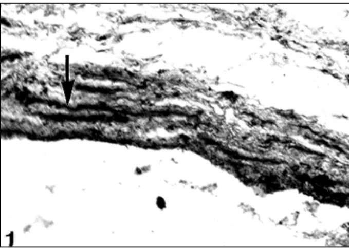
The myocardial fiber bundles were found to originate in the atrial wall and encompass the openings of the pulmonary veins, constituting a type of sphincter (fig. 1). Part of these fiber bundles continues in the walls of the veins in semi-circular and oblique trajectories, in a spiral manner (fig. 2). This behavior was better evidenced in the superior pulmonary venous left atrial junctions than in the inferior ones, in which most fibers are limited to encircling the openings of the pulmonary veins. In both cases, we observed that some of the outermost bundles encompass the veins close to their openings, and return to the atrial wall, constituting true mus-cle loops. However, most fiber bundles end in the venous wall, where they attach to the fibrous elements of the vessel’s
adventitia. These myocardial cuffs are limited to the tunica adventitia, and extend on an average of 10 mm into the supe-rior pulmonary veins and 3 mm into the infesupe-rior ones, being restricted to the intrapericardial part of the vessels.
Discussion
Several authors have proposed that the cardiac mus-culature encompassing the openings of the pulmonary veins has a sphincteral function, which prevents blood from reflowing during atrial systole 5-8,14-16. On the other hand, systolic contractions of the extrapulmonary portions of the pulmonary veins have been shown in the rat and in the mouse, 11,17. Likewise, Carrow and Calhoun 10, in their ex-perimental studies, formulated the hypothesis that the con-duction of the electrical impulse following a trajectory esta-blished by the cardiac muscle in the wall of the pulmonary veins may create a peristaltic or milking action towards the heart, therefore, increasing the venous return and atrial fil-ling. Nathan and Eliakim 6 reported that the presence of stria-ted muscle in the tunica media of the pulmonary veins of the rat has led to the supposition that its contraction associated with the activity of the cardiac muscle facilitates atrial filling during diastole. Likewise, Almeida et al 17 suggest that the mechanical weakening or failure of the myocardial layer in Fig. 1 – Cross-section at the level of the opening of the pulmonary vein in the left atrium. Note the bundles of myocardial fibers with semicircular trajectories around the opening of the left pulmonary vein (arrow). Azam’s trichrome, 45x.
234 234 234 234 234
Cavalcanti & Santos
Left atrium and pulmonary veins junction
Arq Bras Cardiol 2001; 77: 232-4.
the walls of the pulmonary veins could have an increased effect on the pressure of the pulmonary capillaries, favoring stasis and pulmonary edema. Cavalcanti et al 18 reported that the pulmonary venous left atrial junction has a morpholo-gical substrate capable of playing an important role in the pulmonary circulation, not only preventing venous reflow, but also controlling intrapulmonary venous pressure and cardiac performance.
The semicircular myocardial fiber bundles that encom-pass the openings of the pulmonary veins observed in this study are fewer than those reported by Cavalcanti et al 18 in their study analyzing the material from individuals with no pulmonary hypertension. Likewise, the extension of the myocardial cuff of the tunica adventitia of these vessels is, on average, shorter than that reported by Nathan and Elia-kim 6 and by Cavalcanti et al 8 in non- pathological conditi-ons. This is because the myocardial fiber bundles had not reached the pericardial reflection in any of our cases, lea-ding us to assume that the reduction in the amount of myo-cardial fiber bundles encompassing the opening of the pul-monary veins may hinder their possible sphincteric functi-on reported by several authors 6-9,15-18. This might be the cause of blood reflow during atrial systole.
On the other hand, we could assume that a shorter myocardial cuff in the wall of the pulmonary veins would weaken the probable milking function of these fiber bun-dles, as already reported by several authors 7,11,14. This would elevate pressure, causing blood stasis in this vascu-lar area.
It is noteworthy that an important limiting factor of the present study is the nonexistence of clinical data confirming the presence of pulmonary hypertension during life in the patients studied. In addition, our results were compared in a subjective manner with those of the control group, which was constituted of material origi-nating from autopsies of adult female and male patients with different causes of death, those of cardiopulmonary origin excluded 18.
In conclusion, anatomical alterations resulting in a reduction in the amount of myocardial fiber bundles in the pulmonary venous left atrial junction, isolated or in asso-ciation with other factors, may be the cause of disturbance in the pulmonary circulation, resulting in an increase in monary venous pressure, and, consequently, in acute pul-monary edema.
1. Bevilacqua F, Benssonssan E, Silva JMJ, Castro FS, Carvalhaes LP. Manual de Fi-siopatologia Clínica. Rio de Janeiro: Atheneu, 1975: 277.
2. Chazova I, Robbins I, Loyd J, et al. Venous and arterial changes in pulmonary veno-occlusive disease, mitral stenosis and fibrosing mediastinitis. Eur Respir J 2000; 15: 116-22.
3. Veeraraghavan S, Koss MN, Sharma OP. Pulmonary veno-occlusive disease. Curr Opin Pulm Med 1999; 5: 310-3.
4. Stuart R. Hipertensão pulmonar primária. In: Harrison TR edt. Medicina clínica. 12a ed. Rio de Janeiro: Guanabara Koogan, 1992: 60-3.
5. Burch GE, Romney RB. Functional anatomy and throttle valve action of the pul-monary vein. Am Heart J 1954; 47: 58-66.
6. Nathan H, Eliakim M. The junction between the left atrium and the pulmonary veins: a antomic study of human hearts. Circulation 1966; 34: 412-26. 7. Kay JM. Pulmonary vasculature and nerves: comparative morphologic features
of the pulmonary vasculature in mammals. Am Rev Respir Dis 1983; 128: 353-7. 8. Cavalcanti JS, Oliveira ML, Biazotto W, Camargo AM. Morphofunctional study of the left atio-venous junctions in man. Braz J Morphol Sci 1996; 13: 25-30. 9. Hashizume H, Tango M, Ushiki T. Three-dimensional cytoarchitecture of rat
pul-monary venous walls: a light and scanning electron microscopic study. Anat Embryol (Berl) 1998; 198:6: 473-80.
References
10. Carrow R, Calhoun ML. The extent of cardiac muscle in the great veins of the dog. Anat Rec 1964; 150: 249-56.
11. Hooker CW, McAlister HA, Ellis FW. Active contraction of the large thoracic veins in certain mammals. Anat Rec 1964; 148: 2927.
12. Smith JD, Coxe JW. Reactions of isolated pulmonary blood vessels to anoxia, epi-nephine, acetylcholine and histamine. Am J Phisiol 1951; 167: 732-7. 13. Eliakim M, Aviado DM. Effects of nerve stimulation and drugs on the
extrapul-monary portion of the pulextrapul-monary vein. J Pharmacol Exp Therap 1961; 133: 304-12.
14. Rudolph AM, Gootman NL, Golinko, RJ. Observations on a sphincter mechanism at pulmonary venous left atrial junction. Circulation 1961; 24: 1027.
15. Hyman AL. The pulmonary veins. Ann Rev Med 1966; 17: 431-66. 16. Kjelberg SR, Olsson SE. Roentgenologic studies of the sphincter mechanism of
the caval and pulmonary veins. Acta Radiol Stockh 1954; 41: 487-96. 17. Almeida OP, Böhm GM, Carvalho MP, Carvalho AP. The cardiac muscle in the
pulmonary vein of the rat: a morphological and electrophysiological study. J Morph 1975; 4: 409-34.