• Nenhum resultado encontrado

Arq. Bras. Cardiol. vol.73 número5

N/A
N/A
Protected

Academic year: 2018

Share "Arq. Bras. Cardiol. vol.73 número5"

Copied!
5
0
0

Texto

(1)

Instituto do Coração do Hospital das Clínicas - FMUSP.

Mailing address: Fernando Antibas Atik Av. Chibarás, 626/101 04076003 -São Paulo, SP - Brazil.

Received on 2/25/99 Accepted on 8/11/99

Objective - The aim of this work was the follow-up and evaluation of valve replacement in children under 12 years of age.

Methods - Forty-four children less than 12 years old were underwent valve replacement at INCOR-HCFMUSP between January 1986 and December 1992. Forty (91%) were rheumatic, 39 (88.7%) were in functional classes II or IV, 19 (43.2%) were operated upon on an emergency basis, and 6 (13.6%) had atrial fibrillation. Biological prostheses (BP) were employed in 26 patients (59.1%), and mechanical prostheses (MP) in 18 (40.9%). Mitral valves were replaced in 30 (68.7%), aortic valves in 8 (18.2%), a tricuspid valve in 1 (2.3%), and double (aortic and mitral) valves in 5 (11.4) of the patients.

Results - Hospital mortality was of 4.5% (2 cases). The mean follow-up period was 5.8 years. Re-operations occurred in 63.3% of the patients with BP and in 12.5% of those with MP (p=0.002). Infectious endocarditis was present in 26.3% of the BP, but in none of the cases of MP (p=0.049). Thrombosis occurred in 2 (12.5%) and hemorr-hage in one (6.5%) of the patients with a MP. Delayed mor-tality occurred in 5 (11.9%) of the patients over a mean pe-riod of 2.6 years; four had had BP and one had a MP (NS). Actuarial survival and re-operation-free curves after 10 years were respectively, 82.5±7.7 (SD)% and 20.6±15.9%.

Conclusion – Patients with MP required fewer re-operation, had less infectious endocarditis and lower late mortality rates compared with patients with bioprosthe-ses. The former, therefore, appear to be the best valve replacement for pediatric patients.

Key words: cardiac valve replacement, rheumatic fever, children

Arq Bras Cardiol, volume 73 (nº 5), 424-428, 1999

Fernando Antibas Atik, Altamiro Ribeiro Dias, Pablo M. A. Pomerantzeff, Miguel Barbero-Marcial, Noedir Antonio Groppo Stolf, Adib Domingos Jatene

São Paulo, SP - Brazil

Immediate and Long Term Evolution of Valve Replacement in

Children Less than 12 Years Old

The high prevalence of rheumatic fever in infants and adolescents draws attention to the peculiarities of the di-sease, which frequently causes serious valves dysfunction often in association with conditions of rheumatic activity of the cardiac muscle. Due to their important hemodynamic re-percussions, such conditions may at times require urgent surgical intervention 1.

Conservative surgical procedures, a primary approach to these cases, are often not possible due to pathological al-terations present in cardiac valves. On the other hand, valve replacement in infants and adolescents has its own set of difficulties, in particular, which prostheses types and size to use.

The aim of this work is to present the immediate and long-term experience of our valve replacement service with children under 12 years of age, with the aim of identifying the ideal prosthesis for use at this age group.

Methods

Forty-four patients under 12 years of age who under-went to valve replacement at the Heart Institute, Hospital of Clinics, University of São Paulo Medical School between January 1986 and December 1992, were retrospectively analyzed.

Twenty-three (52.3%) were males between 2 and 12 years of age (average age 10 years). Their valve diseases were of rheumatic in origin in 40 (91%), degenerative in 2 (4.5%) and congenital in 2 (4.5%) of the cases. Twenty (45.5%) of the patients were in preoperative NYHA functio-nal class IV, 19 (43.2%) were in functiofunctio-nal class III and 5 (11.3%) were in functional class II of the NYHA scale. Nine-teen (43.2%) had emergency operations, 6 (13.6%) had chro-nic atrial fibrillation and 13 (29.5%) showed laboratory and scintigraphic evidence of rheumatic activity.

(2)

Twelve patients had previously undergone mitral valve replacement, performed twice in two of them, of which one had a tricuspid, the other mitral +aortic prostheses. Previous mitral prostheses had been biological in nine and of dura mater in one patient; patients twice-operated upon had mechanical prosthe-ses implanted only in the second operation. Both the patient with the tricuspid exchange and the one who had a double ex-change received biological prosthesis. The average time interval following the first intervention was 4.3 years for the mitral, 9 years for the tricuspid and 2 years for the double prostheses.

Three patients had undergone previous conservative procedures on the mitral valve whose average durability had been 3.3 years.

Regarding the type of prostheses used in the 44 ope-rations reported in this study, 26 (59.1%) of the patients received biological (Biocor, INCOR, Labcor) and 18 me-chanical (St. Jude, Carbomedics, Omnicarbon, Sorin) pros-theses. In the mitral position, biological (18) predominated over mechanical (12) prostheses. Five biological versus three mechanical prostheses were utilized in the aortic position, one mechanical prostheses were used in the tri-cuspid position and of the 5 patients who underwent to the double exchange, three had biological prostheses.

Associated procedures consisted of tricuspid valve re-pair surgery in 11 patients, mitral valve rere-pair surgery in 4, myocardial biopsies in two, and left atrial thrombectomy in one case. Enlargement of the aortic ring with bovine pericar-dium was required for size 21 prostheses implants in two pa-tients and of size 19 in another.

Sizes of prostheses implanted in the various cardiac valves are shown in table I.

Extracorporeal circulation at 25-28oC hypothermia was

used in all patients operated upon: myocardial protection was afforded by cold crystalloid solution or blood. Average perfusion time was 102.3 minutes and anoxia 70.5 minutes.

Native cardiac valve defects were as follows: retractions in 14 cases, ring dilatation in 9, chord rupture in 8, calcification in 5, prolapse in 5, vegetation in 5, fibrosis in 4, thickening in 4 and chord lengthening in 2. Regarding macroscopic charac-teristics on previously implanted prostheses, calcification was observed in 12, ring dilatation in 5, thickening in 2, rupture in 2, stenosis in 3, retraction in 1 and perivalve escape in one.

Thirty-five (83.3%) of the subjects were followed as out-patients for periods varying between 1 month and 11 years (5.82 years average). Long term follow-up for more than 5 years was possible for 23 (54.8%) patients.

Patients who received mechanical prostheses were kept on the anticoagulant sodium warfarin, INR levels were maintained between 2 and 2.5.

To compare immediate and long-term effects of biologi-cal and mechanibiologi-cal prostheses statistibiologi-cally, Fisher’s exact test and the chi-square test were employed. Values of P<0.05 were considered statistically significant. Estimates of survival and no-re-operation probabilities were perfor-med using the Kaplan-Meier; the descriptive probability le-vel was determined using the log-rank test.

Results

Postoperative periods were characterized by the low cardiac output in 11 patients, cardiac arrhythmia in 5, bron-chopneumonia in 5, bleeding in 4, pericarditis in 4, acute re-nal failure in 3, septicemia in 1 and an ischemic stroke in one. Two (4.5%) of the patients died during the immediate postoperative period: both had been submitted to emergen-cy mitral valve replacement with mechanical prostheses. Death was due to cardiogenic shock in both patients who preoperatively, had evidence of active rheumatic illness, acute pulmonary edema and clinical signs of undernutri-tion. One of the patients had already been operated on and shown development of coagulopathy upon termination of extracorporeal circulation. Table II presents the survey of hospital deaths, according to the type of prostheses used. Due to the small number of patients, no statistically signifi-cant differences were observed.

During the patients’ follow-up periods, the most pro-nounced complications were due to calcification of the biological prostheses in 14 (73.7%) patients, three of whom showed no significant valve dysfunction. The presence of prosthetic infectious endocarditis occurred in 5 (14.3%) patients, all involving biological prostheses, a statisti-cally significant finding (table III). Complications of the mechanical prostheses were caused by faulty anticoa-gulation control, two patients having thrombosis (see

re-Table I - Survey of the size of prostheses implanted in the different cardiac valves

19 21 23 25 27 29 31 33

Mitral 3 1 1 9 11 9 1

Aortic 1 5 5 1 1

Tricuspid 1

Table III - Differences between biological and mechanical prostheses regarding long term morbidity and mortality

Biological Mechanical P

Patients 19 16

-Re-operation 12 (63,2%) 2 (12,5%) 0,002 Infectious endocarditis 5 (26,3%) 0 0,049 Long-term mortality 4 (21,1%) 1 (6,3%) 0,342*

*Statistically non-significant

Table II - Differences between biological and mechanical prostheses in regards to hospital mortality

Biological Mechanical Total

Patients 26 18 44

Preoperative FC

II 4 1 5

III 12 7 19

IV 10 10 20

In-hospital mortality 0 2 2 (4,5%)

(3)

operations) and one with excessive menstrual bleeding leading to hypovolemic shock.

Fourteen patients (40%) were re-operated upon, three (8.6%) of these were re-operated upon twice. The average time interval between the two operations was 4.5 years, and between the second and the third 1.5 years. Biological pros-theses were present in 12 (63.2%) patients, two in the aortic position. Mechanical prostheses were present in 2 (12.5%) of the patients, both in the mitral position. A statistically significant difference between the type of prostheses and the possibility of re-operation is shown in table III.

The reasons for re-operation in cases of biological prostheses were calcification and stenosis in 8 (66.7%) of the patients, calcification and rupture in 2 (16.7%), infec-tious endocarditis in one (8.3%) and faulty closure of the posterior tricuspid due to a manufacturer’s defect in ano-ther. Due to thrombosis, mechanical prostheses required re-placement in both cases.

The size of the prostheses was not the reason for ration of any of the patients, even though 4 of the 14 re-ope-rated patients had received prostheses of a greater diameter.

Operation-free curves (fig.1) demonstrated that the estimated probabilities of maintenance of the original prostheses after 2,5 and 10 years were respectively, 93.7%, 64.2% and 20.6%. When these results were evaluated in accordance with the type of prostheses used (fig.2), mechanical prostheses proved to be superior to biological ones (P=0.022).

Five (11.9%) of the patients died during long-term follow-up, which was an average period of 2.6 years. Four of these had received biological prostheses. One of them died suddenly due to cardiac arrhythmia; at necropsy neither the cause of death nor prosthesis’s abnormalities were reve-aled. Three patients experienced dysfunctions secondary to early calcification and were re-operated upon. One died due to an aortic iatrogenic lesion during median sternoto-my. The other two patients developed infectious endocar-ditis at the site of the prosthesis.

One patient had a mechanical prosthesis at the mitral position and developed thrombosis at this site due to a cere-bral vascular accident. He underwent thrombi removal follo-wed by a renefollo-wed change of the mitral valve with a biological prosthesis, because of infections endocarditis of this pros-thesis accompanied by septicemia and cardiogenic shock.

No statistically significant difference between the type of prosthesis used and delayed mortality occurred (table III). Actuarial survival curves (fig.3), demonstrated that after 2, 5 and 10 years, estimated probabilities were respec-tively, 93.3%, 82.5% and 82.5% (fig.4). The group comprising recipients of mechanical prostheses had better survival cur-ves, although in a statistically non-significant manner, than those with biological prostheses.

Fig. 4 - Estimate of probability of survival (Kaplan-Meier method) - sample stratified according to type of prosthesis.

Follow-up period (years)

probability of survival

Fig. 3 - Estimate of probability of survival (Kaplan-Meier method) - total sample.

Follow-up period (years)

probability of survival

Fig. 2 - Estimate of probability of no re-operation (Kaplan-Meier method) - sample stratified according to type of prosthesis.

probability of no re-operation

Follow-up period (years)

Fig. 1 - Estimate of probability of no reoperation (Kaplan-Meier method) - total sample.

probability of no reoperation

(4)

Of the 25 patients being followed up to the present time, the majority (22 cases) are in functional class I; three of them show signs on echocardiography of calcification without valve dysfunction. The three remaining patients have biological mitral prosthesis and clinically, belong to functional classes II (2 cases) and III (one case). They show significant prosthesis degradation due to calcification accompanied by marked stenosis and insufficiency and await re-operation.

Discussion

Surgical treatment of valve disease in children and adolescents constitutes a challenge for the surgeon. It ac-quires aggravating characteristics due to the current high prevalence of rheumatic disease in our midst. Many affected children have advanced degrees of cardiac failure (functio-nal classes II or IV), of rheumatic myocardial activity, requi-ring at times, emergency operations to correct serious hemo-dynamic disturbances 2.

The existence of previous operations whether conser-vative or not and of varying degrees of malnutrition, are other factors causing this group of patients to exhibit high immediate and delayed rates of mortality, as demonstrated in this study in which 88.7% of the patients were in functional class II/IV, 43.2% had emergency operations, 34.1% had had previous cardiac operations, resulting in a hospital mortali-ty rate of 4.5%. Immediate mortalimortali-ty indexes in the literature vary from zero to 13%. Robbins et al 3 defined less than 2

ye-ars of age, presence of previous operations and double valve replacement as hospital mortality risk factors. Accor-ding to Vosa et al 4, mortality is believed to be related to the

correction of complex congenital anomalies or to the level of myocardial involvement in cases of rheumatic and degene-rative valve pathologies.

In the majority of the cases, serious pathological cardiac valve alterations prevent the use of repair interven-tions, rendering prosthetic valve replacement obligatory. The use of this procedure becomes controversial when per-formed on children as opposed to adults 5.

Due to the reduced size of the valve ring, specially in small children, it is of fundamental importance to implant prostheses large enough to allow for somatic growth and no need for re-operation due to functional stenosis 1.

Ibrahim et al 6 advocate that the smallest initial size should

be 25 mm for the mitral and 21 mm for the aortic position, res-pectively. At times, enlargement of the aortic ring, becomes necessary; in this study it was performed in three patients who had good early and late evolution and no requirement for re-operation. None of the patients in the present study required re-operation directly because of the small size of the prostheses, a result the same as that previously explai-ned 1, by the presence of patients with rheumatic fever, a

condition frequently leading to valve regurgitation associa-ted with ring dilatation.

The type of prostheses to be implanted has given rise to discussion for decades. However, the majority of authors

argue in favor of the use of mechanical prostheses only in patients of this age 7. This tendency is explained by the fact

that long-term follow-up of the evolution of bioprostheses has demonstrated a high incidence of complications con-nected to degradation with precocious calcification, requi-ring re-operation 8-10. Antunes et al 11 found only 19% of the

living patients in their case files to have kept retained their original prostheses after seven years. The rate of degenera-tion was 22.4% in patients per year in comparison with struc-tural faults in 4% of adult patients per year. In our experi-ence, the probability of patients maintaining their original prostheses was 47.5% and 13.2% after follow-up periods of 5 and 10 years, respectively.

This biodegradation process is not innocuous; it leads to the death of many patients during the long period of wai-ting for a new operation, a problem not unusual in our servi-ce. In addition, morbidity and mortality at re-operation are high, as attested by our experience that shows 25% morta-lity, not to mention the high cost of hospitalization.

The causes of bioprostheses calcification have been attributed to repeated rheumatic bursts, leading to incre-ased alkaline phosphatase activity with increincre-ased calcium turnover 12, the process of porcine prostheses preservation

with glutaraldehyde, further promoting calcium deposition. The pathological mechanism is the calcification and fibro-sis of the cusps, leading to deformity and rigidity and valve stenosis.

Due to this, casuistic studies have reflected the actual tendency to use exclusively mechanical prostheses. Long-term follow-up has shown smaller mortality indexes, with ac-ceptable levels of morbidity, related mainly to alterations in coagulation. Anticoagulation methods are a major subject of discussion.

The type of mechanical prostheses has not influenced these favorable results, even though groups investigated have mainly been composed of patients with congenital anomalies 5,6,13,14. Harada et al 5, using St. Judes heart valve,

found no early mortality rates, estimated survival times after 10 years being 90.8%, and free of complications of 84.7% over the same time period. Employing the same prosthesis, Ibrahim et al 6 obtained even better results with no need for

re-operation and/or occurrence of delayed deaths, or either of these. Using different types of mechanical prosthesis in various valve positions, we documented a re-operation-free curve at 5 and 9 years was of 92.3% and 61.55, respectively; the survival curve over the same periods was of 91.6%.

Studies of patients with rheumatic fever 1,2,12, who are

considered by many authors to be more serious patients than those with congenital anomalies, show results similar to the former. Antunes et al 1 in the casuistic study of 352

(5)

statistically similar to those obtained with a group of operated adults over the same period.

According to our patient files, the estimated survival and re-operation-free probabilities at 5 years were 91.6% and 92.3%, respectively.

Which anticoagulation method to use is a controversial issue to be decided. The tendency towards trauma, the desire to achieve pregnancy, the presence of atrial fibrillation and difficulties in maintaining an adequate therapeutic level due to psychological or socio-economic problems of adherence to treatment are the responsible factors. This question is of special importance to us because often no adequate clinical and laboratory evaluation of coagulation levels exists, leading to the occurrence of thromboembolism on the one hand and of hemorrhagic phenomena on the other.

Due to such dangerous irregularities in anticoagulant treatment, Abid et al 12 contraindicate this therapy in a

special group of patients having sinus rhythm and hemo-dynamic stability and have not observed an increased inci-dence of complications. Verrier et al 15 suggest that children

in sinus rhythm, recipients of aortic prostheses, can be safe-ly treated with aspirin, dipyridamole, or both; results are ho-wever controversial among different authors.

Cabalka et al 13, reported relatively high incidence of

early thrombosis (between 3 and 6 months) that occurred only in patients without medication. Therefore, by indicate anticoagulation from 6 to 9 months for recipients of mitral prostheses, preferring to reduce therapeutic levels, since after this time, the tendency toward bleeding supplants the risk of thromboembolism.

Robbins et al 3, however, use oral anticoagulants in all

patients, independently of the valve position. By adopting the same approach, we obtained prosthetic thrombosis in 12.5% and hemorrhage in 6.25% of the patients, all of whom used the medication irregularly.

Dental problems, upper airway infections and trauma in children have influenced us to adopt vigorous prophyla-xis for infectious endocarditis in valve prostheses. In this study, five (14.3%) patients acquired endocarditis, which led to the death of three of them. This serious complication has been reported by others, with slightly lesser frequency, but with a similar course of evolution. Although the incidence is similar regarding the type of prostheses 16, Robbins et al 3

reported a greater incidence with mechanical ones, in contrast to our results. However, samples are too small to allow for final conclusions.

Recent studies 17,18 have reported experiments with

ho-mografts and Ross precedure in aortic valve disease. Re-sults in a selected number of patients are encouraging, sho-wing good early evolution. However, this technique is little known among surgeons, painstaking , and of doubtful re-sults in inexperienced centers.

In conclusion, valve replacement in children is a high-risk procedure, showing considerable rates of immediate and long-term mortality in seriously ill patients. Early calcification, leading to a high rate of re-operations over a short time, renders the use of bioprotheses impraticable for this age group. Mechanical prostheses show a lower rate of re-operation, infectious endocarditis and long-term morta-lity. Rigorous anticoagulation control and prevention of in-fectious endocarditis are the only means of reducing long-term complications. These prostheses seem, therefore, to be the best valve replacement in this age group, even in po-pulations with hazardous anticoagulation control, where al-ternative methods could be used.

Acknowledgments

To Rita Helena Antonelli Cardoso and Carine Sovalli for the statistical analyses performed.

1. Antunes MJ, Vanderdonck KM, Sussman MJ. Mechanical valve replacement in children and teenagers. Eur J Cardiothorac Surg 1989; 3: 222-8.

2. John S, Ravikumar E, Jairaj PS, Chowdhury U, Krishnaswami S. Valve replace-ment in the young patient with rheumatic heart disease. J Thorac Cardiovasc Surg 1990; 99: 631-8.

3. Robbins RC, Bowman FO, Malm JR. Cardiac valve replacement in children: a twenty year series. Ann Thorac Surg 1988; 45: 56-61.

4. Vosa C, Renzulli A, Lombardi PF, Damiani G. Mechanical valve replacement un-der 12 years of age: 15 years of experience. J Heart Valve Dis 1995; 4: 279-83. 5. Harada Y, Imai Y, Kurosawa H, Ishihara K, Kawada M, Fukuchi S. Ten-year

fol-low-up after valve replacement with the St. Jude medical prosthesis in children. J Thorac Cardiovasc Surg 1990; 100: 175-80.

6. Ibrahim M, Cleland J, O’Kane H, Gladstone D, Mullholland C, Craig B. St. Jude medical prosthesis in children. J Thorac Cardiovasc Surg 1994; 108: 52-6.

7. Borman JB, Shimon DV, Deeb M, Simcha A. Valve replacement in children. J Card Surg 1989; 4: 260-81.

8. Snitcowsky R. Evolução tardia do implante de prótese de dura-máter em posição mitral. Estudo de pacientes com idades inferiores a 16 anos. (Tese de doutora-mento). São Paulo: Universidade de São Paulo, 1983.

9. Godoy MF, Branco JNR, Soares HC, et al. Resultados a longo prazo da substitui-ção valvar em crianças. Arq Bras Cardiol 1981; 37: 325-9.

References

10. Salles CA, Christo MC, Stortini MJ, et al. Resultados iniciais com a bioprótese heteróloga de Carpentier-Edwards. Arq Bras Cardiol 1981; 36: 349-52. 11. Antunes MJ. Bioprosthetic valve replacement in children: long-term follow-up

with 135 mitral valve implantations. Eur Heart J 1984; 5: 913-8.

12. Abid F, Mzah N, Euch FE, Ismail MB. Valve replacement in children under 15 ye-ars with rheumatic heart disease. Pediatr Cardiol 1989; 10: 199-204. 13. Cabalka AK, Emery RW, Petersen RJ, et al. Long-term follow-up of the St. Jude

medical prosthesis in pediatric patients. Ann Thorac Surg 1995; 60: S618-23. 14. Guvendik L, Cobanoglu A, Davis NR, Fessler CL, Starr A. Valve replacement in

children under 14 years of age: a review of 22 years of experience. Eur J Cardiotho-rac Surg 1989; 3: 229-34.

15. Verrier ED, Tranbaugh RF, Soifer SJ, Yee ES, Turley MD, Ebert PA. Aspirin anti-coagulation in children with mechanical aortic valves. J Thorac Cardiovasc Surg 1986; 92: 1013-20.

16. Zabal C, Attie F, Barragan R, Buendia A, Duque MP. Resultado tardio del cambio valvular mitral en 155 sujetos menores de 16 años. Estudio comparativo con cua-tro protesis. Arch Inst Cardiol Mex 1992; 62: 333-8.

17. Lupinetti FM, Warner J, Jones TK, Herndon SP. Comparison of human and mechanical prosthesis for aortic valve replacement in children. Circulation 1997; 96: 321-5. 18. Mazzitelli D, Guenther T, Schreiber C, Wottke M, Michel J, Meisner H. Aortic

Imagem

Table I - Survey of the size of prostheses implanted in the different cardiac valves
Fig. 2 - Estimate of probability of no re-operation (Kaplan-Meier method) - sample stratified according to type of prosthesis.

Referências

Documentos relacionados

A mensuração dos efeitos de uma política de estabilização parcial de preços, comparativamente à armazenagem competitiva, serviu para demonstrar os efeitos de se fixar um preço

prostheses have become more accepted and in many cases, preferably, probably due to lower life expectancy than the duration of the bioprosthesis. A mechanical prosthesis

Os consultores externos se destacaram como atores na promoção da inovação, um consultor é responsável pela captação de matéria-prima; o segundo consultor é

Desse modo, o presente artigo tem como objetivo principal, tomando a teoria da estrutura padrão do anúncio publicitário como válida, e a teoria do isomorfismo como decisiva

Neste trabalho o objetivo central foi a ampliação e adequação do procedimento e programa computacional baseado no programa comercial MSC.PATRAN, para a geração automática de modelos

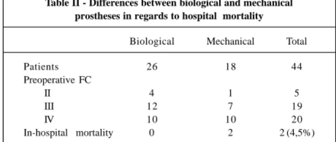
The performed surgical procedures were as follows: mechanical valve replacement in 10 patients, bioprosthetic mitral valve replacement in 5 patients, mechanical mitral valve

The 3 sets were performed keeping the: (i) torso in the upright position on the wheelchair; (ii) torso in the racing position (i.e. trunk bended in the horizontal position) and neck

Supplemental Figure C 2 - ClueGo and CluePedia significance analysis of protein-protein interaction considering common proteins present in epicardial adipose tissue of obese and