Influence of prone position on oxigenation, respiratory rate and muscle
strength in preterm infants being weaned from mechanical ventilation
Influência da posição prona na oxigenação, frequência respiratória e na força muscular nos recém-nascidos pré-termo em desmame da ventilação mecânica
Influencia de la posición prona en la oxigenación, frecuencia respiratoria y en la fuerza muscular en los recién nacidos pre-término en destete de la ventilación mecánica
Rita de Cássia Malagoli1, Fabiana Fagundes A. Santos2, Eduardo Araújo Oliveira3, Maria Cândida F. Bouzada4
ABSTRACT
Objective: To verify the influence of preterm infant positioning on respiratory muscle strength, oxygenation and respiratory rate.
Methods: Cross-sectional study with a paired sample of intubated infants born with gestational age less than 34 weeks, in the final process of weaning from me-chanical ventilation. Infants with malformation, genetic syndromes, neuromuscular diseases, tracheotomies and in the postoperative period of abdominal and thoracic surgery were excluded. Maximum inspiratory pressure measures were checked by a digital manometer; respira-tory rate was visually observed during one minute and oxygen saturation was measured by a pulse oximeter in prone and supine postures. Kruskal-Wallis and Student’s
t-test and Pearson correlation coefficient were applied, being significant p<0.05.
Results: 45 infants with respiratory distress syndrome were evaluated. The mean gestational age was 30.4 weeks and the mean birth weight was 1522g. The oxygen satu-ration was higher in prone position (p<0.001). Values of maximum inspiratory pressure were lower in prone when compared to infants in the supine position (p<0.001). Respiratory rate was similar in the two studied positions (p=0.072).
Conclusions: There was a lower inspiratory pressure and a higher oxygen saturation in prone position when compared to the supine one. Concerning the respiratory rate there was no variation between prone and supine position.
Key-words: posture; infant, premature; oxygenation.
RESUMO
Objetivo: Verificar a influência do posicionamento do recém-nascido prematuro sobre a força da musculatura respiratória, oxigenação e frequência respiratória.
Métodos: Estudo transversal com amostra pareada de recém-nascidos com idade gestacional inferior a 34 semanas, intubados, em processo final de desmame de ventilação mecâ-nica. Foram excluídos aqueles com malformações, síndromes genéticas, doenças neuromusculares, traqueostomizados e em pós-operatório de cirurgias abdominais ou torácicas. As medidas de pressão inspiratória máxima foram aferidas uti-lizando-se manovacuômetro digital; a frequência respiratória através da observação das incursões respiratórias das crianças em um minuto e a saturação de oxigênio por oxímetro, nas posturas prona e supino. Os testes estatísticos aplicados fo-ram Kruskal-Wallis, o teste t de Student e o coeficiente de correlação de Pearson, sendo significante p<0,05.
Instituição: Hospital das Clínicas da Universidade Federal de Minas Gerais (UFMG), Belo Horizonte, MG, Brasil
1Mestre em Ciências da Saúde pela UFMG; Fisioterapeuta do Hospital das
Clínicas da UFMG, Belo Horizonte, MG, Brasil
2Acadêmica de Medicina da UFMG, Belo Horizonte, MG, Brasil
3Doutor em Ciências da Saúde pela UFMG; Professor Titular da UFMG,
Belo Horizonte, MG, Brasil
4Doutor em Ciências da Saúde pela UFMG; Professor Adjunto da UFMG,
Belo Horizonte, MG, Brasil
Endereço para correspondência: Maria Cândida Ferrarez Bouzada
Avenida Professor Alfredo Balena, 190 – sala 2.002 CEP 30130-100 – Belo Horizonte/MG
E-mail: [email protected]
Conflito de interesse: nada a declarar
Resultados: Foram estudadas 45 crianças com síndrome do desconforto respiratório. A idade gestacional média foi de 30,4 semanas e o peso médio ao nascer de 1522g. Os valores de saturação de oxigênio foram mais elevados (p<0,001) e os de pressão inspiratória máxima mais baixos (p<0,001) na posição prona. Os valores de frequência respiratória foram semelhantes nas duas posições estudadas (p=0,072).
Conclusões: Observaram-se menores valores de pressão inspiratória além de aumento da saturação de oxigênio na posição prona quando comparada à supino. Em relação à frequência respiratória, não foi observada variação entre as posturas prona e supino.
Palavras-chave: postura; prematuro; oxigenação.
RESUMEN
Objetivo: Verificar la influencia del posicionamiento del recién-nacido prematuro sobre la fuerza muscular respirato-ria, oxigenación y frecuencia respiratoria.
Métodos: Estudio transversal con muestra pareada de recién nacidos con edad gestacional inferior a 34 semanas, entubados, en proceso final de destete de ventilación mecánica. Se exclu-yeron a aquellos con malformaciones, síndromes genéticos, enfermedades neuromusculares, traqueostomizados y en post-operatorio de cirugías abdominales o torácicas. Las medidas de presión inspiratoria máxima fueron verificadas mediante el uso de manovacuómetro digital; la frecuencia respiratoria, mediante la observación de las incursiones respiratorias de los niños en un minuto y la saturación de oxígeno por oxímetro, en las posturas prona y supina. Las pruebas estadísticas aplicadas fueron Kruskal-Wallis, la prueba t de Student y el coeficiente de correlación de Pearson, siendo significante p<0,05.
Resultados: Se estudiaron 45 niños con síndrome de difi-cultad respiratoria. La edad gestacional mediana fue de 30,4 semanas, y el peso mediano al nacer fue de 1522g. Los valores de saturación de oxígeno fueron más elevados (p<0,001) y los de presión inspiratoria máxima más bajos (p<0,001) en la posición prona. Los valores de frecuencia respiratoria fueron semejantes en las dos posiciones estudiadas (p=0,072).
Conclusiones: Se observaron menores valores de presión inspiratoria, además de aumento en la saturación del oxígeno en la posición prona cuando comparada a la supina. Respecto a la frecuencia respiratoria, no se observó variación entre las posturas prona y supina.
Palabras clave: postura; prematuro; oxigenación.
Introduction
Prone position has been related to higher oxygenation due to the signiicant increase of the movement of the chest wall in this position(1) and due to the better synchronism between thorax and
abdomen, caused by the fact that the incursion and the percent-age of diaphragm shortening are higher in prone position than in supine(2). Some studies indicate that the increase in current volume
is responsible for the higher oxygenation in prone position(3,4),
as it is for the increase of functional residual capacity(5), of the
ventilation-perfusion ratio(6-7) and of alveolar recruitment(8).
Prone position may affect preterm infants’ respiratory mechanisms, leading to changes in gas exchanges. Some studies on the positioning of preterm infants, with or with-out respiratory diseases, show a signiicant improvement in oxygenation in prone position, when compared to supine(9,10).
Prone position is also related to a better sleep pattern(11,12),
less variability in heart rate(11,12), lower frequency of central
and mixed apnea(13,14), lower frequency of bradycardia and
desaturation episodes during apnea periods(13), besides being
related to a higher respiratory rate (RR)(15). However, RR
variation was not reported on other studies(16,17).
Some studies were performed to try to explain why oxygenation is improved when children are in the prone position(3,5-9). Based on these studies, it can be inferred that
the diaphragm acts as the main muscle when preterm infants breathe and that expiratory muscles are responsible for gen-erating more strength in prone position. However, Dimitriou
et al(18) stated that the measurement of the maximum
inspira-tory pressure (MIP) were higher in the supine position than in prone, indicating that the higher oxygenation in the prone position is not due to the greater diaphragm strength in this position. Thus, the effects of positioning on the respiratory mechanisms responsible for the improvement of oxygen-ation in prone position were not yet completely clariied. The population of preterm children of the previous studies varied in relation to gestational age, presence or absence of respiratory compromise, used technique and measured pulmonary function parameters.
This study aimed to investigate and compare the inlu-ence of prone and supine positions on oxygenation, RR and respiratory muscle strength in preterm infants.
Method
gestational age of 34 weeks or less who were in the process of weaning from mechanical ventilation were assessed from June, 2006, to August, 2007. Every admitted child had a term of free and informed consent signed by its parents. The protocol of the study was approved by the Research Ethics Committee of Universidade Federal de Minas Gerais, Brazil. Children with malformations, genetic syndromes or neuromuscular diseases and those who suffered tracheotomy or who were in postoperative from abdominal or thoracic surgery were excluded from the study.
MIP, RR and oxygen saturation (SatO2) measures were obtained when the child showed spontaneous breathing with over 30 breaths per minute (bpm) and a stable condition. The evaluation was performed when children had eaten at least an hour before, after two hours of sedation interruption, and when the children appeared not to be irritated or crying during measurement. For this study, patients were under cardiac monitoring, continuous non-invasive blood pressure monitoring (PNI), oximetry with a Dixtal® monitor and
had their body temperature stabilized around 36–37°C in incubators. No child used steroids or bronchodilator before the measurement of respiratory data. The data were collected in the period up to two hours prior to extubation.
Each child was assessed just once, at the moment its clinical data were collected (birth weight, gestational age, Apgar score, sex, date of birth, type of delivery, use of aminophylline and/or steroids by the child, use of steroids by the mother, date of intubation, mechanical ventilation, post-extubation ventilatory support – continuous positive airway pressure (CPAP) –, extubation weight and occur-rence of bronchopulmonary dysplasia). Soon after, MIP, RR e SatO2 were measured in the prone and the supine positions, randomly chosen, beginning the assessment with the position the child was in at the moment.
The newborn was positioned in prone position over a roller placed longitudinally to the body, which main-tained the chest wall and the abdomen stabilized. Thus, the abdomen remained restricted during breaths. The head was positioned to the right side, upper limb were in 90º abduction, external rotation of the shoulders and a 90º flexion of the elbows. The supine position was performed with head in midline, upper limbs adducted to the side of the chest wall, lower limbs slightly flexed (30–40º) in hips and knees. For both positions, prone and supine, the bed was 15º elevated.
Before measuring MIP, SatO2 and RR, bronchial hygiene and adequate positioning of the child were performed,
followed by a 15 minute wait so the patient calmed down. The RR, MIP and SatO2 were then measured in this order. The child’s position was then changed and another 15 minute wait followed for the patient to stabilize. New measurements of RR, MIP and SatO2 were then taken. The measurements were taken by the same examiner, randomly in relation to positioning.
MIP values were assessed using a MDV 300 (Globalmed®)
digital vacuum manometer connected to a unidirectional valve which allowed expiration but not inspiration, and this valve was connected to the endotracheal tube. The MIP measurements were performed by the occlusion of the device for 20 seconds or at the most 10 spontaneous breath cycles of the child, with no drop in saturation below 85% or drop in heart rate below 90 bpm(19,20). Three series
of occlusions were performed, with a ive-minute interval between them(21,22). The highest value of inspiratory pressure
was elected as MIP.
The saturation measures were collected with the oximeter (Dixtal®). Its sensor was ixed on the child’s left foot. RR
was measured by observing the occurrence of breaths in the children in one minute.
As no data was found in literature about the proportion of children who present MIP alteration in prone and supine position when weaning from mechanical ventilation, a pilot study was performed with 10 preterm infants, following the methodology described in this study. Results showed that 90% of children showed alterations when changing from the supine to the prone position or the reverse, that is, the irst MIP measurement changed when the child was moved from the initial position. From these indings, the sample calculation was made with the computer pro-gram Epi-Info 6. To calculate the sample to be included in the study, two factors were considered: a study power of 80% and a signiicance level of 5%. The ratio of the level of exposed (prone position) and non-exposed (supine position) was considered as 9:10, that is, the frequency of MIP alteration in prone position was 90% and in supine, 100%. By using these parameters a sample of 34 children was calculated.
Results
Forty-five preterm children, born with a mean ges-tational age of 30.4 weeks (26–34 weeks) and a mean birth weight of 1522g (700–2590g) were assessed. Of these, 23 (51%) were male and 25 (56%) were born through vaginal delivery. Twenty-four (53%) mothers had steroids for fetal lung maturation before delivery. Thirty-six (80%) children used surfactants.
The mean RR in the 45 studied newborns was 57bpm in supine position and 53.6bpm in prone (p=0.072). SatO2 in supine was 93.5%, while in prone it was 96.8% (p<0.001). MIP was 53.4cmH2O in supinw and 43.9cmH2O in prone (p<0.001) (Table 1).
Twenty-ive (56%) of the 45 preterm children studied pre-sented MIP in the supine position higher than the MIP average shown in the study that is, 53cmH2O. Fourteen (56%) of these had a postconceptional age greater than or equal to 33 weeks.
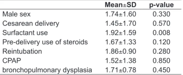
Tables 2 and 3 show there is no correlation between the MIP and RR variation and any of the clinical characteris-tics analyzed: sex, type of delivery, use of surfactant by the child, use of steroids by the mother, use of ventilator support (CPAP) in the post-extubation period, need for reintuba-tion, occurrence of bronchopulmonary dysplasia, gestational age, birth weight, weight at extubation, Apgar score at 5
Mean±SD p-value
Male sex 1.74±1.60 0.330 Cesarean delivery 1.45±1.70 0.570 Surfactant use 1.92±1.59 0.008 Pre-delivery use of steroids 1.67±1.33 0.120 Reintubation 1.86±0.90 0.280
CPAP 1.52±1.38 0.850
bronchopulmonary dysplasia 1.71±0.78 0.450
Table 3 - Variation in oxygen saturation (%) of preterm infants being weaned from mechanical ventilation
SD: standard deviation; CPAP: continuous positive airway pressure
minutes, duration of mechanical ventilation. However, pa-tients who used surfactant had a higher variation in oxygen saturation than the ones who did not use it (p=0.008).
Discussion
This study showed an 87% SatO2 increase in patients in the prone position, a datum conirmed by current literature, which indicates that this position improves oxygenation in children breathing spontaneously(9,16,23,24) and in children under
mechan-ical ventilation(25). This improvement in SatO
2 was not related
to any other variable in the study, with the exception of the use of surfactant. This suggests an intrinsic causal factor, such as the physiological and mechanical changes in the respiratory tract caused by postural change or the use of surfactant.
The better oxygenation in prone position might suggest a higher eficacy of the diaphragm during its contraction, generating more strength, improving ventilation and, thus, optimizing gas exchange. Previous studies show that the prone position increases the tidal volume(3,4), increases the
functional residual capacity(5) and leads to the stabilization
of the chest wall with more synchrony between thorax and abdomen(1). However, it was also shown that MIP is smaller
in prone position in comparison with supine(18). This study
also showed that MIP was smaller in prone in comparison
Supine Prone
p-value Mean±SD Mean±SD
RR (bpm) 57.0±11.4 53.6±12.6 0.072
SatO2 (%) 93.5±11.6 96.8±11.6 <0.001
MIP (cmH2O) 53.4±14.9 43.9± 5.5 <0.001
Table 1 - Measures of central tendency, dispersion and compa-rison of respiratory parameters in the supine and prone position in preterm infants being weaned from mechanical ventilation
SD: standard deviation; RR: respiratory rate; bpm: breaths per minute; SatO2: oxygen saturation; MIP: maximal inspiratory pressure
Respiratory rate MIP SatO2
r p-value r p-value r p-value
Gestational age (weeks) 0.012 0.940 -0.098 0.524 -0.002 0.989 Birth weight (g) -0.107 0.485 -0.151 0.321 -0.107 0.486 Extubation weight (g) -0.067 0.660 -0.104 0.495 -0.096 0.531 Mechanical ventilation (h) 0.001 0.992 0.151 0.321 0.077 0.617
Table 2 - Correlation and determination of coeficients between clinical characteristics and maximal inspiratory pressure, oxygen saturation and respiratory rate of preterm infants being weaned from mechanical ventilation
to supine position (p<0,0001). This decrease in inpiratory muscle strength in prone position might be due to the al-teration in the length-tension relationship of the diaphragm, which occurs with postural change(2).
The data found on respiratory rates are similar to the ones found in the literature, which found no inluence of the prone or supine position on RR(10,16,17).
This study was performed on prone and supine position with a head up tilt of 15º, what might have led to different results from others found. Differences between prone position with a head up tilt of 15º and supine position and a head up tilt of 0º were emphasized(26). Dimitriou et al(18) showed a difference in
MIP and SatO2 when they compared the prone position with a head up tilt of 0º with the supine position with different head up tilts of 0º and 45°. MIP was higher in supine with tilts of 0º and 45° and SatO2 was higher in prone and in supine with tilt of 45°. The position of the head may also affect the respiratory tract. The excessive lexion of the neck in newborns, especially preterm ones, can lead to airway obstruction and cause apnea(27).
Dimitriou et al(18) say there is no signiicant difference between
the supine position with the infant’s head in the midline and supine with the head turned to the right. In this study, prone position with head turned to the right and supine position with head in midline were used.
The time children stay in each position may also in-terfere in results. Some authors report effects of prone position in a short time, i.e., from 2 to 20 minutes(5,10).
Others indicated more benefits of the prone position, when the time children stay in it is longer, from 30 minutes to 48 hours(15,19).
The prone position, in this study, was performed with abdominal support, i.e., with abdominal restriction. Abdominal support in prone position has been described both in adults and in children. However, Wagaman
et al(28), assessing intubated newborns, compared the prone
position with free and restrict abdomen and concluded there is no signiicant difference between both positions related to the assessed parameters.
The MIP measurement technique also might inluence results. This is another controversial topic in literature, since MIP measures are not standardizes for children under ive years old. With regard to respiratory muscle testing, and speciically the MIP measures, the American Thoracic Society/European Respiratory Society (ATS/ERS)(29)
recom-mends, in the item measurement of muscle function in Intensive Care Unit, that the MIP of patients dependent on mechanical ventilation can be measured through a uni-directional valve, in which inspiration is blocked, causing the patient to reach the residual volume of the lung. Thus, the inspiratory pressure measured can be the maximum one. The highest MIP value generated usually occurs after 15–20 efforts or after 15–20 seconds of occlusion. MIP was measured in this study with a unidirectional valve con-nected to the endotracheal tube, which allows the child to inspire, but not to expire, thus increasing its lung volume to near total lung capacity. Some authors describe this technique in preterm infants(15,18). The occlusion for the
MIP measurement was performed throughout a period of 20 seconds, with a series of three occlusions, as reported above(19,20). Adopted procedures for MIP measurement,
period and methodology used to obtain SatO2 and RR measures may also have inluences the results.
In conclusion, smaller values of inspiratory pressure in prone position were found in comparison with supine position, besides and increase in oxygen saturation. Respiratory rate did not present a variation in prone position when compared to supine. However, further studies are needed for this position to be incorporated in the routine care of preterm infants.
References
1. Wolfson MR, Greenspan JS, Deoras KS, Allen JL, Shaffer TH. Effect of position on the mechanical interaction between the rib cage and abdomen in preterm infants. J Appl Physiol 1992;72:1032-8.
2. Rehan VK, Nakashima JM, Gutman A, Rubin LP, McCool FD. Effects of the supine and prone position on diaphragm thickness in healthy term infants. Arch Dis Child 2000;83:234-8.
3. Hutchison AA, Ross KR, Russell G. The effect of posture on ventilation and lung mechanics in preterm and light-for-date infants. Pediatrics 1979;64:429-32.
4. Adams JA, Zabaleta IA, Sackner MA. Comparison of supine and prone noninvasive measurements of breathing patterns in fullterm newborns. Pediatr Pulmonol 1994;18:8-12.
5. Numa AH, Hammer J, Newth CJ. Effect of prone and supine positions on
functional residual capacity, oxygenation, and respiratory mechanics in ventilated infants and children. Am J Respir Crit Care Med 1997;156:1185-9. 6. Richter T, Bellani G, Scott Harris R, Vidal Melo MF, Winkler T, Venegas JG
et al. Effect of prone position on regional shunt, aeration, and perfusion in experimental acute lung injury. Am J Respir Crit Care Med 2005;172:480-7. 7. Mure M, Martling CR, Lindahl SG. Dramatic effect on oxygenation in patients
with severe acute lung insuficiency treated in the prone position. Crit Care Med 1997;25:1539-44.
8. Prisk GK, Yamada K, Henderson AC, Arai TJ, Levin DL, Buxton RB et al. Pulmonary perfusion in the prone and supine postures in the normal human lung. J Appl Physiol 2007;103:883-94.
10. Schwartz FC, Fenner A, Wolfsdorf J. The inluence of body position on pulmonary function in low birthweight babies. S Afr Med J 1975;49:79-81.
11. Goto K, Mirmiran M, Adams MM, Langford RV, Baldwin RB, Boeddiker MA et
al. More awakenings and heart rate variability during supine sleep in preterm infants. Pediatrics 1999;103:603-9.
12. Ariagno RL, Mirmiran M, Adams MM, Saporito AG, Dubin AM, Baldwin RB. Effect of position on sleep, heart rate, variability, and QT interval in preterm infants at 1 and 3 months’ corrected age. Pediatrics 2003;111:622-5. 13. Kurlak LO, Ruggins NR, Stephenson TJ. Effect of nursing position on incidence,
type, and duration of clinically signiicant aponea in preterm infants. Arch Dis Child Fetal Neonatal Ed 1994;71:F16-9.
14. Heimler R, Langlois J, Hodel DJ, Nelin LD, Sasidharan P. Effect of positioning on the breathing pattern of preterm infants. Arch Dis Child 1992;67:312-4. 15. Leipälä JA, Bhat RY, Rafferty GF, Hannam S, Greenough A. Effect of posture
on respiratory function and drive in preterm infants prior to discharge. Pediatr Pulmonol 2003;36:295-300.
16. Antunes LC, Rugolo LM, Crocci AJ. Effect of preterm infant position on weaning from mechanical ventilation. J Pediatr (Rio J) 2003;79:239-44.
17. Levy J, Habib RH, Liptsen E, Singh R, Kahn D, Steele AM et al. Prone versus supine positioning in the well preterm infant: effects on work of breathing and breathing patterns. Pediatr Pulmonol 2006;41:754-8.
18. Dimitriou G, Greenough A, Pink L, McGhee A, Hickey A, Rafferty G et al. Effect of posture on oxygenation and respiratory muscle strength in convalescent infants. Arch Dis Child Fetal Neonatal Ed 2002;86:F147-50.
19. Fitzgerald DA, Mesiano G, Brosseau L, Davis GM. Pulmonary outcome in extremely low birth weight infants. Pediatrics 2000;105:1209-15.
20. el-Khatib MF, Baumeister B, Smith PG, Chatburn RL, Blumer JL. Inspiratory
pressure/maximal inspiratory pressure: does it predict successful extubation in critically ill infants and children? Intensive Care Med 1996;22:264-8. 21. Thiagarajan RR, Bratton SL, Martin LD, Brogan TV, Taylor D. Predictors
of successful extubation in children. Am J Respir Crit Care Med 1999;160:1562-6.
22. Baumeister BL, el-Kahatib M, Smith PG, Blumer JL. Evaluation of predictors of weaning from mechanical ventilation in pediatric patients. Pediatr Pulmonol 1997;24:344-52.
23. Bhat RY, Leipälä JA, Singh NR, Rafferty GF, Hannam S, Greenough A. Effect of posture on oxygenation, lung volume, and respiratory mechanics in premature infants studied before discharge. Pediatrics 2003;112:29-32.
24. Pelosi P, Tubiolo D, Mascheroni D, Vicardi P, Crotti S, Valenza F et al. Effects of prone position on respiratory mechanics and gas exchange during acute lung injury. Am J Respir Crit Care Med 1998;157:387-93.
25. Balaquer A, Escribano J, Roqué M. Infant position in neonates receiving mechanical ventilation. Cochrane Database Syst Rev 2006;18:CDOO3668. 26. Jenni OG, von Siebenthal K, Wolf M, Keel M, Duc G, Bucher HU. Effect
of nursing in the head elevated tild position (15 degrees) on the incidence of bradycardic and hypoxemic episodes in preterm infants. Pediatrics 1997;100:622-5.
27. Thach BT, Stark AR. Spontaneous neck lexion and airway obstruction during apneic spells in preterm infants. J Pediatr 1979;94:275-81.
28. Wagaman MJ, Shutack JG, Mommjian AS, Schwartz JG, Shaffer TH, Fox WW. Improved oxygenation and lung compliance with prone positioning of neonates. J Pediatr 1979;94:787-91.