8 8 Arq Bras Oftalmol. 2014;77(2):88-90
Original Article
The influence of body position on Bielschowsky’s test
A inluência da posição corporal sobre o teste de Bielschowsky
Carlos r. souza-Dias1, Mauro GolDChMit1,2, Fabio P. Moraes2, arthur JaMPolsky3
INTRODUCTION
The compensatory head tilt during certain oculomotor anomalies has the goal of reducing hypertropia, avoiding diplopia, and main taining fusion(1).
Theforced head tilt difference means the diference in the magni tude of vertical deviations when hypertropia is measured with the head tilted toward both shoulders; this primarily occurs in patients with superior oblique paresis and superior rectus contracture (Jam polsky’s syndrome(2)). In 1985, David Robinson(3), after a mathematical analysis of his Robinson’s model of the oculomotor plant with regard to large vertical deviations in the head tilt test, concluded that only a contractured superior rectus muscle could account for such a large
forced tilt difference in some patients with superior oblique palsy. The conventional mechanical explanation for Bieslschowsky’s test(4) is based on the otolith static relex, which is responsible for the counterrolling of eyes induced by head tilting toward the shoulders. When the head is tilted toward the right shoulder, for instance, the right eye tends to incycloduct, which is induced by the otolith system, ABSTRACT
Purpose: To investigate the veracity of Jampolsky’s statement that Bielschowsky’s head tilt test is inverted if performed with the patient in the upsidedown position and to interpret its neuromuscular mechanism.
Methods: We present a series of 10 patients selected from a referred sample who were diagnosed with superior oblique paresis.Hypertropia was measured in the primary position, with the head erect and tilted toward both shoulders with the patient in the erect, supine, and upsidedown positions. The last position was achieved by hanging the patient upsidedown.
Results: As expected, our results showed the veracity of Jampolsky’s statement. The
forced head tilt difference was inverted or significantly decreased when the test was performed in the upsidedown position. Moreover, in all patients, Bielschowsky’s phenomenon was neutralized in the supine body position, in which hypertropia with the head erect tended to vanish. In 3 patients, it disappeared completely.
Conclusions: This study showed that, in patients with superior oblique paresis, differences in the extent of hypertropia in Bielschowsky’s test tended to vanish when the test was performed with the patient in the supine position and invert when it was performed with the patient in the upsidedown position.
Keywords: Strabismus; Body positioning; Troclear nerve; Ophthalmoplegia
RESUMO
Objetivo: Investigar a veracidade da suposição de Jampolsky de que o teste de in-clinação da cabeça de Bielschowsky inverte-se caso seja realizado com o paciente de cabeça para baixo, e tentar interpretar o mecanismo neuromuscular envolvido.
Métodos: Apresentamos uma série de 10 pacientes portadores de paresia do oblíquo superior. Foi medida a hipertropia dos pacientes na posição primária do olhar e com a cabeça inclinada para cada um dos lados nas posições ereta, supina e de cabeça para baixo.
Resultados: Como esperado, nossos resultados confirmaram a suposição de Jampolsky; além disso, e em todos os pacientes, o fenômeno de Bielschowsky foi neutralizado em posição supina. As diferenças da magnitude da hipertropia ao teste de Bielschowsky diminuiram significativamente ou inverteram-se quando o paciente foi testado de cabeça para baixo.
Conclusões: Este estudo demonstrou que, nos pacientes com paresia do oblíquo superior, a hipertropia evidenciada pelo teste de Bielschowsky tende a desaparecer com o paciente na posição supina e a se inverter quando o teste é realizado com o paciente de cabeça para baixo.
Descritores: Estrabismo; Posicionamento do paciente; Nervo troclear; Oftal mo plegia
through the innervation of its intorsional muscles, namely the su perior rectus and superior oblique muscles. These muscles are antagonists in vertical and horizontal actions; therefore, when they are simultaneously innervated, they compensate for each other and result in no vertical or horizontal eye movement. However, they are synergistic when it comes to torsion. When they are simultaneously innervated by right head tilting, as in the case of right superior oblique palsy, the superior rectus muscle overcomes the depressor action of the weakened superior oblique muscle and elevates the eye(5).
Jampolsky stated, in 1994(6), that if Bielschowsky’s test is perfor med with the patient in an upsidedown position, the forced tilt difference
would give a mirror image, that is, an inverted one. This led to inte resting speculations about the otolith mechanism and the muscular mechanics of the forced head tilt difference.
This study aimed to investigate the veracity of this statement and interpret the underlying neuromuscular mechanism. Patients with superior oblique paresis were selected, and the deviation with the head tilted to both sides was measured with the patient in the erect, supine, and upsidedown positions.
Submitted for publication: May 29, 2013 Accepted for publication: October 23, 2013
Study conducted at Faculdade de Ciências Médicas da Santa Casa de Misericórdia de São Paulo, São Paulo, SP, Brazil.
1 Department of Ophthalmology, Faculdade de Ciências Médicas da Santa Casa de Misericórdia de
São Paulo, São Paulo, SP, Brazil.
2 Department of Ophthalmology, Instituto Cema, São Paulo, SP, Brazil.
3 Smith-Kettlewell Eye Research Institute, San Francisco, California, United States of America.
Funding: This study was supported by a grant from the Smith Kettlewell Eye Research Institute. Disclosure of potential conflicts of interest: None of the authors have any potential conflicts of
interest to disclose.
Corresponding author: Carlos R. Souza-Dia. Rua Cincinato Braga, 59 - Cj. 5 B2 - São Paulo (SP) - 01333-011 - Brazil - E-mail: [email protected]
Research Ethics Committee: Approved by the Comitê de Ética e Pesquisa of the Santa Casa de Misericórdia de São Paulo under the number 364/11.
Souza-Dias CR, et al.
8 9
Arq Bras Oftalmol. 2014;77(2):88-90 METHODS
This work was approved by the Ethics Committee of the Faculty of Medical Sciences of the Santa Casa de Misericórdia of São Paulo under the number 364/11. After the procedure was explained to the subjects (or legal guardians for minors) and accepted by free will, informed consent was obtained.
Ten patients [8 unilateral and two asymmetric bilateral (Patients 7 and 10), Table 1] consecutively examined in the Santa Casa and CEMA hospitals who presented with the clinical picture of superior oblique paresis were prospectively examined. The mean age was 31.4 ± 15.3 years (9–52 years).
The data necessary for diagnosing unilateral superior oblique paresis included the following: hypertropia of the afected eye that increased on the contralateral side with the head tilted toward the ipsilateral shoulder and decreased on ipsilateral side with the head tilted toward the contralateral shoulder (Bielschowsky’s test), ex cyclotropia, and a Vanisotropia. Asymmetric superior oblique paresis was diagnosed according to the criteria discussed by SouzaDias in another paper(7).
For this research, we measured the deviations in the primary po sition with the head erect and maximally tilted to both shoulders, with the patient in the erect, supine, and upsidedown positions. Ipsilateral superior rectus contracture was investigated during sur gery with the forced duction test (“knifeedge” maneuver, proposed by Jampolsky in 1978(8)).
After external, anterior segment, and fundus examinations and refractometry, a thorough ocular motility examination was perfor med, with special attention to the alternate prism and cover test in all positions of gaze and in the primary position with the head tilted toward both shoulders. All patients possessed 40” to 50” of stereoa cuity, which was assessed using the Titmus Fly Test®
(Titmus Optical, Petersburg, VI, USA), or were able to recognize the 3 igures on Lang’s test. All patients showed extorsion of the afected eye as diagnosed by the double Maddox test and, in some of them, fundoscopy.
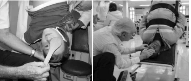
The upsidedown position was achieved in the 2 young patients by an adult hanging them by the legs and in the remaining 8 adults by utilizing a special slanting bed that was speciically built for this investigation (Figure 1).
In order to avoid misunderstanding, let us deine 2 terms. Hyper tropia of an eye indicates that the afected eye is deviated toward the top of the head, and hypotropia of an eye indicates that the afected eye is deviated toward the feet, regardless of the patient’s body position.
RESULTS
In all patients, the forced head tilt difference was neutralized or tended to vanish when the test was performed with the patient in the supine position, while it was inverted or signiicantly decreased in magnitude when the patient was in the upsidedown position.
As seen in table 1, there was a complete inversion (Patient 2) or an almost complete inversion (Patient 3) of the forced head tilt difference
when the test was repeated with the patient in the upsidedown position. In all the other patients, there was a clear tendency for inver sion. In Patient 1, for instance, the left hypertropia with the head tilted toward the right shoulder increased from 8∆ to 25∆, thus increasing by 17∆, while that with the head tilted toward the left shoulder decrea sed from 35∆ to 15∆, thus decreasing by 20∆. The forced tilt difference that measured 27∆ was reduced to 10∆, but in the opposite sense (it was inverted). Therefore, the total change was 37∆. In Patient 10, the hypertropia disappeared in the supine and upsidedown positions.
When the test was repeated with the patients in the supine posi tion, the forced head tilt difference tended to disappear in all patients. It was noteworthy that the hypertropia in the primary position ten ded to vanish when tested with the head erect and the body in the supine position. In Patients 3 and 10, the hypertropia disappeared. In Patient 9, in these body and head positions, there was a tendency for inversion of Bielschowsky’s test, but the hypertropia in the primary position remained (it was only decreased).
The measurement of the forced head tilt difference in the 10 pa tients in the primary position; with the body erect, supine, and upsidedown; and with the head erect and tilted toward both shoulders are listed in table 1.
DISCUSSION
If the otolith mechanism is considered as the explanation for the
forced head tilt difference, it is not diicult to explain the disappearan ce of the hypertropia when the patient is in the supine position. A probable explanation is that if the head is tilted toward the shoulders with the patient in this position, the hypertropia does not appear because the otoliths stimulate other areas inside the utricule; fur thermore, the force G vector (gravity) does not change its relative di rection within the utricle with the change in head position. It is more diicult to explain the tendency for the decrease in the magni tude of the hypertropia in the primary position with the patient in the supine position. Till date, no study regarding this phenomenon has been published. A possible mechanism is relaxation of all the
The influence of body position on Bielschowsky’s test
9 0 Arq Bras Oftalmol. 2014;77(2):88-90
cyclovertical muscles in this position. Its permanence, with only a small decrease in Patient 9, was probably caused by an ipsilateral superior rectus contracture.
However, in the upsidedown position, the sense of the force G vector into the utriculi is inverted, and it is not known if there is such an inversion mechanism in the utriculi. The otoliths stimulate an opposite area. Furthermore, considering the mechanical reasoning for the forced head tilt difference described above, with the patient in the upsidedown position, for the right eye to move toward the, the case of a right superior oblique palsy, there would have to be an imbalance of the vertical forces between the ipsilateral inferior rectus and inferior oblique of this eye (a weakness of the inferior oblique), for the inferior rectus to overcome its elevating action, and depress the eye, which did not exist in this case. On the contrary, in patients with superior oblique palsy, there is generally an ipsilateral inferior oblique overaction that can elevate the eye (to move toward the top of the head) instead of depressing it (to move toward the feet).
It is noteworthy that Wong et al.(9) found diferent data. In patients diagnosed with skew deviation, hypertropia decreased by >50% when measurements performed with the head erect and in the su pine position were compared. This was in contrast with the indings
Table 1. Measurement of hypertropia in the 3 head positions of Bielschowsky’s test (erect and tilted toward each of the shoulders), with the patients in the erect, supine, and upside-down positions
Patients
Bielschowsky’s test Body position
Erect Supine Upside-down Head position
Right Erect Left Right Erect Left Right Erect Left
01 8 25 35 25 15
02 3 25 5 0 +25 3
03 +25 +5 0 0 0 0 0 +20
04 0 10 12 2 6 12 3 5
05 +25 +25 +6 +3 +3 +3 +12 +20 +20
06 3 4 15 6 5 5 12 10 13
07 +15 +8 6 +5 0 +7 0 +2 +5
08 4 15 15 6 5 5 12 12 3
09 0 22 40 12 15 25 15 12 0
10 +15 +8 4 0 0 0 0 0 0
The measurements are in prism diopters. The symbol “+” indicates right hypertropia, while the symbol “” means left hypertropia. The afected eye is indicated by the side of the larger hypertropia in Bielschowsky’s test in the erect body position.
for patients with superior oblique palsy, in whom this decrease was smaller or absent. In all 8 patients who underwent measurements in the supine position, there was a decrease or disappearance in the
forced head tilt difference; the hypertropia in the primary position disappeared in 3 and decreased in the remaining, except in Patient 9 (Table 1).
In conclusion, the behavior of the cyclovertical extraocular mus cles during static head tilt toward the shoulders with the patient in the upsidedown and supine positions remains to be elucidated. This lack of knowledge suggests, as pointed out by Jampolsky in 1994(6), that the neurophysiology of the static vestibuloocular relexes and clinical head tilt test interpretations should be reexamined. The findings also explain the fact that the socalled Parks’ test frequently fails to diagnose diseases of the other cyclovertical muscles other than superior oblique palsy, as pointed out by Bicas & De Sordi(10), PrietoDíaz & SouzaDias(11), and SouzaDias & Goldchmit(12).
ACKNOWLEDGEMENT
The authors acknowledge Dr. Roberto Mitiaki Endo for his assis tance in the submission of this work to the Ethics Committee.
REFERENCES
1. Khawan E, Scott AB, Jampolsky A. Acquired superior oblique palsy. Arch Ophthalmol. 1967;77(6):7618.
2. Khawam E, Ghazi N, Salti H. “Jampolsky Syndrome”: superior rectus overactioncon tracture syndrome: prevalence, characteristics, etiology and management. Binocul Vis Strabismus Q. 2000;15(4):33142.
3. Robinson D. Bielschowsky head tilt test II – Quantitative mechanics of the Bielschowsky head tilt test. Vis Rev. 1985;25(12):19838.
4. Bielschowsky A. Lectures on motor anomalies. Hannover: Dartmouth College Public; 1943. p.73.
5. von Noorden GK, Campos E. Binocular vision and ocular motility. 6th ed. St. Louis: Mosby; 2002. p.417.
6. Jampolsky A. A new look at the head tilt test. In: Fuchs AF, Brandt TH, Buttner U, Zee DS editors. Contemporary ocular motor and vestibular research: a tribute to David A. Robinson. Stuttgart, Ger: Thieme Verlag; 1994. p.4329.
7. SouzaDias C. Asymmetrical bilateral paresis of the superior oblique muscle. JAAPOS. 2007;11(1):126.
8. Jampolsky AS. Surgical leashes and reverse leashes in strabismus surgical manage ment. In: Symposium on Strabismus. Transactions of the New Orleans Academy of Ophthalmol. St. Louis: Mosby; 1978. p.244.
9. Wong AM, Colpa L, Chandrakumar M. Ability of an uprightsupine test to diferen tiate skew deviation from other vertical strabismus causes. Arch Ophthalmol. 2011; 129(12):15705.
10. Bicas HE, De Sordi GB. Contradições nos resultados de testes diagnósticos de dese quilíbrios verticais. In: Anais do XVI Congresso Brasileiro de Oftalmologia, Campinas (SP); 1971. Conselho Brasileiro de Oftalmologia; 1971. p.132.
11. PrietoDíaz J, SouzaDias C. Estrabismo. Buenos Aires, Ediciones Cientíicas Argenti nas; 2005. p.324.