INTRODUCTION
Tumor necrosis factor alpha (TNF-α) is an inlammatory cytokine derived from T cells and macrophages, involved in the regulation of cellular apoptotic activity(1,2). Despite having a critical role in con-trolling inlammatory processes in the human body, raised levels of TNF-α have been associated with cellular and vascular malformations, edemas, and neurovascular degeneration(1,3,4). For instance, patients with non-infectious uveitis demonstrate increased levels of TNF-α, suggesting that TNF-α is a key mediator of uveal inlammation(5-7). Therefore, suppression of TNF-α activity through antagonist drug admi nistration ofers a promising treatment for patients with ocular inlammatory diseases(2,8-10).
ABSTRACT
Purpose: To assess the cytotoxicity and genotoxicity of intravitreal adalimumab treatment in an animal experimental model using cytological and molecular te chniques.
Methods: Eighteen rabbits were randomly assigned to three groups: control, adalimumab treatment, and placebo. Cytotoxicity on retinal cells was evaluated using flow cytometry assays to determine the level of apoptosis and necrosis. Ge-notoxicity was evaluated by comet assays to assess DNA damage, and quantitative real-time polymerase chain reaction (qPCR) was used to evaluate expression of apoptosis-inducing caspases (8 and 3).
Results: No cytotoxicity or genotoxicity was observed in any of the two treatment groups (adalimumab and placebo) following intravitreal administration compared with the control group. Flow cytometry analysis revealed that more than 90% of the cells were viable, and only a low proportion of retinal cells presented apoptotic (~10%) or necrotic (<1%) activity across all groups. Molecular damage was also low with a maximum of 6.4% DNA degradation observed in the comet assays. In addi-tion, no increase in gene expression of apoptosis-inducing caspases was observed on retinal cells by qPCR in both the adalimumab and placebo groups compared with the control group.
Conclusion: The use of adalimumab resulted in no detectable cytotoxicity or ge notoxicity on retinal cells for up to 60 days upon administration. These results therefore indicate that adalimumab may be a safe option for intravitreal application to treat ocular inflammatory diseases in which TNF-α is involved.
Keywords: Intravitreal injections; Retina; Antibodies, monoclonal, humanized/ to xicity; Apoptosis; Tumor necrosis factor alpha; Ani mals; Rabbits
RESUMO
Objetivo: Acessar a citotoxicidade e genotoxicidade do tratamento intravítreo de adalimumabe em um modelo experimental animal utilizando técnicas citológicas e moleculares.
Métodos: Dezoito coelhos foram aleatoriamente selecionados em três grupos:
con trole, tratamento intravítreo com adalimumabe e placebo. Os efeitos tóxicos nas células da retina foram avaliados através de ensaios de citometria de fluxo, para a de -terminação de atividade apoptótica e necrótica. A genotoxidade foi avaliada através de ensaios cometa para determinar danos ao DNA e através de PCR em tempo real para avaliar a expressão genética de caspases (8 e 3) promotoras de apoptose celular. Resultados: Não foram detectadas citotoxicidade e genotoxidade nos dois grupos de tratamento, adalimumabe e placebo, em comparação com o controle. A citometria de fluxo determinou que mais de 90% das células eram viáveis após o tratamento, e uma pequena quantidade de células da retina apresentaram apoptose (~10%) ou necrose (<1%) em todos os grupos. O dano molecular também foi baixo com uma degradação no DNA de no máximo 6,4% detectados nos ensaios cometa. Adicionalmente, não foram observados aumentos na expressão genética das caspases que induzem a apoptose através dos ensaios de PCR em tempo real.
Conclusão: O tratamento intravítreo com adalimumabe não promoveu nenhuma
citotoxicidade e genotoxicidade detectável em células da retina por até sessenta dias. Estes resultados, portanto, indicam que o adalimumabe pode ser uma opção segura para o tratamento de doenças oculares inflamatórias em que o TNF-α está envolvido.
Descritores: Injecções intravítreas; Retina; Anticorpos monoclonais humanizados/ toxicidade; Apoptose; Fator de necrose tumoral alfa; Animais; Coelhos
Adalimumab is a recombinant human monoclonal antibody spe-ciically inhibiting TNF-α activity. Systemic use of TNF-α inhibitors is widely recognized as a treatment for autoimmune(11) and rheumatic diseases(12). Subcutaneous adalimumab administration shows pro-mising clinical improvement in patients with refractive uveitis(13) and macular edema secondary to non-infectious uveitis(14). However, systemic administration of adalimumab and other anti-TNF agents has produced several side efects(15). Thus, the use of intravitreal in-jection is the preferred method for treating ocular diseases because it provides higher drug concentrations with fewer injections of low dosage, preventing the risk of systemic exposure. However, few studies on the safety of intravitreal use of adalimumab have been
Cytotoxicity and genotoxicity of intravitreal adalimumab administration
in rabbit retinal cells
Citotoxicidade e genotoxicidade da administração de adalimumabe intravítreo nas células
da retina de coelhos
Álcio coutinhode Paula1, Marcos Pereirade Ávila1, david leonardo cruvinal isaac1, rodrigo salustiano1, aliny Pereirade liMa2,
Francyelli Mariana Mello2, FlÁviade castro Pereira2, Pedro henriquede Paula silva3, elisângelade Paula silveira lacerda2
Submitted for publication: November 24, 2014 Accepted for publication: February 6, 2015
1 Ophthalmic Center Reference (CEROF), Federal University of Goiás (UFGO), Goiânia, GO, Brazil. 2 Molecular Genetics and Cytogenetics, Institute of Biological Science, Federal University of Goiás
(UFGO), Goiânia, GO, Brazil.
3 Instituto de Ciências Farmacêuticas, Estudos e Pesquisa (ICF) Goiânia, GO, Brazil.
Funding: No specific financial support was available for this study.
Disclosure of potential conflicts of interest: None of the authors have any potential conflict of interest to disclose.
Corresponding author: Álcio Coutinho de Paula. CEROF/UFG. 1a Avenida, s/n - Setor Leste
Uni-versitário - Goiânia, GO - 74605-020 - Brazil - E-mail: [email protected]
published; for these, electroretinography and histological techniques have been used(14,16-18). These techniques cannot detect toxicity at the submicroscopic level and therefore we tested new methods to address intravitreal adalimumab toxicity at the cellular and molecular level in this study.
The purpose of this study was to evaluate, for the irst time, the toxicity of adalimumab intravitreal administration in an animal mo del using cytological and molecular techniques. We administered adali-mumab intravitreously in rabbits to obtain live retina samples, with a view to determine apoptotic activity at the cellular level using flow cytometry. At the molecular level, we assed DNA damage using co met assays and expression of apoptosis-inducing caspases using quantitative real-time polymerase chain reaction (qPCR).
METHODS
M
EDICALPROCEDURESANDEXPERIMENTALDESIGNEighteen New Zealand albino male rabbits weighing 2.5-3.0 kg were used to analyze cytotoxic and genotoxic efects of intravitreal adalimumab injections. The experimental procedures followed the ARVO statement for the Use of Animals in Ophthalmic and Vision Re-search as well as our institutional guidelines. Animals were anestheti-zed before performing any medical procedures. They were subjected to dilated fundus examination to determine overall ocular health at the beginning of the experiment and after 30 and 60 days using eye drops of 10% phenylephrine (Allergan® Pharmaceuticals, San Diego, CA, USA) and 1% tropicamide (Mydriacyl®, Alcon Laboratories, Fort Worth, TX, USA). We used an indirect binocular ophthalmoscope (Welch Allyn, Skaneateles Falls, NY, USA) with 20 diopter lenses (Volk Optical, Mentor, OH, USA) to examine the vitreous cavity, retina, and opacity of the medium. Only animals with healthy eyes were selected for the study.
Animals were randomly allocated to three groups: group 1 was the control (no injections, n=2), group 2 received adalimumab in jec-tions (n=8), and group 3 placebo injecjec-tions (n=8). Animals in group 2 were administered 0.1 mL (0.5 mg) of adalimumab (Humira®, Abbott, Abbott Park, IL, USA) via the right eye. The procedure was performed by inserting a 30-gauge needle via pars plana, 1-mm distant from the limbus. The treatment was reapplied after 30 days. Animals in group 3 were administered the same volume (0.1 mL) of a balanced saline solution (BSS®, Alcon, Fort Worth, TX, USA) using the same method, which was also reapplied after 30 days.
R
ETINADISSECTIONANDANALYSISTo perform the biological assays, the retinas of all study animals were dissected. Immediately after eye enucleation, an ocular dissection was performed and retina samples were obtained by the dislocation method. Retinal cell samples were analyzed by low cytometry, co -met assays, and qPCR at the Molecular Genetics and Cytogenetic Laboratory at the Federal University of Goiás, Goiânia, Brazil.
F
LOWCYTOMETRYDeath of retinal cells was determined using an Annexin V-FITC Apoptosis Detection Kit (Sigma-Aldrich, St. Louis, Mo, USA), according to the manufacturer’s instructions. Detection of apoptosis was based on the binding properties of phosphatidylserine annexin to the cell membrane (Annexin V+ to early and Annexin V- to late apoptotic cells), whereas the detection of necrosis was based on the binding properties of propidium iodide to cell DNA. Isolated cells (100 µL) were suspended in 400 µL of bufer solution and subsequently 5 µL of Annexin and 1 µL of propidium iodide were added. Flow cytometry analyses were performed using a FACSCalibur™ (BD Biosciences, San Jose, CA, USA) low cytometer in conjunction with the software Diva ModFit®.
C
OMETASSAYDNA damage was assessed by comet assays, a single-cell gel elec-trophoresis method, as previously described(19). For the comet assays, retinal samples were placed in Falcon® tubes with 500 µL of 100% trypsin solution (Cultilab, Campinas, SP, Brazil) for 5 min. Subsequen-tly, 500 µL of culture medium supplemented with fetal bovine serum (10%) was added. A 20-µL cell suspension was homogenized with 100 µL of low-melting-point agarose (0.5%) spread onto microscope slides pre-coated with normal-melting-point agarose (1.5%). After 10 min at 4°C, slides were immersed in a cold lysis solution (2.4 M NaCl, 100 mM EDTA, 10 mM Tris, 10% DMSO, and 1% Triton-X, pH 10) for 24 h. After lysis, slides were placed in an electrophoresis chamber and covered with electrophoresis bufer (300 mM NaOH/1 mM EDTA, pH >13) for 20 min to allow for DNA unwinding.
The electrophoresis proceeded for 20 min (25 V and 300 mA). Subsequently, slides were submerged for 15 min in a neutralization bufer (0.4M Tris-HCl, pH 7.5), dried at room temperature, and ixed in 100% ethanol for 5 min. Slide staining was performed immedia-tely before analysis with ethidium bromide (20 µg/mL). Slides were prepared in duplicates and 100 cells were screened per sample (50 cells from each slide) using a luorescence microscope (Leica, Wetzlar, Germany) interfaced with a computer.
Nucleus analysis was performed using the image software Comet Score ver. 15 (TriTek Corp., Sumerduck, VA, USA), according to the migration of DNA fragments as follows: class 0 (no damage), class 1 (little damage with a short tail length smaller than the diameter of the nucleus), class 2 (medium damage with a tail length one or two times the diameter of the nucleus), class 3 (signiicant damage with a tail length between two and a half to three times the diameter of the nucleus), and class 4 (signiicant damage with a long tail of damage greater than three times the diameter of the nucleus)(20). A damage index (DI) value was assigned to each comet assay according to for-mula (1), where n=number of cells in each class analyzed.
DI=(0 x n0) + (1 x n1) + (2 x n2) + (3 x n3) + (4 x n4) (1)
DI may range from 0 (completely undamaged; 100 cells x 0) to 400 (maximum damage; 100 cells x 4)(21).
Q
PCR
The molecular markers used to evaluate the apoptotic activity included the expression of the messenger RNA (mRNA) of caspase 8 (apoptosis initiator) and caspase 3 (apoptosis efector). Retinal cell mRNAs were extracted using TRIzol reagent (Sigma-Aldrich, St. Louis, MO, USA), according to the manufacturer’s instructions. Total mRNA (2 µg) was then used to produce complementary DNA (cDNA) using a random initiator (Applied Biosystems, Foster City, CA, USA) with a total reaction mixture of 20 μL. The primer sequences used were 5’-ACGAAACCTCCGTGGACGCAA-3’ and 5’-AGACCGGGACGACATTC CAGTG-3’ (185 bp) for caspase 3 and 5’-TGGCAGCAGATGATGACAA TGGTG-3’ and 5’-TGGAAGCACTGTCAGAAACAGCAC-3’ (135 bp) for caspase 8. The qPCR was performed using a Line-gene K Real-time PCR Detection System (Bioer Technology, Binjiang, China) in total volumes of 20 μL reaction mixture: 2 μL of cDNA, 10 μL of SYBR Green PCR Master Mix (LGC Biotecnology, Teddington, Middlesex, UK), and 2 μL of each primer (400 nM). The qPCR was initiated at 95°C for 40 thermal cycles of 15 min each, followed by 15 s at 55°C and 30 s at 72°C. All qPCR analyses were performed in triplicates.
S
TATISTICALANALYSISthe comet assays (comet tail length and damage index) was analyzed using one-way ixed efect analyses of variances (ANOVA–SYSTAT Ver. 12). For the ANOVA, assumptions of homogeneity of variance and normality were assessed by scatter plots and normal curves of residuals, respectively(22). Finally, the expression of caspase 8 and 3 as measured by qPCR was also analyzed using PERMANOVA, with the same operational inputs used for the abovementioned cytotoxicity analyses above.
RESULTS
C
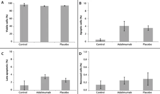
ELLULARAPOPTOSISANDNECROSISANALYSISThe parameters of cell viability, apoptosis, late apoptosis, and ne-crosis were determined by low cytometry analysis. There were no signiicant diferences in any of these parameters between the healthy (control), adalimumab injection, and the placebo injection groups (PERMANOVA, F4,29=0.859, p>0.05). The viability of retinal cells was similar for all groups, with over 90% viable cells detected by low cytometry (Figure 1 A). The mean percentage of early and late cell apoptosis (Figures 1 B and 1 C respectively) was lower for the control group (0.51 ± 0.50 and 1.37 ± 1.16, respectively) than for both the ada-limumab (4.05 ± 1.23 and 3.58 ± 0.52, respectively) and placebo (3.52 ± 0.58 and 2.65 ± 0.51, respectively) groups. However, no signiicant diferences were detected. The proportion of cell necrosis detected by low cytometry was very low, with values of 0.15 ± 0.12% for the controls, 0.26 ± 0.08% for the adalimumab group, and 0.30 ± 0.15 % for the placebo group (Figure 1 D).
DNA
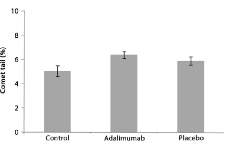
DAMAGEANALYSISLow DNA damage was observed after the intravitreal injections with adalimumab and placebo. The comet assays showed that there was no signiicant diference in DNA damage (tail length) between the control, adalimumab, and placebo groups (ANOVA, F2,18=2.437, p>0.05, Figure 2). The percentage of tail length was similar at 5.0 ± 0.42 for the control group, 6.4 ± 0.28 for the adalimumab group, and 5.0 ± 0.36 for the placebo group (Figure 2). The DI was also low for all groups (<40), with no signiicant diferences (ANOVA, F2,16=3.312, p>0.05) in DI between the controls (30.6 ± 2.3), the adalimumab group (38.6 ± 3.0), and the placebo group (28.5 ± 1.8; Figure 3).
A
POPTOSIS-
INDUCINGCASPASES8
AND3
EXPRESSIONANALYSISTo determine the values of mRNA expression for both caspases (8 and 3) in retinal cells, we used healthy animals (controls) as a calibra-tor group with an absolute genetic expression value of 1, with values over and below representing higher and lower gene expression, res-pectively. There were no signiicant diferences between the control, adalimumab, and placebo groups (PERMANOVA F2,18=0.8482 and p>0.05). Gene expression in the adalimumab group was 1.26 and 0.95 for caspase 8 and 3, respectively (Figure 4). For the placebo group, the values were 1.19 for caspase 8 and 0.48 for caspase 3 (Figure 4).
DISCUSSION
In this study, we detected no signiicant cytological or molecular toxicity to the retinal cells; therefore, these results provide strong evi-dence that adalimumab is safe for intravitreal treatment of non-infec-tious ocular inlammatory diseases. Our data show that ada limumab treatment results in only slight cytotoxic and genotoxic efects on rabbit retinal cells for up to 60 days, comparable with the efects ob-served in the control and placebo groups. Cellular apop to tic activity was very low with >90% viable retinal cells obser ved by low cytome-try. Furthermore, the comet assay and qPCR de mons trated that DNA damage was minimal and expression of the apop to sis-inducing caspases 8 and 3 was low.
The use of intravitreal adalimumab has been evaluated previously in animal models by dose dependence studies. The administration of doses up to 5 mg of adalimumab produced no functional or structu-ral ocular toxicity(16-18). These studies evaluating adalimumab toxicity have, however, used electroretinography and histological methods, which cannot detect at sub-microscopic levels. Therefore, comple-mentary studies using cellular and molecular techniques should be used for early detection of cytotoxic and genotoxic efects such as the onset of cellular apoptosis or expression of caspases. Furthermore, we used a longer time span of 60 days compared with the span of 14 or 42-days previously used. This allows a longer contact between the substance and the retina and more closely resembles the clinical situation.
Based on the systemic adalimumab administration(18), a study suggested that 0.5 mg may be the therapeutically appropriate dose for intravitreal treatment. Using low cytometry analysis, we
conir-Figure 1. Flow cytometry analysis showing the percentage of viable (A), apoptotic (B), late apoptotic (C), and necrotic (D) cells. Error bars (±standard error). Note the change of y-axis scale.
A B
Molecular assays have been increasingly used to determine ge-notoxicity of novel chemicals and pharmaceuticals(26). The comet assay is a microgel electrophoresis technique that measures DNA da mage at single-cell level(19). Single-cell gel electrophoresis (comet assay) has proven to be a highly sensitive method to assess DNA damage induced by several agents(27). For example, TNF-α-induced experiments in pancreatic cells resulted in a 50% increase in comet tail length(28). The use of etanercept, another TNF-α antagonist drug, has also resulted in DNA damage as observed by comet assay(29). In our study, we observed only small increases in comet tail length in the adalimumab and placebo groups compared with the control group. Large molecules, such as monoclonal antibodies, have limited access to the genetic material via cellular membranes and generally promo-te low molecular damage(26). Therefore, the minimal DNA damage observed here shows that adalimumab confers no genotoxicity on retinal cells for up to 60 days.
Evaluation of the molecular mechanisms that induce apoptosis is also an efective strategy for drug genotoxicity evaluation. The process of apoptotic cell death involves the activation of caspase pro-teases. Caspase 8 is an initiator of cellular death signaling pathways, whereas caspase 3 is an efector caspase linked to the process of cellular destruction that accompanies apoptotic signals(24). The use of adalimumab and other anti-TNF agents such as inliximab may increase the expression of apoptosis-inducing caspases in response to drug administration in in vitro studies of human monocytes and
in vivo animal studies(24,30). In this study, however, there were no sig niicant increases in the expression of these apoptosis-inducing caspases in retinal cells in any of the treatment groups (adalimumab and placebo) compared with the control group. These contrasting re-sults may relect diferences in drug administration type and suggest that intravitreal adalimumab treatment is a less toxic administration method than subcutaneous injections.
CONCLUSION
In conclusion, we have demonstrated that the use of adali-mumab results in no detectable cytotoxicity and genotoxicity in rabbit retinal cells for up to 60 days after intravitreal administration, in dica ting that this treatment is a safe option applicable to human cli nical studies that aim to determine the eicacy of adalimumab for various ocular inlammatory diseases where TNF is implicated. Finally, the use of cytological and molecular techniques provides early and more reliable detection of drug toxicity, ofering new approaches in ophtomological research.
REFERENCES
1. McDermott MF. TNF and TNFR biology in health and disease. Cell Mol Biol. 2001; 47(4):619-35.
2. Joussen AM, Poulaki V, Qin W, Kirchhof B, Mitsiades N, Wiegand SJ, et al. Retinal vas cular endothelial growth factor induces intercellular adhesion molecule-1 and en-dothelial nitric oxide synthase expression and initiates early diabetic retinal leukocyte adhesion in vivo. Am J Pathol. 2002;160(2):501-9.
3. Barber AJ, Gardner TW, Abcouwer SF. The signiicance of vascular and neural apopto-sis to the pathology of diabetic retinopathy. Invest Ophthalmol Vis Sci. 2011;52(2): 1156-63.
4. Demircan N, Safran B, Soylu M, Ozcan A, Sizmaz S. Determination of vitreous inter-leukin-1 (IL-1) and tumour necrosis factor (TNF) levels in proliferative diabetic re ti nopathy. Eye (Lond). 2005;20(12):1366-9.
5. Santos Lacomba M, Marcos Martin C, Gallardo Galera JM, Gómez Vidal MA, Collantes Estevez E, Ramirez Chamond R, et al. Aqueous humor and serum tumor necrosis factor-α in clinical uveitis. Ophthalmic Res. 2001;33(5):251-5.
6. Pérez-Guijo V, Santos-Lacomba M, Sánchez-Hernández M, Castro-Villegas MC, Gallar-do-Galera JM, Collantes-Estévez E. Tumour necrosis factor-alpha levels in aqueous humour and serum from patients with uveitis: the involvement of HLA-B27. Curr Med Res Opin. 2004;20(2):155-7.
7. Sugita S, Takase H, Taguchi C, Mochizuki M. The role of soluble TNF receptors for TNF-α in uveitis. Invest Ophthalmol Vis Sci. 2007;48(7):3246-52.
8. Foeldvari I, Nielsen S, Kümmerle-Deschner J, Espada G, Hornef G, Bica B, et al. Tumor necrosis factor-alpha blocker in treatment of juvenile idiopathic arthritis-associated Figure 2. Comet tail length percentage as determined by comet assay. Error bars (±
stan-dard error).
Figure 3. Damage index of retinal cells as determined by comet assay. Error bars (± stan-dard error).
Figure 4. Gene expression of caspases 8 and 3 as determined by quantitative quantitati-ve real-time PCR. Error bars (±standard error).
uveitis refractory to second-line agents: results of a multinational survey. J Rheuma-tol. 2007;34(5):1146-50. Comment in: Nat clin Pract RheumaRheuma-tol. 2007;3(11):608-9. 9. Biester S, Deuter C, Michels H, Haefner R, Kuemmerle-Deschner J, Doycheva D, et al.
Adalimumab in the therapy of uveitis in childhood. Br J Ophthalmol. 2007;91(3):319-24. Comment in: Br J Ophthalmol. 2007; 91(3):274-6.
10. Neri P, Zucchi M, Allegri P, Lettieri M, Mariotti C, Giovannini A. Adalimumab (Humira™): a promising monoclonal anti-tumor necrosis factor alpha in ophthalmology. Int Ophthalmol. 2011;31(2):165-73.
11. Menter A, Tyring SK, Gordon K, Kimball AB, Leonardi CL, Langley RG, et al. Adalimu-mab therapy for moderate to severe psoriasis: a randomized, controlled phase III trial. J Am Acad Dermatol. 2008;58(1):106-15.
12. Weinblatt ME, Keystone EC, Furst DE, Moreland LW, Weisman MH, Birbara CA, et al. Adalimumab, a fully human anti-tumor necrosis factor α monoclonal antibody, for the treatment of rheumatoid arthritis in patients taking concomitant methotrexate: the ARMADA trial. Arthritis Rheum. 2003;48(1):35-45. Comment in: Clin Exp Rheumatol. 2004;22(1):34-5.
13. Diaz-Llopis M, García-Delpech S, Salom D, Udaondo P, Hernández-Garfella M, Bosch-Morell F, et al. Adalimumab therapy for refractory uveitis: a pilot study. J Ocul Pharmacol Ther. 2008;24(3):351-61. Comment in: J Ocul Pharmacol Ther. 2008;24(6): 613-4; author reply 614.
14. Androudi S, Tsironi E, Kalogeropoulos C, Theodoridou A, Brazitikos P. Intravitreal ada-limumab for refractory uveitis-related macular edema. Ophthalmology. 2010;117(8): 1612-6.
15. Scheinfeld N. A comprehensive review and evaluation of the side efects of the tumor necrosis factor alpha blockers etanercept, inliximab and adalimumab. J Dermat Treat. 2004;15(5):280-94. Comment in: J Dermatolog Treat. 2004;15(5):279.
16. Manzano RP, Peyman GA, Carvounis PE, Damico FM, Aguiar RG, Ioshimoto GL, et al. To-xicity of high-dose intravitreal adalimumab (Humira) in the rabbit. J Ocul Pharmacol Ther. 2011;27(4):327-31.
17. Tsilimbaris M, Diakonis VF, Naoumidi I, Charisis S, Kritikos I, Chatzithanasis G, et al. Eva-luation of potential retinal toxicity of adalimumab (Humira). Graefes Arch Clin Exp Oph thalmol. 2009;247(8):1119-25.
18. Myers AC, Ghosh F, Andréasson S, Ponjavic V. Retinal function and morphology in the
mabbit eye after intravitreal injection of the TNF alpha inhibitor adalimumab. Curr Eye Res. 2014;39(11):11106-16.
19. Singh NP, McCoy MT, Tice RR, Schneider EL. A simple technique for quantitation of low levels of DNA damage in individual cells. Exp Cell Res. 1988;175(1):184-91. 20. Kobayashi H, Sugiyama C, Morikawa Y, Hayashi M, Sofuny T. A comparison between
manual microscopic analysis and computerized image analysis in the single cell gel electrophoresis assay. MMS Commun. 1995;3:103-15.
21. Tice R, Agurell E, Anderson D, Burlinson B, Hartmann A, Kobayashi H, et al. Single cell gel/comet assay: guidelines for in vitro and in vivo genetic toxicology testing. Environ Mol Mutagen. 2000;35(3):206-21.
22. Quinn GP, Keough MJ. Experimental design and data analysis for biologists. Cambridge: Cambridge University Press; 2002.
23. Shen C, Assche G, Colpaert S, Maerten P, Geboes K, Rutgeerts P, et al. Adalimumab induces apoptosis of human monocytes: a comparative study with inliximab and eta nercept. Aliment Pharmacol Ther. 2005;21(3):251-8.
24. Shen C, Van Assche G, Rutgeerts P, Ceuppens JL. Caspase activation and apoptosis induction by adalimumab: demonstration in vitro and in vivo in a chimeric mouse model. Inlamm Bowel Dis. 2006;12(1):22-8.
25. Vigna-Pérez M, Abud-Mendoza C, Portillo-Salazar H, Alvarado-Sánchez B, Cuevas-Orta E, Moreno-Valdés R, et al. Immune efects of therapy with Adalimumab in patients with rheumatoid arthritis. Clin Exp Immunol. 2005;141(2):372-80.
26. Robuck PR, Wurzelmann JI. Understanding the drug development process. Inlamm Bowel Dis. 2005;11(1):S13-6.
27. Rojas E, Lopez M, Valverde M. Single cell gel electrophoresis assay: methodology and applications. J Chrom B: Biom Sci App. 1999;722(1):225-54.
28. Delaney CA, Pavlovic D, Hoorens A, Pipeleers DG, Eizirik DL. Cytokines induce deoxyribonucleic acid strand breaks and apoptosis in human pancreatic islet cells 1. En docrinology. 1997;138(6):2610-4.
29. Demirkaya E, Cok I, Durmaz E, Ulutas OK, Ayaz NA, Besbas N, et al. Genotoxicity of an-ti-tumor necrosis factor therapy in patients with juvenile idiopathic arthritis. Arthritis Care Res (Hoboken). 2010;62(1):73-7.
30. Shen C, Maerten P, Van Assche G, Geboes K, Rutgeerts P, Ceuppens J. A fully human anti-TNF mAb adalimumab (D2E7) induces caspase-dependent apoptosis of human peripheral blood monocyte and T cells. Gastroenterology. 2004;126:A153.