Original article
genes in endometrium, myometrium and vagina
of postmenopausal women treated with estriol
Expresión de los genes receptores de estrógeno y progesterona en el endometrio,
miometrio y vagina en mujeres posmenopáusicas tratadas con estriol
Magdalena Bry
ś
1, Krzysztof Szyłło
2, Hanna Romanowicz-Makowska
3, Zbigniew Dobrowolski
4, Izabela Masłowska
5, Wanda Krajewska
6Department of Cytobiochemistry, University of Lodz, Lodz, Poland
1PhD. Associate professor and project leader, Department of Cytobiochemistry, University of Lodz, Lodz, Poland.
2MD. Assistant professor, Department of Surgical Gynecology, Research Institute of the Polish Mothers’ Memorial Hospital, Lodz, Poland. 3MD. Pathologist, Department of Pathology, Research Institute of the Polish Mothers’ Memorial Hospital, Lodz, Poland.
4MD. Gynecologist, Department of Surgical Gynecology, Research Institute of the Polish Mothers’ Memorial Hospital, Lodz, Poland. 5PhD. Researcher, Department of Cytobiochemistry, University of Lodz, Lodz, Poland.
6PhD. Professor and supervisor, Department of Cytobiochemistry, University of Lodz, Lodz, Poland.
ABSTRACT
CONTEXT AND OBJECTIVE: Estriol is an estrogen with considerably weaker stimulatory effects on endometrial proliferation than estradiol. A study was conducted to determine the level of estrogen receptors (ERs) and progesterone receptors (PRs) in women who received 14-day vaginal estriol therapy, compared with those who did not receive this therapy. ER and PR gene expression was analyzed in the endometrium, myometrium and vagina of postmenopausal women treated with estriol.
DESIGN AND SETTING: Analytical cross-sectional study, at the Research Institute of the Polish Mothers’ Memorial Hospital, Lodz, Poland.
METHODS: Twenty-seven postmenopausal women (57-74 years of age) were included in the study. All of them were waiting for per vaginam hysterectomy or plastic surgery on the vagina and perineum because of uterine prolapse. ER and PR gene expression was determined by means of the technique of reverse transcription polymerase chain reaction (RT-PCR).
RESULTS: In the estriol-treated patients, in comparison with the control group, a signiicant increase in ER gene expression was observed in the endometrium and vagina, while enhanced PR gene expression was found in the endometrium. However, under histological examination of the endometrium, estrogen stimulation of low and medium degree was diagnosed for 21.4% and 14.3% of the estriol-treated women, respectively.
CONCLUSION: The results obtained suggest that the women who received 14 days of treatment with vaginal estriol had higher ER and PR mRNA levels. No difference between these groups regarding endometrial proliferation was observed.
RESUMÉN
CONTEXTO Y OBJETIVO: El estriol es un estrógeno con un efecto estimulatorio bastante más débil sobre la proliferación endometrial que el estradiol. Se realizó un estudio para determinar los efectos de una terapia vaginal de 14 días con estriol, sobre el nivel de receptores de estrógeno (ER) y receptores de progesterona (PR), comparado con mujeres sin esa terapia. La expresión de los genes de ER y PR se analizó en el endometrio, miometrio y vagina de mujeres posmenopáusicas tratadas con estriol.
DISEÑO Y UBICACIÓN: Estudio Transversal analítico, en el Instituto de Investigación del Hospital de la Madre Polaca en Lodz, Polonia.
MÉTODOS: Se incluyeron veintisiete mujeres posmenopáusicas (de 57 a 74 años) en el estudio. Todas ellas estaban en espera de una histerectomía per vaginam o de cirugía plástica de la vagina y del perineo debido a un prolapso del útero. La expresión de genes de los receptores ER y PR se estableció por la técnica de RT-PCR.
RESULTS: En las pacientes tratadas con estriol en comparación con el grupo de control, se observó un aumento signiicativo de la expresión del gen de ER en el endometrio y la vagina, mientras que un aumento de la expresión del gen de PR se encontró en el endometrio. De todas formas, en el examen histológico del endometrio, se diagnosticó estimulación estrogénica de bajo y medio grado en el 21.4% y en el 14.3% de las mujeres tratadas con estriol, respectivamente.
CONCLUSIONES: Los resultados obtenidos sugieren que un tratamiento de 14 días con estriol en pacientes, aunque aumentan el nivel de ER y de PR mRNA, tiene muy poco o ningún efecto sobre la proliferación endometrial.
KEY WORDS: Receptors, estrogen. Receptors, progesterone. Estriol.
Hormone replacement therapy. Reverse transcriptase polymerase chain reaction.
PALABRAS-CLAVE: Receptores estrogénicos. Receptores de progesterona. Estriol.
Terapia de reemplazo de hormonas.
INTRODUCTION
Progress in medicine and general improvement in socioeconomic conditions have resulted in longer lifetimes and an increased population of postmenopausal women. he postmenopausal period is a natural pe-riod in women’s lives during which the processes of ageing take place. Hormonal and psychosomatic disturbances intensify. With regard to hormones, hypoestrogenism dominates, together with increased levels of gonadotropins and a drop in the level of androgens.1
Estrogens play a crucial role in regulating the physiology of breast tis-sue and the endometrium.2-4 Furthermore, estrogen has been implicated in the initiation and progression of neoplasms of these tissues. Estrogens mediate their efects through estrogen receptor isoforms and isotypes.5 Both estrogen receptors (ERs) and progesterone receptors (PRs) have two subtypes, i.e. ER-alpha and beta, and PR-A and B, respectively.6 hese subtypes difer in function and expression, and recent reports have corre-lated changes in the normal proportions of these isoforms with neoplas-tic states. he potential clinical relevance of diferential ER-isotype ex-pression has also been discussed with regard to antihormonal therapy.7
Estrogens have neuroprotective and antiapoptotic properties. How-ever, the issue of what involvement the ER-dependent genomic pathway has in these efects still remains controversial.8,9
ERs and PRs are members of the nuclear receptor superfamily of li-gand-dependent transcription factors. It has been found that, in the ab-sence of ligands, ERs and PRs located in target cells are associated with the complex of heat shock proteins and chaperones. Upon ligand bind-ing, the receptors undergo conformational changes that enable spontane-ous dimerization. his event facilitates the interaction of the receptors with speciic DNA enhancer sequences located within the regulatory regions of target genes that regulate estrogen or progesterone-responsive genes.10-13
ER and PR gene expression may be regulated by estriol (E3) in the uterus and other reproductive tissues such as the vagina. Estriol deriva-tives are commonly used in therapy. Estriol has been considered to be an antagonist of estradiol.14-17
he data so far available concerning estriol action on the endometrium, myometrium, vagina and breast are ambiguous, and they provide contra-dictory information.18-21 It seems that diferences in the ainity, speciicity and concentrations of ERs in individual tissues are of much greater im-portance, together with changes in efector tissues that lead to diferent re-sponses.
OBJECTIVE
In the present study, the inluence of 14-day vaginal estriol admin-istration on ER and PR messenger ribonucleic acid (mRNA) expression in the endometrium, myometrium and vagina was investigated within the context of hormonal endometrial stimulation.
METHODS
Patients
Twenty-seven postmenopausal patients were included in the study. All of them were waiting for per vaginam hysterectomy or plastic surgery
on the vagina and perineum because of uterine prolapse. All the patients were treated and operated in the Department of Surgical Gynecology, Research Institute of the Polish Mothers’ Memorial Hospital between 2000 and 2002. hirteen patients were singled out as the control group. he others were treated daily with 0.5 mg of estriol (Ovestin® cream, made by Organon, Poland, applied vaginally with a calibrated applica-tor) for two weeks before the operation. he last dose of the drug was administered around 48 hours before the operation. he presence of oncological diseases was ruled out by means of anamnesis, physical ex-amination and laboratory investigation. Before the operation, all the pa-tients underwent fractionated uterine curettage prior to hormonal treat-ment. hey also underwent endometrial ultrasonography before and af-ter the treatment. Postmenopausal status was deaf-termined on the basis of serum follicle stimulating hormone (FSH) levels. Moreover, for each patient, a cytohormonal examination was conducted, and the matura-tion index was determined before and after estriol treatment.
he operations were performed in accordance with standard proce-dures. During the operation, immediately after the uterus and a small sleeve of vaginal tissue were removed, these tissues were placed in ice and transferred to the Department of Pathology. he body of the uterus was dissected and samples of the endometrium and myometrium were taken. hese, together with samples of vaginal tissues, were stored in three sepa-rate containers at – 70 °C until the biochemical investigation had been completed. Other tissue fragments were placed in a solution of bufered formalin and then subjected to pathological examination, in accordance with standard procedures. he samples taken from the uterus and the vagina were examined by an experienced pathologist under an optical microscope.
RNA extraction and reverse transcription
Total RNA was extracted from the endometrial, myometrial and vag-inal samples of the estriol-treated patients and control group by means of the acid guanidinium thiocyanate-phenol-chloroform method (all chemicals supplied by Sigma-Aldrich, United States). It was quantiied by means of spectrophotometry at 260 nm. RNA with a 260/280 nm ra-tio within the range 1.8-2.0 was considered high quality.22 First-strand complementary deoxyribonucleic acid (cDNA) was synthesized from each RNA pool using the RNA polymerase chain reaction (PCR) kit ver-sion 2.1 (Takara Shuzo Co. Ltd, Japan), in accordance with the manufac-turer’s directions. Briely, 1 µg of RNA was combined with 2.5 pmol of oligo dT-adaptor primer, 4 µl of 25 mM MgCl2, 10 x RNA PCR bufer, 2 µl of 10 mM dNTP mixture, 20 units of RNase inhibitor, ive units of AMV Reverse Transcriptase XL and RNase-free water to a total volume of 20 µl. he mixture was placed in a thermal cycler and incubated using the following condition: 52 °C for 20 minutes, 99 °C for ive minutes, and 5 °C for ive minutes.
PCR ampliication
ARu 5’-AGCTACTCCGGACCTTACG-3’
ARd 5’-AGGTGCCATGGGAGGGTTAG-3’
GAPDHu 5’-GCCACATCGCTCAGACACCA-3’
GAPDHd 5’-GATGACCCTTTTGGCTCCCC-3’
ER-αu 5’-ACTCGCTACTGTGCAGTGTGCAATG-3’
ER-αd 5’-CCTCTTCGGTCTTTTCGTATCCCAC-3’
PR-Au 5’-GGCAGCACAACTACTTATGTGC-3’
PR-Ad 5’-TCATTTGGAACGCCCACT-3’
β2mu 5’-CATCCAGCGTACTCCAAAGA-3’
β2md 5’-GACAAGTCTGAATGCTCCAC-3’
superscript u = upstream primer; superscript d = downstream primer.
AR = androgen receptor; GAPDH = glyceraldehyde 3-phosphate dehydrogenase; ER = estrogen; PR-A = progesterone receptor-A; ß2m = beta-2 microglobulin.
Table 1. Speciic primers for quantitative reverse transcription polymerase chain reaction (RT-PCR)
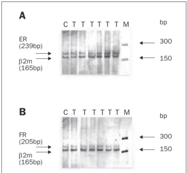
Figure 1. Analysis of the estrogen receptor (ER) (A) and progesterone
receptor (PR) (B) messenger ribonucleic acid (mRNA) expression in the
endometrium, myometrium and vagina of women treated with estriol, taking the ß2-microglobulin (ß2-m) levels as the internal standard. C = tissue from control (untreated) patient; T = tissue from estriol-treated patient; M = molecular weight ladder marker for 50-2000 base pairs (bp) (Sigma, St. Louis, United States).
bp
300
150 ER
(239bp)
2m (165bp)
FR (205bp)
2m (165bp)
bp
300
150
C T T T T T T M
C T
T T T T T M
A
B
To rule out contamination of genomic DNA as a source for ampli-ied products, each reaction was also carrampli-ied out without reverse tran-scriptase. In addition, a control without the template was run for both the RT and PCR stages, for each of the primer sets, and none of these showed any visible PCR products.
Qualitative and quantitative estimations of electropherograms
For qualitative and quantitative analysis of the silver nitrate stained gels, a video densitometer (Biotec-Fischer, Reiskirchen, Germany) with the software program Gel-Pro® Analyzer 3.0 (Media Cybernetics, Unit-ed States) was usUnit-ed. All RT-PCR reactions were done three times for each sample. he integrated optical density (IOD) of the bands on digi-tized images was measured. ER or PR gene expression was expressed as the ratio of ER or PR over β2m. he ratio between the PCR ampliied gene and the ampliied standard was obtained for each sample. A con-ventional cutof of 15 fmol/mg protein for ER status corresponded to an ER/β2m ratio of 0.5 and, according to the regression equation, the conventional cutof of 15 fmol/mg protein for PR status corresponded to a PR/β2m ratio of 0.03.23
Histological examination of the endometrium
Each patient’s endometrium was examined under an optical micro-scope for histological evidence of estrogen stimulation. When exposed to excessive long-term estrogen action, the endometrium normally re-acts by stimulating proliferation processes, and thus the following cri-teria were taken into consideration: 1) glandular surface/stroma ratio; 2) distribution, size and shape of glandular tubes; 3) cell distribution in tubes; 4) cell stratiication in tubes; 5) mitotic activity; and 6) cel-lular atypia.
he endometrial samples evaluated were scored within a possi-ble range of 0-6 points; 1-2 points signiied weak estrogen action; 3-4 points, medium-strength action; and 5-6 points, strong action.
Statistical analysis
None of the parameters recorded in the tumor material passed the test for normal distribution (Kolmogorov-Smirnov test). Hence, non-parametric statistical tests were used throughout, for analyzing the re-sults. P-values < 0.05 were considered to be signiicant.
RESULTS
ER and PR mRNA expression
ER and PR gene expression in the endometrium, myometrium and vagina of women treated with estriol, in comparison with the control group, was investigated by means of RT-PCR. A representative electro-pherogram from the RT-PCR analysis of ER and PR gene expression is shown in Figure 1.
In the group of estriol-treated women,ERmRNA was detected in 85.71% (12/14), 78.57% (11/14) and 100% (14/14) of the endometri-al, myometrial and vaginal specimens, respectively. In the control group, ER mRNA expression was observed in 61.54% (8/13) of the endome-trial, 61.54% (8/13) of the myometrial and 69.23% (9/13) of the vagi-nal specimens.
cDNA. he ER and PR gene expressions were determined following co-ampliication and normalization by means of an internal control se-quence, i.e. using β2-microglobulin (β2m). he speciic primers for RT-PCR are shown in Table 1.
ER mRNA was found to be overexpressed in 78.57% (11/14), 42.86% (6/14) and 92.86% (13/14) of the endometrial, myometrial and vaginal samples obtained from the estriol-treated women, respectively. Statistically signiicant diferences (Mann-Whitney U test, P< 0.001) were recorded between ER gene expression in the endometrium and va-gina of the estriol-treated women and control group (Table 2).
In the tissues obtained from estriol-treated women,PRmRNA was detected in 85.71% (12/14) of the endometrial, 85.71% (12/14) of the myometrial and 50% (7/14) of the vaginal samples. In the group of control tissues, PR mRNA expression was observed in 46.15% (6/13), 53.85% (7/13) and 46.15% (6/13) of the endometrial, myometrial and vaginal samples, respectively.
PR mRNA was overexpressed in 50% (7/14) of the endometrial, 42.86% (6/14) of the myometrial and 21.43% (3/14) of the vaginal samples obtained from the estriol-treated women. Statistically signif-icant diferences (Mann-Whitney U test, P< 0.001) were noted be-tween PR gene expression levels in the endometrium of the estriol-treat-ed women and control group (Table 2).
Cytohormonal examination
All of the estriol-treated patients showed a higher maturation index, with a shift towards supericial cells. he smears from the control group were atrophic. Histological evaluation showed atrophic features for al-most all the patients included in the study. In only one case, ultrasonog-raphy (USG) showed an expanded endometrium (12.5 mm). However, histopathological examination did not provide any evidence of endome-trial proliferation. After the estriol treatment, a small degree of prolifera-tion was detected in 5/14 cases (36%) at the postoperative assessment. he endometrium of these ive patients showed symptoms of hormonal stimulation, with three low-degree cases (21.4%) and two medium-de-gree cases (14.3%). Out of the 14 estriol-treated patients, nine did not show estrogen stimulation of the endometrium: seven cases were de-ined as atrophic and two as typical. he endometrium from the con-trol group showed no evidence of estrogen stimulation. It was deined as atrophic in 12 cases, and typical in one case (Table 3).
DISCUSSION
Regulation of gene transcription is central both to tissue speciic-gene expression and to regulation of speciic-gene activity in response to speciic hormonal stimuli. In order to produce efects, transcription factors will require the ability to bind to DNA and then to inluence transcription either positively or negatively.25,26
ERs and PRs as transcription factors can be regulated at two levels, i.e. the levels of transcription factor synthesis and transcription factor activity. Furthermore, they are regulated through synthesis in one par-ticular tissue or cell type and not in other tissues.27-29
Although the regulation of transcription factor synthesis is an im-portant control point, it cannot be the only regulatory mechanism con-trolling transcription factor activity. If this were the case, enhanced syn-thesis of a transcription factor in response to a particular stimulus would be controlled by enhanced transcription of its corresponding gene, which in turn would require de novo synthesis of further transcription factors, thus resulting in the need for new transcription of these genes, and so on.25
In a series of experiments, we demonstrated the expression levels of ER and PR mRNA in the endometrium, myometrium and vagina of postmenopausal women treated with estriol. Our data showed difer-ences between the three tissues studied. Signiicantly higher ER and PR mRNA levels were found in the endometrium of estriol-treated women, compared with the control group. In the case of vaginal tissue, only ER gene expression was enhanced after estriol therapy. here were no statis-tically signiicant diferences in ER and PR mRNA levels between the myometrial tissues obtained from the estriol-treated women and control patients. It was observed that the endometrium and vagina were sensi-tive to estriol stimuli, but the precise mechanism of this phenomenon is unknown. Although the general mechanism through which estrogens and progestins increase their activity in the female reproductive system is well established, the ways in which transcriptional regulation takes place and steroid receptors act remain elusive.
ERs have at least two regions that are required for maximum tran-scriptional activity: activating function-1 and 2 (AF-1 and AF-2). hese AFs have been shown to function in a cell-speciic manner and it has been found that the requirement for these activation functions varies depending on the cell and promoter context.30 he fact that either AF-1 or AF-2 activity is suicient for ER action in some contexts, whereas both are required in others, suggests that the role of ERs in diferent cells and on diferent promoters is not the same. hus, it has been pro-posed that ERs are components of two distinct signaling pathways with-in the cell: the classical ER/ERE (estrogen receptor/estrogen response
el-Tissues
Patients Estriol-treated group
n = 14
Control group n = 13 ER (IOD ER/IODβ2m)
Endometrium* 1.24 ± 0.04 0.57 ± 0.06
Myometrium 0.64 ± 0.06 0.58 ± 0.07
Vagina* 0.97 ± 0.04 0.48 ± 0.04
PR (IOD PR/IODβ2m)
Endometrium* 0.079 ± 0.008 0.032 ± 0.003
Myometrium 0.057 ± 0.004 0.036 ± 0.004
Vagina 0.050 ± 0.011 0.061 ± 0.006
*statistically signiicant differences.
ER = estrogen receptor; PR = progesterone receptor; IOD = integrated optical density; β2m = beta-2
microglobulin
Table 2. Relative estrogen and progesterone receptor messenger ribonucleic acid (mRNA) levels
Patients Endometrial evaluation
No stimulation Low degree of stimulation Medium degree of stimulation High degree of stimulation
Estriol-treated group 9/14 (64.3%) 3/14 (21.4%) 2/14 (14.3%) 0
Control group 13/13 (100%) - -
ement) - mediated signal transduction pathway and/or the non-classical pathway of interaction with AP1 complexes.31
It is generally accepted that PR induction is a speciic response by an estrogen target tissue to an estrogenic stimulation. PR concentration is hormonally regulated. PRs induced by estrogens and downregulated by progestins.32,33 Analysis of the 5’ lanking region, 5’ untranslated region and exon 1 of rat PR genes has identiied four estrogresponsive en-hancers. When linked together as an artiicial cassette, these enhancers form a strong estrogen-responsive transcription regulator.34
Our results are concordant with the data published by Haaften et al.21 hese authors measured ERs and PRs through protein levels in patients treated for three weeks with 0.5 mg of estriol daily. hey postu-lated that in human postmenopausal endometrium, ER and PR protein levels increase signiicantly following vaginal application of estriol. he increases of ER and PR mRNA levels observed in our study may relect transcription per se or posttranscriptional modiication, which may alter the stability of the mRNA and/or receptor protein.
he action efected by estriol on the endometrium is unclear. Any stimulation of the endometrium that may occur depends directly on the pharmacokinetic and pharmacodynamic properties of estriol, i.e. the speed of absorption, conjugation and secretion, and on rapid disso-ciation of nuclear receptors.35,36
Endometrial atrophy prior to treatment had not been assessed by other authors and, in the case of one study, only the results from the progesterone test were taken into consideration.37 In a long-term study, Boseli et al. evaluated the endometrium 3, 6, 12, 18 and 24 months after a vaginal estriol preparation in 0.5 mg doses had been applied, and they observed no proliferative changes.38 Furthermore, Weiderpass et al. found that vaginal treatment with low-potency es-trogen formulations had no impact on the relative risk of endometrial neoplasia.39
No proliferative changes were observed in our study after 14 days of estriol application. Only a low degree of endometrial stimulation was noted. he answer to the question of whether 14-day estriol apy before the operation is safe and efective was airmative. he ther-apy was efective, considering that the atrophic condition of the vagi-nal tissue improved in all of the patients. his was conirmed by the increased maturation index and enhanced ER expression. he healing of the wound was regular for all the patients, and the postoperative pe-riod did not exceed nine days. he therapy was safe, considering that none of the patients showed any symptoms of atypical endometrium proliferation, even though their ER and PR mRNA levels were over-expressed. hese results are concordant with those of other authors, who demonstrated that noticeable proliferative changes to the endo-metrium appeared after at least three weeks of administering doses of 0.5 mg of estriol.21
CONCLUSION
he results obtained suggest that although 14 days of estriol treat-ment enhanced the patients’ ER and PR mRNA levels, it had very little or no efect on the proliferation status of the endometrium.
REFERENCES
1. World Health Organization. Research on the menopause in the 1990s. Report of a WHO Scientiic Group. Technical Report Series, No 866. Geneva: World Health Organization; 1996.
2. Rich RL, Hoth LR, Geoghegan KF, et al. Kinetic analysis of estrogen receptor/ligand interac-tions. Proc Natl Acad Sci U S A. 2002;99(13):8562-7.
3. Peck JD, Hulka BS, Poole C, Savitz DA, Baird D, Richardson BE. Steroid hormone levels during pregnancy and incidence of maternal breast cancer. Cancer Epidemiol Biomarkers Prev. 2002;11(4):361-8.
4. Fujimoto J, Sakaguchi H, Aoki I, Toyoki H, Tamaya T. Clinical implications of the expression of estrogen receptor-alpha and -beta in primary and metastatic lesions of uterine endometrial cancers. Oncology. 2002;62(3):269-77.
5. Ito K, Utsunomiya H, Yaegashi N, Sasano H. Biological roles of estrogen and progesterone in human endometrial carcinoma—new developments in potential endocrine therapy for endometrial cancer. Endocr J. 2007;54(5):667-79.
6. Hanstein B, Beckmann MW, Bender HG. Charakterisierung und Bedeutung verschiedener Ostrogenrezeptortypen unter besonderer Berücksichtigung des Endometriumkarzinoms [Role of estrogen receptor isoforms in the pathogenesis and treatment of endometrial can-cer]. Zentralbl Gynakol. 2002;124(1):17-9.
7. Jazaeri AA, Nunes KJ, Dalton MS, Xu M, Shupnik MA, Rice LW. Well-differentiated endometrial adenocarcinomas and poorly differentiated mixed mullerian tumors have altered ER and PR isoform expression. Oncogene. 2001;20(47):6965-9.
8. Gräser T, Koytchev R, Müller A, Oettel M. Comparison of the eficacy and endometrial safety of two estradiol valerate/dienogest combinations and Kliogest for continuous combined hor-mone replacement therapy in postmenopausal women. Climacteric. 2000;3(2):109-18. 9. Kajta M, Budziszewska B, Marszal M, Lason W. Effects of 17-beta estradiol and estriol on
NMDA-induced toxicity and apoptosis in primary cultures of rat cortical neurons. J Physiol Pharmacol. 2001;52(3):437-46.
10. Parker MG, Arbuckle N, Dauvois S, Danielian P, White R. Structure and function of the estro-gen receptor. Ann N Y Acad Sci. 1993;684:119-26.
11. Le Marchand L, Donlon T, Kolonel LN, Henderson BE, Wilkens LR. Estrogen metabolism-related genes and breast cancer risk: the multiethnic cohort study. Cancer Epidemiol Bio-markers Prev. 2005;14(8):1998-2003.
12. Yu J, Yu J, Cordero KE, et al. A transcriptional ingerprint of estrogen in human breast cancer predicts patient survival. Neoplasia. 2008;10(1):79-88.
13. Klein-Hitpass L, Schwerk C, Kahmann S, Vassen L. Targets of activated steroid hormo-ne receptors: basal transcription factors and receptor interacting proteins. J Mol Med. 1998;76(7):490-6.
14. Kano H, Hayashi T, Sumi D, et al. Estriol retards and stabilizes atherosclerosis through an NO-mediated system. Life Sci. 2002;71(1):31-42.
15. Taylor M. Unconventional estrogens: estriol, biest, and triest. Clin Obstet Gynecol. 2001;44(4):864-79.
16. Teede HJ, Liang YL, Kotsopoulos D, Zoungas S, Cravent R, McGrath BP. A placebo-controlled trial of long-term oral combined continuous hormone replacement therapy in postmeno-pausal women: effects on arterial compliance and endothelial function. ClinEndocrinol (Oxf). 2001;55(5):673-82.
17. Pardini D. Terapia hormonal da menopausa [Menopausal hormone therapy]. Arq Bras Endo-crinol Metabol. 2007;51(6):938-42.
18. Estevão RA, Baracat EC, Logullo AF, Oshima CT, Nazário AC. Eficacy of estriol in inhibiting epithelial proliferation in mammary ibroadenoma: randomized clinical trial. Sao Paulo Med J. 2007;125(6):343-50.
19. Estevão RA, Nazário AC, Baracat EC. Effect of oral contraceptive with and without associated estriol on ultrasound measurements of breast ibroadenoma: randomized clinical trial. Sao Paulo Med J. 2007;125(5):275-80.
20. Henriksson L, Stjernquist M, Boquist L, Alander U, Selinus I. A comparative multicenter study of the effects of continuous low-dose estradiol released from a new vaginal ring versus estriol vaginal pessaries in postmenopausal women with symptoms and signs of urogenital atrophy. Am J Obstet Gynecol. 1994;171(3):624-32.
21. van Haaften M, Donker GH, Sie-Go DM, Haspels AA, Thijssen JH. Biochemical and histolo-gical effects of vaginal estriol and estradiol applications on the endometrium, myometrium and vagina of postmenopausal women. Gynecol Endocrinol. 1997;11(3):175-85. 22. Chomczynski P, Sacchi N. Single-step method of RNA isolation by acid guanidinium
thiocya-nate-phenol-chloroform extraction. Anal Biochem. 1987;162(1):156-9.
24. Bassam BJ, Caetano-Anollés G, Gresshoff PM. Fast and sensitive silver staining of DNA in polyacrylamide gels. Anal Biochem. 1991;196(1):80-3.
25. Latchman DS. Eukaryotic transcription factors. 2nd ed. London: Academic Press; 1995. 26. Latchman DS. Transcription factors: an overview. Int J Biochem Cell Biol. 1997;29(12):
1305-12.
27. McDonnell DP, Clemm DL, Hermann T, Goldman ME, Pike JW. Analysis of estrogen re-ceptor function in vitro reveals three distinct classes of antiestrogens. Mol Endocrinol. 1995;9(6):659-69.
28. McDonnell DP, Clemm DL, Imhof MO. Deinition of the cellular mechanisms which distin-guish between hormone and antihormone activated steroid receptors. Semin Cancer Biol. 1994;5(5):327-36.
29. Pardini D. Terapêutica de reposição hormonal na osteoporose da pós-menopausa [Hormo-nal replacement therapy in osteoporosis of postmenopausal]. Arq Bras Endocrinol Metab. 1999;43(6):428-32.
30. Tzukerman MT, Esty A, Santiso-Mere D, et al. Human estrogen receptor transactivational capacity is determined by both cellular and promoter context and mediated by two functio-nally distinct intramolecular regions. Mol Endocrinol. 1994;8(1):21-30.
31. Webb P, Lopez GN, Uht RM, Kushner PJ. Tamoxifen activation of the estrogen receptor/AP-1 pathway: potential origin for the cell-speciic estrogen-like effects of antiestrogens. Mol En-docrinol. 1995;9(4):443-56.
32. Graham JD, Clarke CL. Physiological action of progesterone in target tissues. Endocr Rev. 1997;18(4):502-19.
33. Wlaźlak E, Surkont G, Suzin J. Estradiol – nowość czy standard w terapii dopochwowej. Przegląd Menopauzalny. 2004;5(2):50-6. Available from: http://www.termedia.pl/maga-zine.php?magazine_id=4&article_id=2640&magazine_subpage=ABSTRACT. Accessed in 2009 (Jun 15).
34. Kumar MV, Tindall DJ. Transcriptional regulation of the steroid receptor genes. Prog Nucleic Acid Res Mol Biol. 1998;59:289-306.
35. Gerbaldo D, Ferraiolo A, Croce S, Truini M, Capitanio GL. Endometrial morphology after 12 months of vaginal oestriol therapy in post-menopausal women. Maturitas. 1991;13(4): 269-74.
36. Ciszko B, Zdrojewicz Z. Znaczenie endogennego i egzogennego estriolu w praktyce klini-cznej [Compliance endogenous and exogenous estriol in clinical practice]. Ginekol Pol. 2006;77(7):559-65.
37. Englund DE, Johansson ED. Endometrial effect of oral estriol treatment in postmenopausal women. Acta Obstet Gynecol Scand. 1980;59(5):449-51.
38. Van Gorp T, Neven P. Endometrial safety of hormone replacement therapy: review of literatu-re. Maturitas. 2002;42(2):93-104.
39. Weiderpass E, Baron JA, Adami HO, et al. Low-potency oestrogen and risk of endometrial cancer: a case-control study. Lancet. 1999;353(9167):1824-8.
Sources of funding: Not declared
Conlict of interest: Not declared
Date of irst submission: April 2, 2008
Last received: June 23, 2009
Accepted: June 24, 2009
Address for correspondence:
Magdalena Bryś
Department of Cytobiochemistry, University of Lodz Banacha 12/16, 90-237