Márcia Ferreira da Silva, Maria do Carmo Gouveia Pelúzio, Paulo Roberto dos Santos Amorim, Vitor Neiva
Lavorato, Natália Pereira do Santos, Luiz Henrique Marchesi Bozi, Arlete Rita Penitente, Daniel Luciano Falkoski,
Felipe Gomes Berfort, Antonio Jose Natali
Universidade Federal de Viçosa, Viçosa, MG - Brazil
Mailing address: Antonio Jose Natali •
Av. PH Rolfs, s/n - Departamento de Educação Física - Campus Universitário da Universidade Federal de Viçosa - 36570-000 - Viçosa, MG - Brazil E-mail: [email protected]
Manuscript received August 06, 2010; revised manuscript received October 29, 2010; accepted on December 21, 2010.
Abstract
Background: Experimental diabetes promotes contractile dysfunction in cardiomyocytes, but the effects of swimming in this disorder are not known.
Objective: To test the effects of a swimming training program (STP) on cardiomyocyte contractile dysfunction in rats with experimental diabetes.
Methods: Wistar rats (age: 30 days; mean body weight: 84.19 g) with diabetes induced by streptozotocin (60 mg/kg body weight; glucose > 300 mg/dl) were divided into sedentary diabetic rats (SD, n = 10) and exercised diabetic rats (ED, n = 13). Animals of same age and weight served as sedentary controls (SC, n = 10) and exercised controls (EC, n = 06). Animals and ED and EC underwent a STP (05 days/week, 90 min/day) for 08 weeks. Left ventricular (LV) myocytes were isolated and electrically stimulated at 3.0 Hz at room temperature (~ 25° C).
Results: Diabetes reduced contractile function in cardiomyocytes of animals compared to controls (i.e., lower amplitude of contraction, longer duration of contraction and relaxation). The STP attenuated the reduced amplitude of contraction (SC, 11 ± 0.2% vs ED, 11.6 ± 0.2%), time to peak contraction (SC, 319 ± 5.8 ms vs ED, 333 ± 4.8 ms) and time to 50.0% of relaxation (SC, 619 ± 22.2 ms vs ED 698 ± 18.6 ms) of cardiomyocytes of diabetic rats. Diabetes reduced the size of cardiomyocytes, however, the STP minimized the reduction of cell volume and width, without changing length.
Conclusion: The swimming training program attenuated the contractile dysfunction of the LV myocytes of rats with experimental diabetes. (Arq Bras Cardiol. 2011; [online].ahead print, PP.0-0)
Keywords: Swimming; physical exertion; myocytes, cardiac; rats; diabetes mellitus.
as reduction in systolic volume, end-diastolic volume and ejection fraction, cardiac output and fractional shortening7,11,12. At the cellular level, however, few studies on the effects of chronic exercise on the contractile function of cardiomyocytes of diabetic rats were performed and, in addition to using only the treadmill, showed controversial results. For example, continuous running programs on a treadmill with low intensity (09 m/min, 0.0% inclination, 30 min/day) or moderate intensity (18 m/min, 0.0% inclination) did not affect the contraction of rat cardiomyocytes13. Another program of continuous running with high intensity (18 m/ min, 5.0% inclination, 60 min/day) led to negative changes, as it prolonged cell contraction time and did not alter the amplitude of contraction of rat cardiomyocytes14. However, running programs on a treadmill with higher intensity (20-25 m/min, 5.0% inclination, 60 min/day) were able to restore the contractile function of rat and mice cardiomyocytes7,15. So far, the effects of continuous exercise lasting over 60 minutes daily, especially swimming on the contractile function of cardiomyocytes of diabetic rats are not known.
This study aims to test whether a swimming program, lasting 90 minutes daily, alters the contractile function of
Introduction
Diabetes mellitus type 1 is a risk factor for cardiovascular events, including the development of diabetic cardiomyopathy. The inability to maintain glucose homeostasis in the myocardium compromises cardiac structure and function in humans and animals with experimental diabetes1-4. At the cellular level, it was shown that diabetes impairs the contractile function of cardiomyocytes, mainly for causing structural and functional changes in the regulation of intracellular calcium (Ca2+)5-7.
isolated cardiomyocytes of the left ventricle of rats with experimental diabetes.
Methods
Experimental animals and treatments
Wistar rats (age 30 days; mean body weight of 84.19 g) were divided into 04 groups: Sedentary controls (SC, n = 10); Exercised control (EC, n = 10); Sedentary diabetic (SD, n = 20); and Exercised diabetic (ED, n = 20) and were maintained at average temperature of 22° C and inversed lighting regime of 12/12h light/dark with water and commercial diet ad libitum.
This study followed the standards established in the Guide for the Care and Use of Laboratory Animals (Institute of Laboratory Animal Resources, National Academy of Sciences, Washington, D.C., 1996) and respected the Ethical Principles in Animal Experiments of the Brazilian College of Animal Experimentation (COBEA). The study was approved by the Ethics Committee of Universidade Federal de Viçosa (proceeding No. 03/2009).
Induction of diabetes
After fasting for 12 hours, the animals in groups ED and SD received an intraperitoneal injection (60 mg/kg body weight) of streptozotocin (STZ, Sigma, St. Louis, USA), diluted in 1.0 ml of sodium citrate buffer (0.1 M, pH 4.5). Animals in groups SC and EC received the same dose of sodium citrate buffer (0.1 M, pH 4.5) without STZ. Seven days after application of STZ and fasting for 12 hours, blood glucose was measured at rest (One Touch Ultra - Johnson & Johnson, Mexico). Animals with levels of fasting blood glucose above 300 mg\dl were considered diabetic (SD, n = 10, ED, n = 13). Fasting blood glucose and body weight were monitored weekly over the experimental period.
Swimming training program
After 45 days of hyperglycemia, the animals in group ED and EC were subjected to a swimming training program (adapted from Medeiros et al16) for 8 weeks. In the first week, the animals exercised in the water, no overload, over 10-50 min, the duration increased by 10 min/day. In the second week, the animals exercised with a load of 1.0% of their body weight and exercise duration was increased by 10 min/day until 90 min of continuous swimming. From the third week, the load was increased weekly (0.5% of body weight) up to 4.0% of body weight in 8 weeks. During the swimming sessions, the animals in groups SD and NC were placed in a polypropylene box with warm water (28-30° C) and 10 cm depth.
Four animals in the group EC died from drowning.
Isolation of cardiomyocytes
After euthanasia, hearts were removed and left ventricular myocytes were isolated as described by Natali et al17. Briefly, hearts were cannulated via the aorta in a Langendorff system and perfused with isolation solution [composition (mM): 130 Na+; 5.4 K+; 1.4 Mg+; 140 Cl-; 0.75 Ca2+; 5.0 Hepes; 10 glucose; 20 taurine; and 10 creatine; pH = 7.3 at room
temperature]. Then the heart was perfused with calcium-free solution containing 0.1 mM ethylene glycol-bis (ß-aminoethyl ether)-N, N, N’, N’-tetraacetic acid (EGTA), for a period of 4 -6 min. Then, the heart was perfused with a solution containing 1.0 mg.ml-1 collagenase type 2 (Worthington, USA) and 100.0 mM CaCl2 for 20 to 25 min. The solutions were oxygenated (100% O2 - White Martins, Brazil) and maintained at a temperature of 35° C. After perfusion, ventricles were separated from the atria and weighed. Then, the left ventricle was placed in a vial containing 5.0 ml of enzyme solution (collagenase) and bovine serum albumin (10.0%). The vial was shaken moderately for 05 min in a water bath at 37° C, after which the tissue was removed from the vial and the remainder was centrifuged (3.000 rpm) for 30 s. The supernatant was removed and the cardiomyocytes were suspended in isolation solution and stored in refrigerator (5° C) until use.
Contractile function of cardiomyocytes
Cell contractions were measured using the technique of changing the length of cardiomyocytes using the edge detection system (Ionoptix, Milton, MA, USA) mounted on an inverted microscope (Nikon Eclipse - TS100, Japan) as described previously18. Briefly, myocytes were accommodated in an experimental chamber with a glass base and bathed in buffer solution with the following composition (in mM): 136.9 NaCl; 5.4 KCl; 0.37 NaH2PO4; 0.57 MgCl2; 5.0 Hepes = 5; 5.6 Glucose and 1.0 CaCl2 (pH = 7.4 at room temperature). Myocytes were viewed on a monitor via a camera (Myocam, Ionoptix, frequency of 240 Hz) attached to the microscope using an image detection program (Ionwizard, Ionoptix). Cardiomyocytes were stimulated externally at a frequency of 3.0 Hz (10 Volts, lasting 5 min) using a pair of steel electrodes and an electric field (Myopacer, Ionoptix). The movements of the longitudinal edges of the myocytes were captured by the edge detection system (Ionwizard, Ionoptix) and stored for later analysis. Contraction measurements employed only those cardiomyocytes that were in good condition, with the edges and well-defined sarcomeric striations, at rest, with no voluntary contractions. The contractions were analyzed as described previously19.
Dimensions of cardiomyocytes
The length and width of myocytes were measured using a system for capturing images, from images of cardiomyocytes displayed horizontally on the monitor of a microcomputer, as described17. The cell length was determined by measuringthe cell image generated on the monitor from the right edge to the left edge, at the midpoint of the cardiomyocyte width. The width of cells was determined by measuring the image generated on the monitor, from the top edge to the bottom edge, at the midpoint of the length of cardiomyocytes. The cell volume was calculated using the formula: [Volume (pL) = Length (mm) x width (mm) x (7.59 x 10- 3 pL/mm2)], according to Satoh et al20.
Statistical analysis
used analysis of variance with two inputs (two-way ANOVA) and Tukey’s post hoc for multiple comparisons. This analysis was performed using the Sigma Stat software, version 3.0. We adopted alevel of significance of up to 5.0% (p ≤ 0.05).
Results
Before the application of STZ, there was no statistical difference in blood glucose between the groups (Table 1). Forty-five days after the application of STZ (beginning of the year) and at the end of the experiment, diabetic animals had blood glucose higher than control animals. The blood glucose levels were not altered by exercise in both diabetic animals (ED vs SD) as in control animals (EC vs SC). There was no interaction among exercise and diabetes (two-way ANOVA, p > 0.05) for this or for other variables analyzed.
The initial body weights were not different among the four groups (Table 1). Forty-five days after application of STZ, the animals in groups SD and ED had body weights lower than controls SC and EC. The same happened at the end of the experiment. Likewise, the swimming program did not alter these parameters in animals of both ED group as compared to SD, and in EC as compared to SC.
The SD animals had lower ventricular weights (p < 0.05) than SC (Table 1). The swimming program did not affect the ventricular weight of diabetic animals (SD vs ED). However, in control animals, ventricular weight was higher in EC than in SC animals. Ventricular weight related to body weight, ventricular hypertrophy index, was higher in the SD group than in the SC group. Among diabetic rats, those in the ED
group presented higher relative ventricular weight than those in the SD group. Among controls, EC animals exhibited a higher relative ventricular weight than SC.
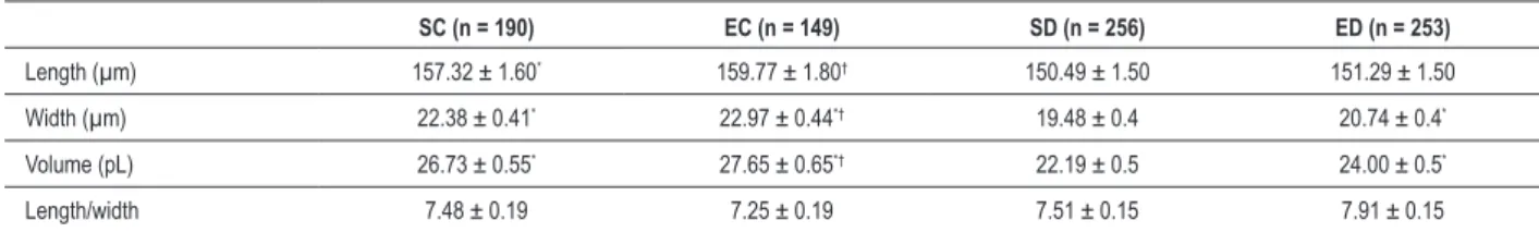
Independently, diabetes reduced the length of cardiomyocytes in sedentary animals (SD vs SC) and trained animals (ED vs CE; Table 2). However, the swimming program has not changed the length of cardiomyocytes in diabetic rats (ED vs SD) and in non-diabetic rats (SC vs CE). Diabetes reduced the width of myocytes in sedentary animals (SD vs SC) and trained animals (ED vs CE). However, the swimming program has increased the width of cardiomyocytes of diabetic rats (ED vs SD). This did not occur in control animals (EC vs SC). There was a reduction of cell volume in the SD group compared to the SC group. The swimming program increased the cell volume in diabetic animals (ED vs SD), but not in control animals (EC vs SC). It is observed that the length/width ratio of cardiomyocytes was not affected by diabetes or by the swimming program.
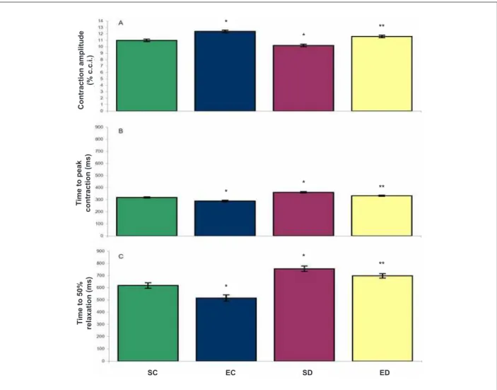
The analysis of the contractile function of cardiomyocytes showed that the amplitude of cell contraction was reduced by diabetes (SC, 11.0 ± 0.2% vs SD, 10.2 ± 0.2%, p < 0.001) (Figure 1 A). The swimming program increased the contraction amplitude of cardiomyocytes in these animals (SD, 10.2 ± 0.2% vs ED, 11.6 ± 0.2%, p < 0.001). Among the control animals, the swimming program increased the amplitude of contraction (SC, 11.0 ± 0.2% vs EC, 12.4 ± 0.2%, p < 0.001). The cardiomyocytes of animals in the SD group had time to peak contraction longer than the SC group (361 ± 5.7 ms
vs 319.0 ± 5.8 ms, respectively, p < 0.001) (Figure 1 B). The swimming program reduced the time to peak contraction in diabetic animals (SD, 361 ± 5.7 ms vs ED, 333.0 ± 4.8 ms,
Table 1 - Body and ventricular weights and blood glucose levels of diabetic and control rats
SC (n = 10) EC (n = 06) SD (n = 10) ED (n = 13)
Initial BW (g) 83.51 ± 1.9 82.72 ± 1.8 87.80 ± 2.0 82.71 ± 1.8
BW after 45 days (g) 353.93 ± 11.3* 352.12 ± 11.3* 193.72 ± 11.9 186.91 ± 10.7
End BW (g) 443.50 ± 18.1* 410.81 ± 25.7* 198.82 ± 18.1 204.25 ± 18.1
Initial BG (mg/dl) 82.4 ± 4.2 84.0 ± 4.2 89.0 ± 4.5 93.0 ± 4.0
BG after 45 days (mg/dl) 87.8 ± 11.3 76.2 ± 11.3 525.1 ± 11.3† 520.1 ± 11.9†
Final BG (mg/dl) 88.3 ± 32.1 86.8 ± 45.5 475.8 ± 32.1† 483.7 ± 32†
VW (mg) 1,590.00 ± 0.08* 1,930.00 ± 0.11* 1,120.00 ± 0.08 1,330.00 ± 0.08
End VW/BW (mg/g) 3.59 ± 0.4 4.72 ± 0.6‡ 5.98 ± 0.4 † 7.97 ± 0.4†§
Data in mean ± SEM. n - number of animals; SC - sedentary controls; EC - exercised controls; SD - sedentary diabetics; ED - exercised diabetics; BG - blood glucose; BW - body weight; VW - ventricular weight; * - different from SD and ED; †- different from SC and EC; ‡- different from SC; §- different from SD (p < 0.05).
Table 2 - Dimensions of cardiomyocytes of diabetic and control rats
SC (n = 190) EC (n = 149) SD (n = 256) ED (n = 253)
Length (µm) 157.32 ± 1.60* 159.77 ± 1.80† 150.49 ± 1.50 151.29 ± 1.50
Width (µm) 22.38 ± 0.41* 22.97 ± 0.44*† 19.48 ± 0.4 20.74 ± 0.4*
Volume (pL) 26.73 ± 0.55* 27.65 ± 0.65*† 22.19 ± 0.5 24.00 ± 0.5*
Length/width 7.48 ± 0.19 7.25 ± 0.19 7.51 ± 0.15 7.91 ± 0.15
p < 0.001). The same effect was observed in cardiomyocytes of control animals (EC, 289.0 ± 6.8 ms vs SC, 319.0 ± 5.8 ms, p < 0.001).
For 50.0% of relaxation, the time was higher in cardiomyocytes of sedentary diabetic rats than in sedentary controls (SD, 756 ± 22.1 ms vs SC, 619.0 ± 22.2 ms, p < 0.001) (Figure 1C). The swimming program reduced the time for 50.0% of relaxation in the cardiomyocytes of these animals (SD, 756 ± 22.1 ms vs
ED, 698 ± 18.6 ms, p = 0.044). The same effect was observed in cardiomyocytes of control animals (EC, 516.0 ± 26.1 ms vs
SC, 619.0 ± 22.2 ms, p = 0.003).
Discussion
Our data demonstrate that the contractile dysfunction of cardiomyocytes caused by diabetes was moderated by swimming training program lasting 90 minutes. Furthermore, we found increased width and volume of cardiomyocytes, without altering the cell length in response to chronic exercise.
The reduced contraction amplitude observed in this study reflects important changes in the myocardium of diabetic rats
in vivo, such as reduced fractional shortening in end diastolic diameter, left ventricular end systolic diameter and cardiac input7,11. Although we have not tested mechanisms, it is knownthat the decreased sensitivity of contractile myofilaments to Ca2+ and reduced intracellular concentration of Ca2+ are involved21. However, cardiomyocytes of Goto-Kakizaki rat showed increased amplitude of contraction associated with transient reduction of Ca2+. This suggests that the sensitivity of myofilaments to Ca2+ was increased, which could be a compensatory mechanism to preserve the mechanical function of the heart in diabetes22. There is evidence that the intracellular concentration of Ca2+ is reduced in cardiomyocytes of animals with experimental diabetes23. This occurs due to the increased activity of sodium and calcium exchanger (NCX) and decreased uptake of Ca2+ by the sarcoplasmic reticulum (SR) via calcium ATPase of the SR (SERCA2)15 and reduced release of Ca2+ of SR, via ryanodine receptors (RyR2)24. Moreover, it is possible that the reduced density of transverse tubules in cardiomyocytes of diabetic animals can change the space between the L-type calcium channels and RyR2, which reduces the efficiency of excitation-contraction coupling15.
Figure 1 -Contractile function of cardiomyocytes of control and diabetic rats. A - contraction amplitude; B - time to peak contraction; C - time to 50.0% relaxation. SC, sedentary controls (106 cells). EC - exercised controls (78 cells). SD - diabetic sedentary (109 cells). ED - trained diabetics (153 cells). Data are mean ± SEM*, different from SC**, which is different from SD (p < 0.05).
T
im
e
to
5
0
%
re
la
x
a
ti
o
n
(m
s
)
C
o
n
tr
a
c
ti
o
n
a
m
p
li
tu
d
e
(%
c
.c
.i
.)
T
im
e
to
p
e
a
k
c
o
n
tr
a
c
ti
o
n
(m
s
)
SC EC SD ED
*
*
**
*
** *
*
*
In contrast, chronic exercise increased the contraction amplitude in diabetic animals and controls. For diabetic animals, this fact indicates that exercise promoted positive changes in cardiomyocytes that contribute to alleviating some of the mechanical abnormalities of diabetic myocardium observed in
vivo7,11,12. Some mechanisms have been proposed as responsible
for increasing the contraction amplitude of cardiomyocytes of diabetic rats in response to chronic exercise: there is evidence that chronic exercise can normalize the operation of NCX and calcium calmodulin kinase II (CaMKII), reduce the leakage of Ca2+ of SR and increase the content of Ca2+ of SR7,15.
Experimental diabetes prolonged the time required for peak cell contraction. This indicates that cardiomyocytes of diabetic rats contract more slowly than their controls. This change has a negative impact on the cardiac function in these animals. The speed of contraction of cardiomyocytes is controlled by proteins that regulate intracellular Ca2+ movement and the rate of ATP hydrolysis which, in turn, regulates the rate of formation of crossed bridges21. Cardiomyocytes of diabetic animals reduce the expression of regulatory proteins such as CaMKII, NCX, RyR2, SERCA2 and phospholamban (PLB) 5,7,15,24-26, which may delay the availability of Ca2+ for cell contraction.
However, the swimming program reduced the time to peak contraction in diabetic animals. Adaptations to regular exercise, which accelerate the availability of cytosolic Ca2+ and increase the rate of ATP hydrolysis, contributed to this reduction. The speed of availability of cytosolic Ca2+ is regulated mainly by the output speed of SR Ca2+ via RyR221. There is evidence that regular exercise increases the expression and/or the activity of RyR2 and sensitivity of RyR2 and contractile myofilaments to Ca2+ in diabetic animals7. Furthermore, exercise can increase the density and responsiveness of beta-adrenergic receptors in diabetic rats12, which can affect the speed of cell contraction. We have also demonstrated that experimental diabetes has prolonged cell relaxation time. The relaxation of cardiomyocytes depends on the removal of Ca2+ from the cytosol to the SR (via SERCA2, PLB) to the extracellular medium (via NCX, sarcolemmal Ca2+ ATPase) and to the mitochondria (via transport of mitochondrial Ca2+)21. The expression and function of these cellular structures are reduced in cardiomyocytes of diabetic animals12,25-28. This fact reduces the rate at which Ca2+ is removed from the cytosol. These changes are also associated with depression of protein kinase A (PKA) and CaMKII, responsible for phosphorylation of PLB. Furthermore, non phosphorylation of PLB by CaMKII reduced the affinity of SERCA2 for Ca2+ and inhibits reuptake of Ca2+ by SR, which helps to slow down cell relaxation26. These findings at the cellular level are consistent with the diastolic dysfunctions observed in diabetic hearts in vivo7,11.
The swimming program applied, in turn, reduced the relaxation time of cardiomyocytes of diabetic animals. This effect has been attributed to the ability of regular exercise to increase the speed of removal of Ca2+ from cytosol via increased expression of SERCA2 and PLB7,15, normalization of NCX expression and function, reduction in the phosphorylation of CaMKII and restoration of the density of transverse tubules15. The swimming program applied did not affect fasting plasma glucose in diabetic and control animals at rest. In
diabetic rats, STZ induces the apoptosis of ß-pancreatic cells29, which inhibits the secretion of insulin. It is also possible that there has been an increase in glucagon secretion in such animals30 and its counterregulatory action has helped in the maintenance of hyperglycemia. Our results are consistent with those of other studies7,11-14,31, although these have used different exercise protocols (i.e., treadmill). On the other hand, some studies have shown that exercise was able to improve glucose metabolism in diabetic rats32,33. Probably a lack of consensus between the results of these studies is due to the use of different methodological procedures.
Diabetic animals showed polyuria and polydipsia characteristic of diabetes, but despite feeding normally, without food restriction [e.g., weekly consumption of food (diabetics: 199.47 ± 3.55 g vs controls: 194.36 ± 4.4 g)], moving freely within the box housing (SD group) and exercising (ED group), they did not gain as much weight as non-diabetic controls. The lower body and ventricular weights of diabetic animals indicate that they had impaired growth. In rats with diabetes induced by STZ, in addition to insulin, the secretion of hormones such as the growth hormone, glucagon, pancreatic polypeptide and, consequently, growth factor similar to that of insulin, are altered and affect the growth34-36. It is also known thatdiabetes induces increased use of fatty acids and accelerates protein catabolism37.
Yet, the swimming program used was unable to significantly alter the body weight of diabetic or nondiabetic animals, but increased the absolute ventricular weight in non-diabetic animals. However, more importantly, both diabetes and the swimming program increased the relative ventricular weight of diabetic animals and the swimming program increased this parameter in nondiabetic control animals, which shows ventricular hypertrophy. Cardiac hypertrophy induced by experimental diabetes (pathological) and chronic exercise (physiological) have been documented in previous studies7,14,17,31.
The reduction in the dimensions of cardiomyocytes of diabetic rats compared to controls, as observed in this study, is consistent with the smaller ventricular weight presented by the diabetic animals. However, the swimming program used increased the volume of cardiomyocytes of diabetic rats. This suggests that inhibition of cell growth caused by diabetes was affected by the exercise applied and denotes cell hypertrophy. In fact, the relative ventricular weight in animals of group ED was more pronounced than in animals of group EC (33.3%
vs 31.5%, respectively).
Conclusion
We concluded that the swimming training program attenuated the contractile dysfunction of the LV cardiomyocytes of rats with experimental diabetes. These findings are relevant for the understanding, at the cellular level, of the benefits of exercise training on the contractile function of cardiac muscle of individuals with diabetes type I.
Potential Conflict of Interest
Sources of Funding
This study was funded by FAPEMIG.
Study Association
This article is part of the thesis of master submitted by Márcia Ferreira da Silva, from Universidade Federal de Viçosa.
References
1. Fang ZY, Prins JB, Marwick TH. Diabetic cardiomyopathy: evidence, mechanisms, and therapeutic implications. Endocr Rev. 2004;25(4):543-67.
2. Jweied EE, McKinney RD, Walker LA, Brodsky I, Geha AS, Massad MG, et al. Depressed cardiac myofilament function in human diabetes mellitus. Am J Physiol Heart Circ Physiol. 2005;289(6):H2478-83.
3. Lacombe VA, Viatchenko-Karpinski S, Terentyev D, Sridhar A, Emani S, Bonagura JD, et al. Mechanisms of impaired calcium handling underlying subclinical diastolic dysfunction in diabetes. Am J Physiol Regul Integr Comp Physiol. 2007;293(5):R1787-97.
4. Reuter H, Gronke S, Adam C, Ribati M, Brabender J, Zobel C, et al. Sarcoplasmic Ca2+ release is prolonged in nonfailing myocardium of diabetic patients. Mol Cell Biochem. 2008;308(1-2):141-9.
5. Kim HW, Ch YS, Lee HR, Park SY, Kim YH. Diabetic alterations in cardiac sarcoplasmic reticulum Ca2+-ATPase and phospholamban protein expression. Life Sci. 2001;70(4):367-79.
6. Bracken N, Howarth FC, Singh J. Effects of streptozotocin-induced diabetes on contraction and calcium transport in rat ventricular cardiomyocytes. Ann N Y Acad Sci. 2006;1084:208-22.
7. Shao CH, Wehrens XH, Wyatt TA, Parbhu S, Rozanski GJ, Patel KP, et al. Exercise training during diabetes attenuates cardiac ryanodine receptor dysregulation. J Appl Physiol. 2009;106(4):1280-92.
8. Lehmann R, Kaplan V, Bingisser R, Bloch KE, Spinas GA. Impact of physical activity on cardiovascular risk factors in IDDM. Diabetes Care. 1997;20(10):1603-11.
9. Searls YM, Smirnova IV, Fegley BR, Stehno-Bittel L. Exercise attenuates diabetes-induced ultrastructural changes in rat cardiac tissue. Med Sci Sports Exerc. 2004;36(11):1863-70.
10. Monteiro P, Gonçalves L, Providencia LA. Diabetes and cardiovascular disease: the road to cardioprotection. Heart. 2005;91(12):1621-5.
11. Loganathan R, Bilgen M, Al-Hafez B, Zhero SV, Alenezy MD, Smirnova IV. Exercise training improves cardiac performance in diabetes: in vivo demonstration with quantitative cine-MRI analyses. J Appl Physiol. 2007;102(2):665-72.
12. Bidasee KR, Zheng H, Shao CH, Parbhu SK, Rozanski GJ, Patel KP. Exercise training initiated after the onset of diabetes preserves myocardial function: effects on expression of beta-adrenoceptors. J Appl Physiol. 2008;105(3):907-14.
13. Howarth FC, Almugaddum FA, Qureshi MA, Ljubisavijevic M. Effects of varying intensity exercise on shortening and intracellular calcium in ventricular myocytes from streptozotocin (STZ)-induced diabetic rats. Mol Cell Biochem. 2008;317(1-2):161-7.
14. Howarth FC, Almugaddum FA, Qureshi MA, Ljubisavijevic M. The effects of heavy long-term exercise on ventricular myocyte shortening and intracellular Ca2+ in streptozotocin-induced diabetic rat. J Diabetes Complications. 2009;24(4):278-85.
15. Stolen TO, Hoydal MA, Kemi OJ, Catalucci D, Ceci M, Aasum E, et al. Interval training normalizes cardiomyocyte function, diastolic Ca2+ control, and SR Ca2+ release synchronicity in a mouse model of diabetic cardiomyopathy. Circ Res. 2009;105(6):527-36.
16. Medeiros A, Gianolla RM, Kalil LMP, Bacurau RFP, Rosa LFBC, Negrão CE, et al. Efeito do treinamento físico com natação sobre o sistema cardiovascular de ratos normotensos. Rev paul Educ Fís. 2000;14(1):7-15.
17. Natali AJ, Wilson LA, Peckham M, Turner DL, Harrison SM, White E. Different regional effects of voluntary exercise on the mechanical and electrical properties of rat ventricular myocytes. J Physiol. 2002;541(Pt 3):863-75.
18. Prímola-Gomes TN, Campos LA, Lauton-Santos S, Balthazar CH, Guatimosim S, Capettini LS, et al. Exercise capacity is related to calcium transients in ventricular cardiomyocytes. J Appl Physiol. 2009;107(2):593-8.
19. Roman-Campos D, Duarte HL, Sales PA, Natali AJ, Ropert C, Gazzinelli RT, et al. Changes in cellular contractility and cytokines profile during Trypanosoma cruzi infection in mice. Basic Res Cardiol. 2009;104(3):238-46.
20. Satoh H, Delbridge LM, Blatter LA, Bers DM. Surface:volume relationship in cardiac myocytes studied with confocal microscopy and membrane capacitance measurements: species-dependence and developmental effects. Biophys J. 1996;70(3):1494-504.
21. Bers DM. Calcium cycling and signaling in cardiac myocytes. Annu Rev Physiol. 2008;70:23-49.
22. Howarth FC, Qureshi MA. Myofilament sensitivity to Ca2+ in ventricular myocytes from the Goto-Kakizaki diabetic rat. Mol Cell Biochem. 2008;315(1-2):69-74.
23. Ren J, Bode AM. Altered cardiac excitation-contraction coupling in ventricular myocytes from spontaneously diabetic BB rats. Am J Physiol Heart Circ Physiol. 2000;279(1):H238-44.
24. Bidasee KR, Nallani K, Yu Y, Cocklin RR, Zhang Y, Wang M, et al. Chronic diabetes increases advanced glycation end products on cardiac ryanodine receptors/calcium-release channels. Diabetes. 2003;52(7):1825-36.
25. Bidasee KR, Zhang Y, Shao CH, Wang M, Patel KP, Dincer UD, et al. Diabetes increases formation of advanced glycation end products on Sarco(endo) plasmic reticulum Ca2+-ATPase. Diabetes. 2004;53(2):463-73.
26. Choi KM, Zhong Y, Hoit BD, Grupp IL, Hahn H, Dilly KW, et al. Defective intracellular Ca2+ signaling contributes to cardiomyopathy in Type 1 diabetic rats. Am J Physiol Heart Circ Physiol. 2002;283(4):H1398-408.
27. Hattori Y, Matsuda N, Kimura J, Ishitani T, Tamada A, Gando S, et al. Diminished function and expression of the cardiac Na+-Ca2+ exchanger in diabetic rats: implication in Ca2+ overload. J Physiol. 2000;527(Pt 1):85-94.
28. Vasanji Z, Cantor EJ, Juric D, Moyen M, Netticadan T. Alterations in cardiac contractile performance and sarcoplasmic reticulum function in sucrose-fed rats is associated with insulin resistance. Am J Physiol Cell Physiol. 2008;291(4):C772-80.
29. Konrad RJ, Mikolaenko I, Tolar JF, Liu K, Kudlow JE. The potential mechanism of the diabetogenic action of streptozotocin: inhibition of pancreatic beta-cell O-GlcNAc-selective N-acetyl-beta-D-glucosaminidase. Biochem J. 2001;356(1):31-41.
30. Ponery AS, Adeghate E. Distribution of NPY and SP and their effects on glucagon secretion from the in vitro normal and diabetic pancreatic tissues. Peptides. 2000;21(10):1503-9.
31. Howarth FC, Marzouqi FM, Al Saeedi AM, Hameed RS, Adeghate E. The effect of a heavy exercise program on the distribution of pancreatic hormones in the streptozotocin-induced diabetic rat. JOP. 2009;10(5):485-91.
32. Broderick TL, Poirier P, Gillis M. Exercise training restores abnormal myocardial glucose utilization and cardiac function in diabetes. Diabetes Metab Res Rev. 2005;21(1):44-50.
33. Hall JL, Sexton WL, Stanley WC. Exercise training attenuates the reduction in myocardial GLUT-4 in diabetic rats. J Appl Physiol. 1995;78(1):76-81.
35. de Almeida Leme JA, de Araújo MB, de Moura LP, Gomes, RJ, de Moura RF, Rogatto GP, et al. Effects of physical training on serum and pituitary growth hormone contents in diabetic rats. Pituitary. 2009;12(4):304-8.
36. Menon RK, Stephan DA, Rao RH, Shen-Orr Z, Downs LS, Roberts CT, et al. Tissue-specific regulation of the growth hormone receptor
gene in streptozocin-induced diabetes in the rat. J Endocrinol. 1994;142(3):453-62.