207
Low-Intensity Swimming Training Does not Protect the
Skeletal Muscle Against Exhaustive Exercise-Induced
Injuries in Rats
EXERCISE AND SPORTS SCIENCES
ORIGINAL ARTICLE Edson da Silva1,2
Izabel Regina dos Santos Costa Maldonado1
Sérgio Luís Pinto da Matta1
Giselle Carvalho Maia1
Luíz Henrique Marchesi Bozi1
Karina Ana da Silva3
Cynthia Aparecida Castro3
Antônio José Natali3
1. Department of General Biology. Federal University of Viçosa – UFV – Viçosa, Minas Gerais.
2. Department of Basic Health Sciences. Federal University of the Jequitinhonha and Mucuri Valleys – UFVJM – Diamantina, Minas Gerais.
3. Department of Physical Education. Federal University of Viçosa – UFV – Viçosa, Minas Gerais.
Mailing address:
Antônio José Natali
Departamento de Educação Física Universidade Federal de Viçosa UFV - Rua P.H. Rolfs, s.n - 36570 000 Viçosa - MG - Brasil
E-mail: [email protected]
ABSTRACT
While regular aerobic exercise promotes beneficial adaptations to the skeletal muscle, acute exhaustive exercise causes structural damage to the skeletal muscle cells. The aim of this study was to verify whether a low-intensity swimming program protects the skeletal muscles against dam-age induced by exhaustive exercise. Male Wistar rats (weight: 376.50 ± 4.36g; dam-age: 90 days) were randomly divided into four groups: control sedentary (CS, N=8); sedentary submitted to exhaustive test (ES, N=7); swimming trained (ST, N=7); swimming trained submitted to exhaustive test (STE, N=7). Animals of TN and TNE groups were submitted to a swimming regimen without overload for 90 min/day, 5 days/wk, during 17 weeks. Forty-eight hours after the last training session, animals from SE and TNE groups were submitted to an exhaustive exercise protocol. At sacrifice, fragments of soleus and rectus femoris muscles were collected and submitted to histological analysis and heat shock protein (HSP70) expression measurement. The results showed that the time until exhaustion was greater in the STE than in SE group (125.0 6.00 vs. 90.0 8.48 min, respectively, P<0.05). The levels of blood lactate during exhaustive exercise were lower in animals from TNE than SE (5.31 ± 0.22 vs. 8.76 ± 0.59 mmol/L, respectively, P<0.05)The frequency of damaged fibers in the muscles was greater in SE (soleus: 34.86±0.04; rectus femoris: 37.57 ± 0.07) and STE (soleus: 41.57±0.08; rectus femoris: 39.57 ± 0.05), compared to groups SC (soleus: 13.88±0.81; rectus femoris: 16.75 ± 0.79) and ST (soleus: 24.14±0.06; rectus femoris: 24.0 ± 0.05), respectively, (P<0.05). There was no significant difference in the HSP70 levels of the analyzed muscles among the four groups (P>0.05). In conclusion, although a low-intensity swimming training increased the animals’ performance in the exhaustive exercise test, it did not protect their skeletal muscles against damage induced by exhaustive exercise.
Keywords: HSP70, physical exercise, muscle damage.
INTRODUCTION
Exhaustive exercise can induce to lesions in the muscle fibers(1,2). Among the factors proposed as responsible for lesions are mechanical stress, disturb in the intracellular calcium homeostasis, pH and body temperature alteration, and inflammatory response(3,4). Mechanical and oxidative factors trigger the lesion and some inflammatory mediators exacerbate this situation in the days subsequent to the exercise(5). Injured fibers present cytoplasmic vacuolization, dislocation of the cellular nuclei from the fiber periphery to the center, onset of pyknotic nuclei, segmental necrosis and phagocytes invasion(1,2). The stress caused by exhaustive exercise may determine cellular death in skeletal muscles by two distinct mechanisms: necrosis and apoptosis(6). In necrosis, there is increase in the to-tal volume of the cell and the organelles, followed by autolysis, which involves dissolution of its subproducts, which stimulate the exudative inflammation(7). In the apoptosis process, there is a variety of intra and extracelular signs which regulate the ex-pression of specific genes which will be able to express proteins which trigger apoptosis (eg.: Bax, Fas, p53) and proteins which
inhibit apoptosis (eg.: Bcl2, Bcl-XL)(8), as well as protein protease (eg.: caspases)(9).
However, multicellular organisms are provided with a group of highly conserved proteins, the so called heat shock proteins (HSPs), which help in the maintenance of the functions and in the cells survival facing different types of stress, including pH and temperature alterations, and oxidative stress(10). The HSPs families with molecular weight of 70kDa (HSP 70) are found in the cytoplasm and nucleus and share the function of interacting with other proteins during their maturation and keep them in the cytoplasm, stabilizing and/or preventing hence premature folding until their subsequent importation by the appropriate organelles(11). There is evidence that the HSP70 may inhibit the cellular death both by the caspases-dependent way and by the caspases-independent way(12).
Regular moderate physical activity, on the other hand, is able to increase the antioxidant defense capacity of the body(13), as well as to increase the Hsp72 expression, the inducible HSP70
form(14,15). Thus, physical training has been associated with
208
attenuation of cellular death in cardiac and skeletal muscles(19,20). The aim of this study was to verify whether a swimming program with low intensity is able to protect the skeletal muscles against lesions induced by exhaustive exercise.
METHODS
Animals – Adult male Wistar rats (Rattus norvegicus, albinus), aged 90 days were used. The animals were placed in collective cages (four animals per cage), receiving water and food ad libi-tum and were kept in environment with mean temperature of 24oC and luminosity regimen of 12 hours dark by 12 hours light. The animals were randomly divided into eight groups with eight animals each: control sedentary (CS); sedentary submitted to exhaustion test (ES); swimming trained (ST); swimming trained submitted to exhaustion test (STE). The SE, ST and STE lost one animal each by drowning.
All procedures were performed according to the Ethical Principles in Animal Experimentation designed by the Brazilian College of Animal Experimentation (COBEA), follow the specific resolutions (Law number 6,638, from May 8 ,1979; and decree number 24,645 from July 10, 1934) and were approved by the ethics in animal experimentation committee of the Federal Uni-versity of Viçosa.
Swimming program – swimming was performed in a brick tank (width: 65cm, length: 75cm, height: 85cm), water level at 45cm. Water temperature was kept at 30°C ± 1°C. Eight animals swam simultaneously in each tank. The training program was adapted from Bolter and Gordon(21). The training sessions started with duration of 10 minutes, being increased 10 minutes each day, ending up in 90 minutes at day nine, and was kept this way until the end of the experiment. The program was composed of a daily session, five days per week, with duration of 17 weeks. The animals swam freely, with no addition of overload. This model is characterized as an aerobic exercise of low intensity since the animals exercise below the lactate threshold (22). The animals from group ES were placed in the swimming tank for 10 minutes once a week for water familiarization.
Exhaustion test – Forty-eight hours after the last training session, the animals from groups ES and STE were submitted to a swimming exhaustion test. The animals swam until exhaus-tion, bearing load of 4% of their body weight attached to their bodies(23). Exhaustion was defined as the point at which the rat remained 10 seconds under water(24) and the exhaustion time was timed.
At the exhaustion test, blood samples (one drop) were col-lected from the tail tip of the animals in three moments: before the test (rest) and after 15 minutes of exertion. In order to avoid dilution in water, the animals were quickly dried with a towel for blood collection. Lactate concentration was determined using the automatic blood lactate analyzer Accutrend Check (Accus-port, Roche).
Samples collection – Forty-eight hours after the last training session, for groups CS and ST, and 48 hours after the exhaus-tion test for groups ES and STE, the animals suffered euthanasia (CO2). One muscle of slow contraction (soleus) and one of fast contraction (rectus femoris, white portion) of the right and left
limbs of each animal were removed and stored for histological analysis and of HSP70. Histological analysis was performed with the samples being placed in paraformaldehyde at 4% in phos-phate buffer 0.1M, pH 7.2-7.4. HSP70 analysis was performed with the samples being involved in aluminum foil, frozen in liquid nitrogen and stored at – 80°C.
Morphometric analysis – After dehydration, the muscles were included in hydroxyethyl methacrylate (Historesin®
Lei-ca) and sectioned with 4µm thickness in rotative microtome (Reichert-Jung 2045 Multicut, Germany). The sections obtained were stained with toluidine blue-pironine and assessed in op-tical microscope (Olympus BX-50). Morphometric analysis was performed using the image analysis program (Image Pro Plus, version 4.5 for Windows 98). The frequency of the injured fibers was determined where for each parameter 10 images of each animal were taken, using the SPOT program, version 3.5.9, and objective with 20x increase and zoom of 1.25 xs.
HSP70 analysis – The samples were removed from the freez-er and aftfreez-er having been weighed, they wfreez-ere crushed for acquisi-tion of a tissue extract to which the respective buffer was added (composition: 15mM Tris HCL; 600mM of NaCl; 1mM PMSF, pH 7.5). In 60mg of extract 1,200µl of extraction buffer were added (1:20 ratio). Subsequently, the samples were added to Eppen-dorfs, which were identified and kept in ice for approximately five minutes. After this incubation period, the cellular leftovers were discarded by centrifuging at 14,000g, at 4ºC, during 20 minutes and the supernatant was stored at –20° C. The total proteins extracts were quantified according to the Bradford method(25), using the three recommended replicas.
Polyacrylamide gels electrophoresis (PAGE) containing sodi-um dodecyl sulfate detergent (SDS) was essentially performed according to Laemmli(26). The protein extract was incubated for five minutes, at 100°C, in sample buffer [glycerol 10% (v/v), SDS 2.3%, bromophenol blue 0.25%, 2-mercaptoetanol 5% (v/v) and Tris-HCl 0.0625mol/L, pH 6.8] before being applied in the gel. The electrophoresis was conducted for approximately 16 hours, at 48V, in the running buffer (Tris-HCl 0.025mol/L, glycine 0.2mol/L, EDTA 1mmol/L and SDS 3.5mmol/L). This procedure was per-formed for acquisition of three gels. One of these was stained with staining solution [methanol 40% (v/v), CH3COOH 7.5% (v/v) and Coomassie Brilliant Blue R-250 0.01%], for approximately 12 hours, and unstained in unstaining solution [methanol 10% (v/v) and acetic acid 7.5% (v/v)]. The other two gels were submitted to the immunoblotting technique in order to verify if the Mono-clonal Anti-Heat Shock Protein 70 antibody against HSP70 of rats was able to recognize the protein under interest.
209
Figure 1. Microphotographs representative of fibers of the soleus muscle of Wistar rats, in transversal cut, stained with toluidine blue-pironine. A: Control sedentary; B: sedentary submitted to exhaustion test; C: swimming trained; D: swimming trained submitted to exhaustion test. Asterisk: infiltra-ted inflammatory; Arrow: nuclei dislocainfiltra-ted to the center of the muscle fiber; Star: muscle fibers with cytoplasmic vacuolization; Arrow head: perimysium; Dotted arrow: endomysium. Bar: 20µm.
A
B
C
D
Figure 2. Microphotographs representative of muscle fibers of the soleus muscle of Wistar rats, in transversal cut, stained with toluidine blue-pironine. A: control sedentary; B: sedentary submitted to exhaustion test; C: swimming trained; D: swimming trained submitted to exhaustion test. Asterisk: infiltra-ted inflammatory; Arrow: nuclei dislocainfiltra-ted to the center of the muscle fiber; Star: muscle fibers with cytoplasmic vacuolization; Arrow head: perimysium; Dotted arrow: endomysium. Bar: 20µm.
A
B
C
D
hours under agitation. After the incubation period, the mem-brane was washed with TBS-T four times, for 15 minutes each and later incubated with the Anti-Mouse IgG antibody (alkaline phosphatase – SIGMA), in 1:10.000 dilution, for approximately two hours. The membrane was extensively washed with TBS-T, again for four times of 15 minutes each and subsequently incubated with enzyme buffer (Tris-HCl 0.1mol/L, pH 9.8, NaCl 0.1mol/L, MgCl2 0.5mol/L) for 10 minutes. The alkaline phosphatase activ-ity was detected using the NBT (nitro-blue tetrazolium, GIBCO/ BRL) and BCIP (5-bromo-4-chloro-3-indolium-phosphate, GIBCO/ BRL) substrates. The immunoblotting bands were quantified by computer densitometry using the Personal Densitometer equipped with the Image Quant program, version 5.2 (Molecu-lar Dynamics –USA).
Statistical analysis– The means concerning the values of the variables were compared between groups through analysis of variance (ANOVA), followed by the Duncan’s test. The software Statistica for Windows 3.11 was used , and significance level was up to 5% (P < 0.05).
RESULTS
The results showed that the trained animals remained ex-ercising for a longer time until exhaustion than the sedentary animals (table 1). The blood lactate concentration was not dif-ferent between the sedentary and trained animals at rest, but at 15 minutes the one from trained animals was lower than the one from sedentary animals. We highlight that during the ex-periments three rats died, one of each group (ES, ST and STE).
DISCUSSION
This study investigated whether a swimming training pro-gram with low intensity, below the lactate threshold, is able to protect the skeletal muscles against lesions induced by exhaus-tive exercise in rats. We observed that the applied training pro-gram improved performance of the animals, but did not promote protection to the skeletal muscles against lesions induced by exhaustive exercise.
The muscles analysed of the animals from group CS pre-sented predominance of fibers with preserved morphology, of polygonal aspect in transversal cut, involved by the endomysium and organized in fascicles involved by the perimysium (figure 1A, figure 2A). However, in the other groups the lesions may be visualized in figure 1 B, C, and D and figure 1 B, C and D.
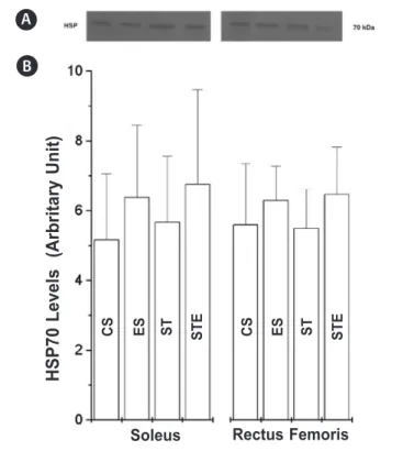
The frequency of lesions in the soleus and rectus femoris muscles of the animals from the control groups (CS and ST) were not statistically different (table 2). Nevertheless, the frequency of injured fibers after the exhaustion test in group ES was statistically higher than in group CS in both analysed muscles. Likewise, in group STE the frequency of lesions was higher than in group ST. There was a rare presence of inflammatory infiltrates, but the frequency was not different between groups (data not shown). Figure 3 shows the build-up of HSP70 in the soleus and rectus femoris muscles. The animals from groups ES and STE presented tendency (P = 0.09) of higher build-up of HSP70 (soleus:12.52 and 17.66%; rectus femoris: 23.45 and 19.26%, re-spectively), compared to their controls (CS and ST, rere-spectively), however, the differences have not reached statistical significance .
Table 1. Time until exhaustion and blood lactate concentration of the animals from the experimental groups.
Group Lactate at rest (mmol/L)
Lactate at 15min. (mmol/L)
Exhaustion time (min)
ES (n = 7) 2.86 ± 0.08 8.76 ± 0.59 90.0 ± 8.48
STE (n = 7) 2.90 ± 0.04 5.31 ± 0.22* 125.0 ± 6.00#
Data are presented as means ± S.E.M. *, P<0.05 vs. ES, in the same column. ES, sedentary submitted to exhaustion. n, number of animals.
Table 2. Frequency of animal injured fibers of the experimental groups.
Groups Rectus femoris Soleus
CS (n = 8) 16.75 ± 3.79 13.88 ± 3.81
ES (n = 7) 37.57 ± 0.07* 34.86 ± 0.04*
ST (n = 7) 24.0 ± 0.05 24.14 ± 0.06
STE (n = 7) 39.57 ± 0.05# 41.57 ± 0.08#
210
We demonstrated that the swimming program applied was efficient in causing adaptations to the animals’ organisms since the trained animals swam for a longer period of time until ex-haustion, if compared to the sedentary animals. Moreover, these animals performed exertion with less lactate build-up than the sedentary controls(27). These data indicate that the animals from groups ST and STE were trained.
However, after the exhaustion test the muscles assessed presented lesions in response to the exertion performed. The main lesions observed in this study were: cytoplasmic vacuoliza-tion; light interstial inflammatory reactions and onset of nuclei with more volume and dislocated to the central position of the fiber (2). These results show that the exhaustion test was sufficient to cause muscular lesions.
In the sedentary animals, both in soleus – muscle of oxida-tive features – and in the white portion of the rectus femo-ris – muscle of glycolytic features –, the frequency of injured fibers was higher in the animals submitted to exhaustion than in the control ones (soleus: 151%; rectus femoris: 126%). Dur-ing intense physical exercise there is increase of 10 to 20 times in the total oxygen consumption of the body and increase of 100 to 200 times in the oxygen uptake by the muscle tissue(1). This kind of exercise may trigger different ways of formation of oxygen reactive species as well as muscle injuries such as the ones observed in this study(3).
In the same way, in the trained animals the frequency of injured fibers was higher in the animals submitted to exhaustion (STE) than in the control ones, group ST (soleus: 64.00%; rectus femoris: 155.00%). These data show that the training adaptations
did not protect the muscles against injuries induced by exhaus-tive exercise. One of the adaptations to physical training which could play the protective role is the increase of HSP70 expres-sion by its antiapoptotic function(19). The increase of HSP in the skeletal muscle after exercise is able to restore the homeostasis, promote cellular remodeling and protect against the recurrent cellular insult occurred during physical activity(28).
Our findings show that the HSP70 levels, despite being higher in the trained animals than in the control ones after exhaus-tion test, were not statistically different. The Hsp72 is exclusively expressed by stress events(11), being highly induced by physical exercise in skeletal and cardiac muscles(16), while the Hsp73 is constitutive(29). Thus, the higher values HSP70 values found in the trained animals when compared to the control animals (CS), despite of not being statistically different, reflect the induction of the Hsp72 expression in the assessed muscles (figure 3).
In agreement with the results of the present study, Smolka et al.(4), reported that exercise until exhaustion did not result in the HSP70 induction or decrease in activity of antioxidant enzymes in trained rats. According to the authors, exhaustion was less stressing to the trained animals due to the high activity of the pre-existing antioxidant enzymes in the soleus muscle of these animals. It is possible that reactive oxygen species attack muscle proteins of the sedentary rats and signal to the induc-tion of HSP70 expression after exhausinduc-tion. On the other hand, if the pre-existing antioxidant enzymatic system is sufficient to avoid or minimize the attack of the reactive oxygen species, the induction of the HSP 70 expression may not be initiated or slightly initiated.
Another possibility is that the training intensity applied in the present study has not been sufficient to increase the HSP70 expression in the skeletal muscles of these animals. The mechanisms responsible for the induction of the HSP70 synthesis, in response to chronic exercise, involve many physiological alterations and factors such as increase of inner and muscle temperature, increase of the lactate concentration, increase of catecholamine activity, decrease of pH, glycogen and ATP, increase of free radicals and alterations in the cytosolic calcium. Such alterations could, independently or collectively, explain the consequent increase of HSP70. However, these factors are present in a differentiated manner, if we consider volume, intensity and level of stress caused by the exercise model(14,29). Some studies have given credit to the high levels of HSP70 expression in response to exercise to its intensity(14,30).
In conclusion, although the swimming program with low intensity increases performance of the animals in the exhaustion test, it did not promote protection to the skeletal muscles studied against lesions induced by exhaustive exercise.
ACKNOWLEDGEMENTS
Project sponsored by the Support to Research Foundation of Minas Gerais State (FAPEMIG-CBB 1851/05). A.J. Natali has a productivity scholarship from CNPq.
All authors have declared there is not any potential conflict of interests concerning this article.
Figure 3. Build-up of heat shock protein (HSP70) in the soleus and rectus femoris muscles of Wistar rats. The same amounts of proteins of the muscles were separated by SDS-PAGE and the immunoblotting was done with the monoclonal antibody HSP70. A: control sedentary; B: sedentary submitted to exhaustion test; C: swimming trained; D: swimming trained submitted to exhaustion test.
A
B
H
SP7
0
L
e
v
e
ls
(A
rb
ri
ta
ry
U
n
it)
Soleus Rectus Femoris
C
S
211
REFERENCES
1. Clebis NK, Natali MRM. Lesões musculares provocadas por exercícios excêntricos. R Bras Ci e Mov
2001;9:47-53.
2. Duarte JA, Mota MP, Neuparth MJ, Apeel HJ, Soares JMC. Miopatia do exercício. Anatomopatologia
e fisiopatologia. Rev Port Cien Desp 2001;1:73-80.
3. Koury JC, Donangelo CM. Zinco, estresse oxidativo e atividade física. Rev Nutr 2003;16:433-41.
4. Smolka MB, Zoppi CC, Alves AA, Silveira LR, Marangoni S, Perreira-da-Silva L, et al. HSP72 as a comple-et al. HSP72 as a comple-. HSP72 as a comple-HSP72 as a
comple-mentary protection against oxidative stress induced by exercise in the soleus muscle of rats. Am J Physiol Regul Integr Comp Physiol 2000;2791539-45.
5. Clarkson PM, Sayers SP. Etiology of exercise-induced muscle damage. Can J Appl Physiol 1999;24:234-48.
6. Arends MJ, Wyllie AH. Apoptosis: mechanisms and roles in pathology. Int Rev Exper Pathol 1991;32:223-54.
7. Sanchez-Torres LE, Vargas FD. Apoptosis: el fenómeno y su determinación. Téc Pecu Méx 2003;41:49-62.
8. Guimarães CA, Linden R. Programmed cell death. Apoptosis and alternative deathstyles. Eur J
Biochem 2004;271:1638-50.
9. Nakagawa T, Yuan J. Cross-talk between two cysteine protease families. Activation of caspase-12 by
calpain in apoptosis. J Cell Biol 2000;150:887-94.
10. Kiang JG, Tsokos GC. Heat shock protein 70 kDa: molecular biology, biochemistry, and physiology. Pharmacol Ther 1998;80:183-201.
11. Snoeckx LH, Cornelussen RN, Van Nieuwenhoven FA, Renemam RS, Van Der Vusse GJ. Heat shock proteins and cardiovascular pathophysiology. Physiol Rev 2001;81(4):1461-97.
12. Adhihetty PJ, Hood DA. Mechanisms of apoptosis in skeletal muscle. Basic Appl Myol 2003;13:171-9. 13. Ji LL. Exercise-induced modulation of antioxidant defense. Ann NY Acad Sci 2002;959:82-92. 14. Milne KJ, Noble EG. Exercised-induced elevation of HSP70 is intensity dependent. J Appl Physiol
2002;93:561-8.
15. Lunz W, Oliveira EC, Neves MT, Fontes EP, Dias CM, Natali AJ. Anabolic steroid and exercise induced cardiac stress protein (HSP72) in the rat. Braz J Med Biol Res 2006;39:889-93.
16. Whitham M, Fortes MB. Heat shock protein 72: release and biological significance during exercise. Front Biosci 2008;13:1328-39.
17. Radak Z, Taylor AW, Ohno H, Goto S. Adaptation to exercise-induced oxidative stress: from muscle to brain. Exerc Immunol Rev 2001;7:90-107.
18. Bupha-Intr T, Wattanapermpool J. Cardioprotective effects of exercise training on myofilament calcium
activation in ovariectomized rats. J Appl Physiol 2004;96:1755-60.
19. Siu PM, Bryner RW, Martyn JK, Alway SE. Apoptotic adaptations from exercise training in skeletal and cardiac muscles. Faseb J 2004;18:1150-2.
20. Siu PM, Bryner RW, Murlasits Z, Alway SE. Response of XIAP, ARC, and FLIP apoptotic supressors to 8 wk of treadmill running in rat heart and skeletal muscle. J Appl Physiol 2005;99:204-9.
21. Bolter CP, Gordon BL. Cholinergic responses of isolated atria from swim-trained rats. Eur J Appl Physiol 1983;51:231-6.
22. Voltarelli FA, Gobatto CA, Mello MAR. Determination of anaerobic threshold in rats using the lactate minimum test. Braz J Med Biol Res 2002;35:1389-94.
23. Vendetti P, Di Meo S. Antioxidants, tissue damage, and endurance in trained and untrained young male rats. Arch Biochem Biophys 1996;331:63-8.
24. Dawson CA, Horvarth SM. Swimming in small laboratory animals. Med Sci Sports Exerc 1970;2:51-78. 25. Bradford MM. A rapid and sensitive method for the quantitation of microgram quantities of protein
utilizing the principle of protein-dye binding. Anal Biochem 1976;72:248-54.
26. Laemmli UK. Cleavage of sctructural proteins during the assembly of the head of bacteriophage T4. Nature 1970;227:680-5.
27. Gobatto CA, Mello MAR, Sibuya CY, Azevedo JRM, Santos LA, Kokubun E. Maximal lactate steady state in rats submitted to swimming exercise. Comp Biochem Physiol 2001;130:21-7.
28. Morton JP, MacLaren DP, Cable NT, Bongers T, Griffiths RD, Campbell IT, et al. Time course and dif-ferential responses of the major heat shock protein families in human skeletal muscle following acute nondamaging treadmill exercise. J Appl Physiol 2006;101:176-82.
29. Skidmore R, Gutierrez JA, Guerriro V, Kregal KC. HSP70 induction during exercise and heat stress in rats: role of internal temperature. Am J Physiol 1995; 268:92-7.