Biochemistry
Physicochemical characterization of two
deproteinized bovine xenografts
Abstract: Calcium phosphate salts, or more speciically hydroxyapatite, are products of great interest in the ields of medical and dental science due to their biocompatibility and osteoconduction property. Deprotein-ized xenografts are primarily constituted of natural apatites, sintered or not. Variations in the industrial process may affect physicochemical properties and, therefore, the biological outcome. The purpose of this work was to characterize the physical and chemical properties of depro-teinized xenogenic biomaterials, Bio-Oss (Geistlich Biomaterials, Wol-huser, Switzerland) and Gen-Ox (Baumer S.A., Brazil), widely used as bone grafts. Scanning electron microscopy, infrared region spectrosco-py, X-ray diffraction, thermogravimetry and degradation analysis were conducted. The results show that both materials presented porous gran-ules, composed of crystalline hydroxyapatite without apparent presence of other phases. Bio-Oss presented greater dissolution in Tris-HCl than Gen-Ox in the degradation test, possibly due to the low crystallinity and the presence of organic residues. In conclusion, both commercial materi-als are hydroxyapatite compounds, Bio-Oss being less crystalline than Gen-Ox and, therefore, more prone to degradation.
Descriptors: Dental implantation; Hydroxyapatite; Chemical analysis; Physical analysis.
Thais Accorsi-Mendonça(a) Márcio Baltazar Conz(b) Teresa Cristina Barros(c) Lídia Ágata de Sena(d) Glória de Almeida Soares(e) José Mauro Granjeiro(f)
(a)MSc in Endodontics, Department of
Endodontics, School of Dentistry of Bauru, University of São Paulo, Bauru, SP, Brazil.
(b)PhD, Metallurgy and Materials Engineering
Program (COPPE), Federal University of Rio de Janeiro, RJ, Brazil.
(c)PhD, Health Science Institute, Paulista
University, Bauru, SP, Brazil.
(d)PhD, Scientific Metrology Directory
(Inmetro), Duque de Caxias, RJ, Brazil.
(e)PhD, Metallurgy and Materials Engineering
Program (COPPE), Federal University of Rio de Janeiro, RJ, Brazil.
(f)PhD in Cell and Molecular Biology, Biology
Institute, Fluminense Federal University, RJ, Brazil.
Corresponding author:
José Mauro Granjeiro
Outeiro de São João Baptista, s/n.
Campus do Valonguinho Niterói - RJ - Brasil CEP: 24020-150
E-mail: [email protected]
Introduction
Calcium phosphate salts, in particular hydroxy-apatite (HA), are materials of great interest for treatment of osseous deiciencies due to their bio-compatibility and osteoconduction property.1 Grafts of xenogenic origin have been widely studied due to abundant sources and accessible processing costs. However, bovine bone processing should be quite well controlled in order to avoid disease transmis-sion,2 and to assure adequate physicochemical prop-erties for the intended use.3
It is important to mention that the structure of apatite is similar among different species.4 Several papers in the literature have shown the biocompat-ibility of deproteinized bovine matrix as bone graft material,5-7 which, besides providing a supportive structure for osteoconduction, releases calcium and phosphate ions essential for osteogenesis.8
The presence of pores in bone graft biomaterials has been shown as a requisite of great importance for repairing osseous deiciency, favoring the osteo-conduction through osseous growth also inside the pores.9,10 In order to allow the penetration of repair-ing tissue and osseous neoformation in the pores, they should be greater than 100Pm.11-13
Bio-Oss® (Geistlich Biomaterials, Wolhusen, Switzerland) is a widely studied bovine xenograft consisting of a mineral osseous matrix obtained af-ter the removal of the organic components of me-dullar bovine bone during a thermal treatment at 300qC.14 Another bovine xenograft available in the Brazilian market is Gen-Ox® (Baumer S.A., São Paulo, Brazil) obtained through deproteinization at high temperatures (between 950 and 1,000qC). The increased processing temperature enhances mate-rial crystallinity4,15 being also concurrent with the decomposition of the hydroxyapatite in other phases like tricalcium phosphate (TCP). The most crystal-line biomaterial will be the least degradable, but the presence of other phases (usually more soluble than HA) may compensate or null this effect.16 The cor-rect indication of a biomaterial shall depend on its physicochemical properties. Thus, the aim of this study was the physicochemical characterization of the products Bio-Oss and Gen-Ox, which are both deproteinized xenografts used as bone grafts.
Material and Methods
Materials
The two commercial biomaterials used in this study derived from inorganic medullar bovine bone: 1) Bio-Oss, consisting of granules (250 to 450Pm) deproteinized at 300qC, according to the manu-facturer’s data; 2) Gen-Ox, consisting of granules (with particles from 250 to 1,000Pm) deproteinized at 950 – 1,000qC, according to the manufacturer’s data (Baumer S.A. – Brazil, Registration at the Min-istry of Health No. 10345500001).
Physicochemical characterization
Morphological characterization of the materials was carried out through scanning electron micros-copy (Zeiss, model DSM 940A, Oberkochen, Baden-Württemberg, Germany) operating at 20 kV of elec-trons acceleration. The presence of residues from the organic phase or different species of phosphate was assessed through the use of thermogravimetry and infrared spectroscopy, respectively. Thermogravim-etry was performed from 50 to 950qC with a heat-ing rate of 10qC/minute in a dynamic atmosphere of nitrogen (50 ml min-1) (TA Instruments, model TGA 2950, coupled to a thermal analyzer of TA Instru-ments, Model TA 2000), while Fourier-transformed infrared spectroscopy (FTIR) was obtained in a Per-kin-Elmer (model 1000, Waltham, Massachusetts, USA) equipment.
In order to identify the crystalline phases in the xenografts, an X-ray diffractometer (DRX-Mini-lex, Rigaku, Tokyo, Japan) was operated at 30 kV, 15 mA and radiation CuKD, and the data was col-lected from 5-100q 2T. The crystallinity of the mate-rials was determined from the DRX patterns accord-ing to the methodology proposed by Landi17 (2000); whereas the rate of degradation was determined threefold using protocol ISO/FDIS 10993-14:2001 (in buffer Tris-HCl, pH 7.3 for 120 hours).
Results
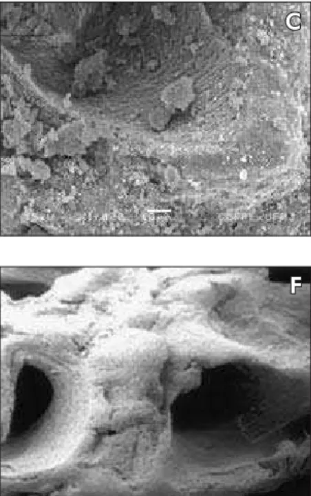
Inorganic Gen-Ox (Figure 1, E – H) also looks po-rous (Figure 1, E and F); when magniied (Figure 1, G and H), the coalescence of the grains taken place during the calcination of the material at high tem-peratures can also be seen.
In the Fourier Transform infrared spectroscopy (FTIR), bands can be identiied in the wavelength ranges of 1,554-1,569, 1,400-1,420 and 850 cm-1 corresponding to the functional group CO3-2 of HA for Bio-Oss (Graph 1A). The phosphate group ap-pears in both materials, but Gen-Ox (Graph 1B) presents better-deined bands between 600 and 1,100 cm-1. Bands around 3,571 cm-1 are also high-lighted indicating the presence of the structural O-H stretch, as well as bands in 1,172, 1,088, 1,025 and 963 cm-1, indicating the presence of PO
4 -3
groups.
Bio-Oss and Gen-Ox thermogravimetric analysis (Graph 1, C and D, respectively) showed the materi-als’ loss of mass due to the increase in temperature. Bio-Oss’ loss of mass was about 2.6, 0.5 and 2.4% between 30qC to 420qC, 420qC to 635qC (tem-perature range between loss 1 and 2) and 630qC to 950qC, respectively. The residual mass at 950qC was 94.3%. Gen-Ox presented loss of mass at about 0.35% between the initial temperature and 384qC, increasing up to 0.6% from 665qC to 950qC, result-ing in a residual mass of 99.4%.
In the patterns obtained for X-ray diffraction (Graph 1, E – F), the major peaks identiied were re-lated to synthetic HA that according to card data JCPDS 9-432 (JCPDS-ICDD1, 1992) are placed at 2T= 32, 33 and 26q, with relative intensity of 100, 60 and 40%, respectively. From X-ray diffraction
B
C
D
Figure 1 - Electronmicrographs of the studied Biomaterials. Bio-Oss (A-D). Particles with regular dimensions covered with nanoparticles, and showing different pore sizes (arrows). Inorganic Gen-Ox (E-H). Porous particles showing the fusion of hydroxyapatite crystals. Magnification: A (70 X), B-C (1,000 X), D (10,000 X), E (50 X), F (100 X), G (10,000 X), H (20,000 X).
A
G
E
F
patterns the crystallinity was determined17 for each material, resulting in 40% and 96% for Bio-Oss and Gen-Ox, respectively.
The degradation test in Tris-HCl (120 h, pH 7.3,
37qC) indicated the loss of mass, the average of 4.7% (r1.61) for Bio-Oss; however, no alteration of Gen-Ox mass was detected after 120 hours of deg-radation assay.
Trans
mittance
(%)
0 25 50 75 100
3,500 3,000 2,500 2,000 1,500 1,000 500 Wave number (cm–1)
CO3–2 PO4–3
H-O-H P-O
OH–
A
Trans
mittance
(%)
Wave number (cm–1) 3,500
0
2,500
3,000 2,000 1,500 1,000 500 25
50 75 100
B
R
es
idu
al
Mas
s
(%)
Temperature (°C) 99
99.5 100
1,000 0 200 400 600 800
D
R
es
idu
al
Mas
s
(%)
Temperature (°C) 94
96 98 100
0 200 400 600 800 1,000
C
R
elativ
e
I
nte
ns
ity
2Q 1,000
800
600
400
200
0
20 40 60
26 32
33
E
R
elativ
e
I
nte
ns
ity
2Q 1,000
800
600
400
200
0
20 26
32
33
40 60
F
Discussion
Both materials analyzed are classiied as xenografts from inorganic bovine bone, and are indicated as bone defect illing materials18,19 due to their osteoconduc-tive properties.5,7 The study about synthetic and natu-ral apatites has shown that the material degradation rate affects the biological response to the material.20-22 Recent studies have shown that fast absorption of the biomaterial may jeopardize adequate repair.20 On the other hand, non-resorbable materials can make some clinical procedures like grafts unfeasible.23 Thus, the processing way used by the manufacturers of natural and synthetic apatites affects both the physicochemi-cal properties and the degradation rate of these bio-materials, possibly affecting bone repair.10,24
It was observed through scanning electron mi-croscopy that both materials presented pores on their surfaces (Figure 1). The degree of microporos-ity depends on the compaction method of the start-ing powder and the temperature of synthetic materi-als or the use of natural materimateri-als on which the pores were biologically designed. The presence of pores in the granules increases the surface area of the xeno-graft material, favoring osteoconduction, enabling bone growth also in the pores.10 Porosity was simi-lar in both materials, but the effect of increased tem-perature of sintering over the coalescence of apatite crystals is clear for Gen-Ox (Figures 1G and 1H).
Infrared spectral analysis has shown that both Bio-Oss and inorganic Gen-Ox have typical HA bands.3,24,25 Bands resolution is better in Gen-Ox, sug-gesting high crystallinity, and no bands related to oth-er compounds are obsoth-erved. Preliminary data about material crystallinity may be obtained through infra-red spectrum, especially from the bands that are in the 590-610 cm-1 and 1,000 cm-1 regions.3,16 Diffracto-grams of the materials assessed conirm the presence of hydroxyapatite (according to ICDD data, 9-432); however, again, the bands have better resolution for Gen-Ox. Wide bands observed in the Bio-Oss diffrac-togram may be related to the different characteristics of the material, like the presence of non-crystalline phases (like organic residues), nanometric dimensioned crystals or defective powder structure.15,24,25
The loss of mass (nearly fourfold higher than that of Gen-Ox) observed in the thermogravimetric assay
suggests the presence of residual organic material in Bio-Oss graft (around 3%). As it is expected for calci-nated apatites (treatment temperature above 900qC), Gen-Ox showed a loss of mass above 700qC only; however, this loss is virtually none, about 0.3% of the sample. No relections related to other phases of calcium phosphates were detected. It was conirmed through Landi’s17 (2000) method that Gen-Ox pre-sented higher crystallinity than Bio-Oss, which con-irmed the results of infrared and morphology of the crystals through scanning electron microscopy. This result is coherent with the calcination process to which it was submitted, since crystallinity is directly proportional to the processing temperature of the biomaterial. According to the manufacturers’ data, Bio-Oss is processed in a lower temperature than Gen-Ox, 300 and 1,000qC, respectively. The effect of processing temperature is related to the elimina-tion of carbonate content in the bone, which hap-pens at temperatures above 400qC only.4,15
Data about materials crystallinity is important, as the bioabsorption rate shall be compatible with the re-pairing process of the recipient tissue. Calcium phos-phates solubility depends on the relation Ca/P and, for the same composition, it depends on the crystallinity degree; so less crystalline materials are more soluble.4 Therefore, the presence of second phases (usually more soluble than HA) may compensate or null this effect.15 In this case there was no composition effect of the HA, prevailing the effect of crystallinity in in vitro degra-dation. Thus, the degradation test indicated the loss of mass, at an average of 4.7% (r1.61) for Bio-Oss, while no alteration of mass was detected in Gen-Ox.
The results suggest that the content of non-crys-talline phases, probably organic matter and carbon-ate, identiied through thermogravimetric, infrared and X-ray diffraction analyses, generates a biomate-rial of lower crystallinity and, consequently, more prone to degradation. Thus, the processing tempera-ture may be a way to change the physicochemical properties of the material, producing materials with different reabsorption levels.
Conclusions
and is, therefore, more prone to degradation.
Acknowledgments
The authors would like to acknowledge the
i-nancial support received from CNPq (CT-Saude, process number 504.808/2004-4), Capes, Fapesp (Grant no99/10655-5 and 01/10707-7), Faperj and FINEP (Grant no. 01.04.0469.00).
References
1. Kostopoulos L, Karring T. Regenerations of the sagittal su-ture by GTR and its impact on growth of the cranial vault. J Craniofac Surg. 2000;11(6):553-61.
2. Wenz B, Oesch B, Horst M. Analysis of the risk of transmitting bovine spongiform encephalopathy through bone grafts derived from bovine bone. Biomaterials. 2001;22(12):1599-606. 3. Conz MB, Granjeiro JM, Soares GA. Physicochemical
character-ization of six commercial hydroxyapatites for medical-dental ap-plications as bone graft. J Appl Oral Sci. 2005;13(2):136-40. 4. Le Geros RZ. Calcium phosphates in oral biology and
medi-cine. Basel: Karger; 1991. Monographs in oral science. v. 15. 5. Masters DH. Implants. Bone and bone substitutes. CDA J.
1988:16(1):56-65.
6. Oliveira RC, Sicca CM, Silva TL, Cestari TM, Oliveira DT, Buzalaf MAR et al. Efeito da temperatura de desproteinização no preparo de osso cortical bovino microgranular. Avaliação microscópica e bioquímica da resposta celular em subcutâneo de ratos. Rev FOB. 1999;7(3/4):85-93.
7. Taga MLL. Análise histológica e radiográfica do potencial os-teopromotor da membrana de cortical óssea bovina no reparo de defeito ósseo de tamanho crítico na calvária de cobaia ( Ca-via porcellus) [Dissertação de Mestrado]. Bauru: Faculdade de Odontologia da USP; 2004.
8. Sciadini MF, Dawson JM, Johnson KD. Evaluation of bo-vine-derived bone protein with a natural coral carrier as a bone-graft substitute in a canine segmental defect model. J Orthop Res. 1997;15(6):844-57.
9. Kalkura SN, Anee TK, Ashok M, Betzel C. Investigations on the synthesis and crystallization of hydroxyapatite at low temperature. Biomed Mater Eng. 2004;14(2):581-92. 10. Werner J, Linner-Krcmar B, Friess W, Greil P.
Mechani-cal properties and in vitro cell compatibility of hydroxy-apatite ceramics with graded pore structure. Biomaterials. 2002;23(21):4285-94.
11. Carotenuto G, Spagnuolo G, Ambrosio L, Nicolais L. Mac-roporous hydroxyapatite as alloplastic material for dental ap-plications. J Mater Sci Mater Med. 1999;10(10/11):671-6. 12. Misch CE. Contemporary implant dentistry. 2nd ed. St. Louis:
CV Mosby; 1999.
13. Tampieri A, Celotti G, Sprio S, Delcogliano A, Franzese S. Porosity-graded hydroxyapatite ceramics to replace natural bone. Biomaterials. 2001;22(11):1365-70.
14. Thaller SR, Hoyt J, Dart A, Borjeson K, Tesluk H. Repair of experimental calvarial defects with Bio-Oss particles and collagen sponges in a rabbit model. J Craniofac Surg. 1994;5(4):242-6.
15. Tadic D, Epple M. A thorough physicochemical characterisation of 14 calcium phosphate-based bone substitution materials in comparison to natural bone. Biomaterials. 2004;25(6):987-94. 16. Tadic D, Peters F, Epple M. Continuous synthesis of amor-phous carbonated apatites. Biomaterials. 2002;23(12):2553-9.
17. Landi E. Densification behavior and mechanisms of synthetic hydroxyapatite. J Eur Cer Soc. 2000;(20):2377-87.
18. Blank BS, Levy AR. Combined treatment of a large peri-odontal defect using GTR and DFDBA. Int J Periodontics Restorative Dent. 1999;19(5):481-7.
19. Zitzmann NU, Naef R, Scharer P. Resorbable versus non-resorbable membranes in combination with Bio-Oss for guided bone regeneration. Int J Oral Maxillofac Implants. 1997;12(6):844-52.
20. Burg KJ, Porter S, Kellam JF. Biomaterials development for bone tissue engineering. Biomaterials. 2000;21(23):2347-59.
21. Houser BE, Mellonig JT, Brunsvold MA, Cochran DL, Mef-fert RM, Alder ME. Clinical evaluation of inorganic bovine xenograft with a bioabsorbable collagen barrier in the treat-ment of molar furcation defects. Int J Periodontics Restorative Dent. 2001;21(2):161-9.
22. Yamada S, Shima N, Kitamura H, Sugito H. Effect of porous xenographic bone graft with collagen barrier membrane on periodontal regeneration. Int J Periodontics Restorative Dent. 2002;22(4):389-97.
23. Su-Gwan K, Hak-Kyun K, Sung-Chul L. Combined implanta-tion of particulate dentine, plaster of Paris, and a bone xeno-graft (Bio-Oss) for bone regeneration in rats. J Craniomaxil-lofac Surg. 2001;29(5):282-8.
24. Le Geros RZ. Calcium phosphate biomaterials: preparation, properties and biodegradations. In: Wise DL [editor]. En-cyclopedic Handbook of Biomaterials and Bioengineering. New York: Marcel Dekker; 1995. v. 1, pt. A, p. 1429-63. 25. Elliot JC. Structure and chemistry of the apatite and other