Maria Cristina Ramos Lima PADOVANI(a)
Maria Teresa Botti Rodrigues SANTOS(b)
Giselle Rodrigues de SANT’ANNA(a)
Renata Oliveira GUARÉ(a)
(a)Department Pediatric Dentistry, School of
Dentistry, Universidade Cruzeiro do Sul - UNICSUL, São Paulo, SP, Brazil.
(b)Special Needs Individuals, School of
Dentistry, Universidade Cruzeiro do Sul - UNICSUL, São Paulo, SP, Brazil.
Prevalence of oral manifestations in
soft tissues during early childhood in
Brazilian children
Abstract: This study aimed at assessing the prevalence of soft tissue oral
manifestations in children during early childhood, according to age group, gender, and site in the oral cavity, and at correlating these oral manifes-tations with systemic alterations. A cross-sectional study was conducted involving 586 children from 0 to 3 years of age (12.4 ± 11.8 months), 316 (53.9%) male and 270 (46.1%) female, in the city of Mauá, SP, Brazil. Exami-nation was performed by a single examiner (Kappa Index = 0.90) accord-ing to World Health Organization criteria (WHO, 1997).The prevalence of oral manifestations in the soft tissues of children during early childhood was 34.8%. The age group showing statistical signiicance was 0-1 months old (56.4%). Epstein’s pearls were signiicantly present (43.2%) in 0-1-month-old babies, and gingivitis in 12-24-month-0-1-month-olds (15.9%). The palate was the most affected region (16.7%). Infectious alterations were the most preva-lent systemic alteration (20%). An association was observed between the presence of systemic alterations and the occurrence of oral manifestations. The prevalence of oral manifestations was 34.8%, regardless of gender, and was manifested mostly in 0-1-month-old babies. The palate was the most prevalent region, and the majority of oral manifestations were associated with systemic alterations.
Keywords: Child; Diagnosis, Oral; Pathology; Prevalence.
Introduction
In order to achieve and maintain oral health during early childhood, it is important for the pediatric dentist to know how to diagnose and treat oral manifestations, whenever necessary. A baby presents unique and transitional anatomical structures that are typical of this age of life. Aside from the characteristic physiological alterations occurring in this age group, there are speciic developmental alterations and diseases.1,2,3,4
Numerous studies have proven the importance of the dentist during the diagnostic process of many systemic diseases. The irst manifestation of systemic alterations often occurs in the oral cavity.5,6
There are many reports in the literature regarding dental caries7,8 and
periodontal disease;9,10 however, there are few studies related to the
preva-lence of soft tissue manifestations, and they address varied methodological criteria. The effort being made to offer an appropriate system for collecting data is insuficient, resulting in a series of methodological problems that often yield incorrect and inconsistent results. Epidemiological studies conducted in Declaration of Interests: The authors
certify that they have no commercial or associative interest that represents a conflict of interest in connection with the manuscript.
Corresponding Author:
Maria Cristina Ramos Lima Padovani Email: [email protected]
DOI: 10.1590/1807-3107BOR-2014.vol28.0036 Epub XXX XX, 2014
Submitted: May 09, 2013
the past have shown a considerable discrepancy in the prevalence of soft tissue lesions in the oral cavity.11,12,13,14
The purpose of this study was to assess the preva-lence of oral manifestations in the soft tissues of Bra-zilian children from 0 to 36 months old, according to age group, gender, and location, and to correlate these manifestations with the presence of systemic alterations.
Methodology
This cross-sectional survey was irst conducted with a sample composed of 706 children treated at the Unidade de Saúde (Municipal Health Unit - HU) of Mauá, State of São Paulo, Brazil, from May to December 2007, following approval by the Research Ethics Committee (EC/CSU- 072/06).The inclusion criteria were children from 0 to 36 months old, of both genders and all ethnicities. The exclusion criteria were children with nasal/labial cleft, children undergoing dental restorative treatment who were not participating in the orientation groups, chil-dren with dental urgencies, and chilchil-dren whose legal guardians did not agree to sign the consent term. The inal sample resulted in 586 children (12.4 ± 11.8 months), 316 (53.9%) male and 270 (46.1%) female. Diagnosis of systemic alterations was determined by a pediatrician.
The clinical examination of babies under 12 months old was conducted in the knee-to-knee position, under the operating light of the dental chair, whereas the remaining children were examined in the dental chair. In the maternity ward, the procedure was conducted in the maternity crib, using a penlight. The examination was conducted by a single, previously trained exam-iner (Kappa=0.90). The spreadsheet data from the epi-demiological survey and the photos of the oral mani-festations, duly authorized by the legal guardians, were collected by the HU dental nurse, and the data from the maternity were collected by the neonatal nurse.
The WHO clinical diagnostic criteria were used to determine oral mucosal alterations,15,16 as used by
other authors.12,17,18 The sequence of examinations of
the oral mucosa includes: 1- upper lip (vermilion); 2- upper labial mucosa; 3- upper alveolar mucosa; 4- upper gingival/alveolar ridge; 5- hard palate; 6- soft palate; 7- oropharynx; 8- dorsum of the tongue; 10- ventral tongue; 11- loor of the mouth; 12, lower gingival/alveolar ridge; 13- lower alveolar mucosa; 14-
right and left buccal mucosa; 15- lower labial mucosa; 16- lower lip (vermillion); and 17- labial commissures. For data analysis purposes, the 586 children exam-ined were divided into 4 groups according to age: 0 to 1 month old (34%), 1 to 12 months old (19%), 12 to 24 months old (24.8%) and 24 to 36 months old (22.2%). The criteria for dividing the sample into age groups were based on major changes in the oral cavity and the appearance of oral manifestations: 0-1 months, newborn; 1-12 months, a period of neurophysiologic changes and feeding accompanied by tooth eruption; 12-24 months, eruption of primary molars; 24-36 months, a period in which the primary dentition is complete and stable.
Age groups, gender, oral manifestations and systemic conditions were compared using the Chi square and Fisher Exact tests (Minitab statistic soft-ware, version 14.2, Minitab Inc., State College, USA), together with a binary logistic regression model to identify predictors of oral manifestations, considered as independent variables (PASW Statistics v. 18.0.0, SPSS Inc., Chicago, USA).
All the tests were analyzed with a signiicance of p < 0.05.
Results
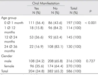
The oral manifestation distribution according to age and gender (Table 1) showed a statistically sig-niicant difference related to age. In the irst month of life, a higher percentage of oral manifestations are observed, probably related to inclusion cysts.
Table 1. Distribution of oral manifestation cases according to age and gender.
Oral Manifestation Yes
N (%)
No N (%)
Total
N (%) p
Age group
0 Ø 1 month 111 (56.4) 86 (43.6) 197 (100) < 0.001 1 Ø 12
months
18 (15.8) 96 (84.2) 114 (100)
12 Ø 24 months
53 (36.6) 92 (63.4) 145 (100)
24 Ø 36 months
22 (16.9) 108 (83.1) 130 (100)
Gender
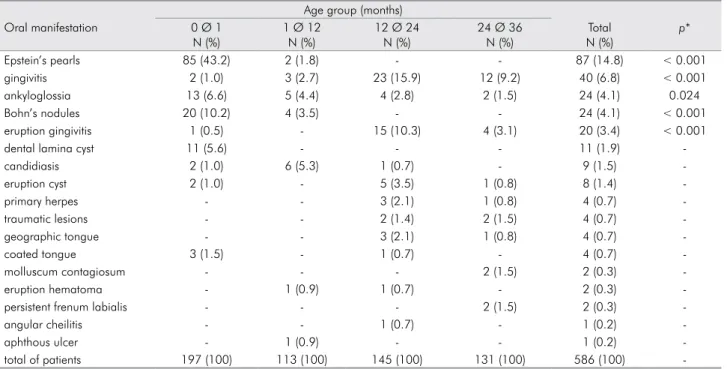
Accordingly, Epstein’s pearls (a subtype of inclu-sion cysts) were more prevalent in the irst month of life (Table 2), insofar as they are typical of this age group. However, gingivitis was prevalent in the age group of 12-24 months, the period of tooth eruption. Inclusion cysts are frequently found in the oral cavities of newborn infants and are sub-grouped according to their location (alveolar or palatal) and origin. Epstein’s pearls are cystic, keratin-filled nodules observed along the mid palatine raphe, derived from entrapped epithelial remnants. Bohn’s nodules are mucous-gland cysts, on the alveolar ridges in the vestibular and lingual sur-faces. Dental lamina cysts are observed on top of the alveolar ridge of newborns, originating from remnants of the dental lamina.4,19
No signiicant difference in the prevalence of oral manifestations was observed related to the child’s sex.
Concerning the region of the manifestations, the palate presented the most oral manifestations (16.7%), followed by gingiva (11.4%), alveolar ridge (8.9%) and tongue (7.8%).
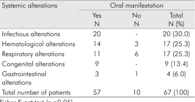
The oral manifestation distribution, according to the systemic alterations, is presented in Table 3,and theprevalence of systemic alterations, according to the oral manifestation, in Table 4.
Table 5 shows the results for the logistic regression, together with the odds ratio values for each variable, in relation to oral manifestations. The results of this regression established an independent association between age group (1-12 months, 12-24 months, and 24-36 months), systemic change and oral manifesta-tions. Children aged 1-12 months, 12-24 months and 24-36 months were 8.2%, 30.4% and 11.3% more likely, respectively, to present oral manifestations than babies between 0-1 months. Furthermore, children who pre-sented systemic changes showed a 17.4-fold greater risk of exhibiting oral manifestations, compared with chil-dren who presented no systemic change. The inclusion of all these variables in the model, including gender, did not effect any change in the associations between oral manifestations, systemic changes and age group identiied in the univariate analyses.
Table 2. Prevalence of oral manifestations according to age group.
Age group (months)
Oral manifestation 0 Ø 1
N (%)
1 Ø 12 N (%)
12 Ø 24 N (%)
24 Ø 36 N (%)
Total N (%)
p*
Epstein’s pearls 85 (43.2) 2 (1.8) - - 87 (14.8) < 0.001
gingivitis 2 (1.0) 3 (2.7) 23 (15.9) 12 (9.2) 40 (6.8) < 0.001
ankyloglossia 13 (6.6) 5 (4.4) 4 (2.8) 2 (1.5) 24 (4.1) 0.024
Bohn’s nodules 20 (10.2) 4 (3.5) - - 24 (4.1) < 0.001
eruption gingivitis 1 (0.5) - 15 (10.3) 4 (3.1) 20 (3.4) < 0.001
dental lamina cyst 11 (5.6) - - - 11 (1.9)
-candidiasis 2 (1.0) 6 (5.3) 1 (0.7) - 9 (1.5)
-eruption cyst 2 (1.0) - 5 (3.5) 1 (0.8) 8 (1.4)
-primary herpes - - 3 (2.1) 1 (0.8) 4 (0.7)
-traumatic lesions - - 2 (1.4) 2 (1.5) 4 (0.7)
-geographic tongue - - 3 (2.1) 1 (0.8) 4 (0.7)
-coated tongue 3 (1.5) - 1 (0.7) - 4 (0.7)
-molluscum contagiosum - - - 2 (1.5) 2 (0.3)
-eruption hematoma - 1 (0.9) 1 (0.7) - 2 (0.3)
-persistent frenum labialis - - - 2 (1.5) 2 (0.3)
-angular cheilitis - - 1 (0.7) - 1 (0.2)
-aphthous ulcer - 1 (0.9) - - 1 (0.2)
-total of patients 197 (100) 113 (100) 145 (100) 131 (100) 586 (100)
-* Chi square test (p < 0.05).
Table 3. Distribution of oral manifestations according to cases of systemic alterations.
Oral manifestation Systemic
alterations
Yes N (%)
No N (%)
Total
N (%) p
Yes 57 (85.1) 10 (14.9) 67 (100)
No 147 (28.3) 372 (71.7) 519 (100) <0.001 Total 204 (34.8) 382 (65.2) 586 (100)
Discussion
In order to achieve and maintain oral health dur-ing early childhood, it is important for the pediat-ric dentist to know how to diagnose and treat oral manifestations in this age group. This study helps identify the oral soft tissue manifestations that are more prevalent in early childhood, mainly related to systemic changes, and determine the appropri-ate approach. The prevalence of oral manifestations in the soft tissue of children, as found in our study, was very similar to that reported in other studies; however, studies involving the age group from 0 to 36 months old are uncommon. In Spain,20 38.9% of 6
year-old children presented oral manifestations in soft tissues, whereas a prevalence of 32.9% was reported in 18 to 80 month-old children in South Africa.17 In
Argentina,21 33.8% of oral manifestations occurred in
the age group from 0 to 15 years old, and 39%, from 4 to 13 years old, whereas the frequency of children age 0 to 12 years presenting oral mucosal diseases was 28.4% in Italy.22
In other regions, an increase was observed in the prevalence of oral manifestations in soft tissues, from 52.57% in the age group from 0 to 12 years old23 to
24.9% in the age group from 0 to 4 years old.18 In
Bra-zil, the age group from 0 to 24 months old19 showed
a prevalence of 21% for oral manifestations, whereas the prevalence in the age group from 0 to 5 years old was 2.30%.24 In other countries, the prevalence was 4%
in the age group from 5 to 17 years old,12 compared
with other studies, like NHANES III, where the prev-alence for children from 2 to 17 years old was 9.11%.25
Among cystic manifestations, the most prevalent were newborn cysts, with 20.8% in the age group from 0 to 36 months old. In relation to newborn cysts (Epstein’s pearls, Bohn’s nodules and dental lamina cysts), Epstein’s pearls were the manifestation with the highest prevalence (14.8%). Similarly, Baldani et al.19 also
observed that the newborn inclusion cysts (23.62%) were more prevalent than the other cystic lesions.
Notably, some authors observed a higher preva-lence of gingival cysts than that observed in this study. One such case was Flinck et al.,4 who determined a
prevalence of 74.9% for all the children assessed in the same age group from 0 to 1 month old. Corrêa
et al.26 determined a prevalence rate of 42.9% in the
age group from 0 to 1 month old, similar to the rate found in the present study; however, Baldani et al.19
veriied gingival cysts in 7.5% of the children in the age group from 0 to 24 months old.
Among the infectious manifestations, chronic gingivitis was confirmed as the most prevalent (6.8%) among all the children assessed, in that the most commonly affected age group was that of 12-to-24-month-olds (10.3%). However this manifestation was observed only in 1.5% of 0 to 24-month-olds,19
and the most commonly affected age group was also that of 12-to-24-month-olds (2.86%).
When considering candidiasis, 1.5% presented this infectious manifestation, in that the most com-monly affected age group was 1-to-12-month-olds (5.3%). Another study25 reported that 1.23% of the
chil-dren assessed presented candidiasis in the age group from 0 to 7 years old. Candidiasis was slightly more prevalent among males (1.9%) than females (1.1%). Baldani et al.19 reported candidiasis in 2.50% of the
age group from 0 to 24 months old, and males were Table 4. Prevalence of systemic alterations according to cases
of oral manifestation.
Systemic alterations Oral manifestation Yes
N
No N
Total N (%)
Infectious alterations 20 - 20 (30.0)
Hematological alterations 14 3 17 (25.3) Respiratory alterations 11 6 17 (25.3)
Congenital alterations 9 - 9 (13.4)
Gastrointestinal alterations
3 1 4 (6.0)
Total number of patients 57 10 67 (100) Fisher Exact test (p<0.05)
Table 5. Results of the logistic regression using oral manifes-tations as the dependent variable and the other variables as independent variables.
Variables Odds Ratio
(95% CI) p value
Systemic alterations 17.426 8.880- 34.197
<0.001
Sex (female) 1.044
0.700- 1.556
0.833
1 - 12 months 0.082
0.041- 0.162
<0.001
12 - 24 months 0.304
0.186- 0.497
<0.001
24 - 36 months 0.113
0.062- 0.206
slightly more affected (2.78%), similar to the results found in the present study. Crivelli et al.21 observed
candidiasis at a frequency similar to that determined in the present study; however, Kleinman et al.12
deter-mined a prevalence of only 0.01% in cases of children from 5 to 17 years of age. This is signiicantly lower than the percentage determined in the present study, although the age groups differed. The prevalence of candidiasis within a broad age group in some stud-ies ranges from 12%13 to 0.57%.24
It should be emphasized that few studies exist reporting or even including cases of molluscum con-tagiosum, a viral disease that affects the skin and mucosa of the entire body, with the exception of the palm of the hands and the sole of the feet.5,20,27,28 The
present study observed that 0.3% of the children assessed presented this disease, in that the most affected age group was that of 24-to-36-month-olds (1.5%), although no statistically signiicant differ -ences were observed in regard to gender.
Regarding herpetic gingivostomatitis, the study revealed that, among all the children assessed, 0.7% presented this disease, in that the most affected age group was 12-to-24-month-olds (2.1%). Santos et al.23
determined a 0.6% prevalence of primary herpes, whereas Bessa et al.18 reported 0.4%. With respect to
this manifestation, males were slightly more affected (1%). The absence of recurring lip herpes cases in this study can probably be attributed to the fact that the age group studied was very young. It is important to highlight that similar studies focused on differ-ent age groups, making direct comparison with the present study results dificult.
Among all the children examined, 4.1% presented ankyloglossia, and males were more frequently affected (5.4%). Messner et al.29 observed a similar
result of 4.8% cases among all the children assessed in the age group from 0 to 1 month old, whereas other authors reported a lower prevalence.4,11,19
Gar-cia-Pola et al.20 also reported that males were slightly
more affected.
The present study observed that 0.7% of the total number of children assessed presented traumatic lesions, in that the most affected age groups were those 12 to 24 months old (1.4%) and 24 to 36 months old (1.5%). Similar results were observed in some
studies,21 whereas a slightly higher prevalence was
seen in others.13,17,18,23
In relation to all the types of non-traumatic lesions that affect the mucosa, aphthous ulceration was the most prevalent.30 In our study, 0.2% of all the children
examined presented aphthous ulceration, in that the most affected age group was that of 1 to 12 months old (0.9%), and males were the most affected (0.3%). Similar results were observed in some studies,12,23
whereas the prevalence was higher in others,11,13,20
and, in one, females were slightly more affected than males.20
According to the literature, a wide range of ter-minology is used in relation to the data observed: lesions,2,6,11,13,17,20,22,25 diseases,14,16 abnormalites,1,26 oral
indings,4 conditions,16,23,24 oral pathologies,21 oral
alterations,18 and oral changes.19 The term “oral
man-ifestations” generally encompasses all of these clas-siications, and was used in this study.
Few epidemiological studies have correlated oral manifestations with systemic alterations. In the pres-ent research, 67 (11.4%) of the 586 children assessed presented systemic alterations. Notably, a very strong association was observed between the existence of a systemic alteration and the occurrence of oral mani-festations (p<0.001). A large percentage of mucosal lesions were related to systemic changes (85.1%), and were diagnosed following dental and medical evalu-ation, in contrast with another study,18 in which oral
alterations were observed following the use of anti-biotics. After diagnosis, speciic prescriptions were given for systemic changes.
Immunosuppression (systemic alteration) pre-disposes a child to candidiasis, recurring aphthous ulceration, periodontal disease, herpes simplex and glandular alterations.22
Crivelli et al.11 and Arendorf and Vander Ross17
reported angular cheilitis in children of low socio-economic conditions, resulting from malnutrition and folic acid, ribolavin and iron deiciencies. However, the research by Garcia-Pola et al.20 found that
The limitations of this study were that it was a convenience sample and had a cross-sectional design. Although based on the indings of the present study and those of other authors, it can be asserted that oral manifestations in newborns are not as rare as the sparse literature might suggest. However, it is neces-sary to standardize research methodologies, in order to enable the comparison and discussion of all the results obtained. Dental professionals who treat children in this age group must be able to diagnose and treat the oral manifestations whenever necessary, and to refer the child to a pediatrician for effective treatment, if the
presence of any systemic alteration is suspected. Thus, it is fundamental to understand the child in a holistic and dynamic manner, acknowledging the constant changes during his/her development and growth.
Conclusions
The prevalence of oral manifestations in the soft tissues of children during early childhood was 34.8%, and could be associated with systemic alterations. Sys-temic alterations are indicators of risk of oral manifes-tations, and children aged 12 to 24 months old show a greater chance of presenting these oral manifestations.
1. Sedano HO, Carreon Freyre I, Garza de la Garza ML, Gomar Franco CM, Grimaldo Hernandez C, Hernandez Motoya ME, et al. Clinical orodental abnormalities in Mexican children. Oral Surg Oral Med Oral Pathol. 1989 Sep; 68(3):300-11. 2. Flaitz CM, Baker KA. Treatment approaches to common
symptomatic oral lesion in children. Dent Clin North Am. 2000 Jul;44(3):671-96.
3. Mattos-Graner RO, Moraes AB, Rontani RMP, Birman EG. Relation of oral yeast infection in Brazilian infants and use of a pacifier. ASDC J Dent Child. 2001 Jan-Feb;68(1):33-6,10. 4. Flinck A, Paludan A, Matsson L, Holm AK, Axelsson I. Oral
findings in a group of newborn Swedish children. Int J Pae-diatr Dent. 1994 Jun;4(2):67-73.
5. Pérez VMF, Mata M, Vielma H, Oliver M.[Molluscum con-tagiosum on lip unusual presentation: a case report]. Acta Odontol Venez. 2000 [cited Aug 10];38(3):36-8. Avaliable from: http://www.actaodontologica.com/ediciones/2000/3/ molusco_contagioso_labio.asp. Spanish.
6. Guerra ME, Tovar V, Garrido E, Carvajal A. [Oral lesions and immune status in children HIV/AIDS]. Acta Odontol Venez [Internet]. 2007 [cited Aug 10];45(2):225-8. Avaliable from: http://www.actaodontologica.com/ediciones/2007/2/ pdf/lesiones_bucales_estatus_inmunologico.pdf Spanish. 7. Maxim DC, Danila I, Dascalu CG, Balcos C. [Early childhood
caries. Epidemiological considerations]. Rev Med Chir Soc Med Nat Iasi. 2010 Jul-Sep;114(3):866-9. Romanian.
8. Arora A, Scott JA, Bhole S, Do L, Schwarz E, Blinkhorn AS. Early childhood feeding practices and dental caries in pre-school children: a multi-centre birth cohort study. BMC Pub-lic Health. 2011 Jan 12;11:28.
9. Foglio-Bonda PL, Gabriele M, Graziani F, De Andrea M, Mon-dini M, Gariglio M. High prevalence of human cytomegalo-virus in a population of periodontally healthy subjects. Med Oral Patol Oral Cir Bucal. 2010 Mar 1;15(2):e292-6.
10. Vidal F, Figueredo CM, Cordovil I, Fischer RG. Higher prevalence of periodontitis in patients with refractory ar-terial hypertension: a case-control study. Oral Dis. 2011 Sep;17(6):560–3.
11. Crivelli MR, Aguas S, Adler I, Quarracino C, Bazerque P. In-fluence of socioeconomic status on oral mucosa lesion preva-lence in schoolchildren. Community Dent Oral Epidemiol. 1988 Feb;16(1):58-60.
12. Kleinman DV, Swango PA, Pindborg JJ. Epidemiology of oral mucosal lesions in United States schoolchildren: 1986-87. Community Dent Oral Epidemiol. 1994 Aug;22(4):243-53. 13. Haberland CM, Allen CM, Beck FM. Referral patterns, lesion
prevalence, and patient care parameters in a clinical oral pathology practice. Oral Surg Oral Med Oral Pathol Oral Radiol Endod. 1999 May;87(5):583-8.
14. Rioboo-Crespo MR, Planells-del Pozo P, Rioboo-Garcia R. Epidemiology of the most common oral mucosal diseases in children. Med Oral Patol Oral Cir Bucal. 2005 Nov-Dec;10(5):376-87.
15. World Health Organization. Oral health surveys: basics
methods. 4th ed. Geneva: WHO; 1997.
16. World Health Organization. Guide to epidemiology and di-agnosis of oral mucosal diseases and conditions. Community Dent Oral Epidemiol. 1980 Feb;8(1):1-26.
17. Arendorf T, Van der Ross R. Oral soft tissue lesions in a black pre-school South African population. Community Dent Oral Epidemiol. 1996 Aug;24(4):296-7.
18. Bessa CFN, Santos BPJ, Aguiar MCF, Carmo MAV. Prevalence of oral mucosal alterations in children from 0 to 12 years old. J Oral Pathol Med. 2004 Jan;33(1):17-22.
19. Baldani MH, Lopes CML, Scheidt WA. Prevalence of oral changes in children attending public pediatric dental clinics in Ponta Grossa, PR, Brazil. Pesqui Odontol Bras. 2001 Oct-Dec;15(4):302-7. Portuguese.
20. Garcia Pola MJ, Garcia Martin J, Gonzáles-Garcia M. Preva-lence of oral lesions in the 6-year-old pediatric population of Oviedo (Spain). Med Oral. 2002 May-Jun; 7(3):184-91. 21. Crivelli MR, Muhlmann M, Adler I, Cornicelli JC. [Prevalence
of oral pathology in children]. Rev Asoc Odontol Argent. 1986 June;74(3):80-2. Spanish.
22. Majorana A, Bardellini E, Flocchini P, Amadori F, Conti G, Campus G. Oral mucosal lesions in children from 0 to 12 years old: ten years’ experience. Oral Surg Oral Med Oral Pathol Oral Radiol Endod. 2010 Jul; 110(1):e13-8.
23. Santos BPJ, Bessa CF, Aguiar MCF, Carmo MAVC. Cross-sectional study of oral mucosal conditions among a central Amazonian Indian community, Brazil. J Oral Pathol Med. 2004 Jan;33(1):7-12.
24. Bezerra S, Costa I. Oral conditions in children from birth to 5 years: the finding of a children’s dental program. J Clin Pediatr Dent. 2000 Fall;25(1):79-81.
25. Shulman JD. Prevalence of oral mucosal lesions in children and youths in the USA. Int J Paediatr Dent. 2005 Mar;15(2):89-97
26. Corrêa MSNP, Villena RS, Frascino SMV. Avaliação clínica das características da cavidade bucal e da ocorrência de anomalias em recém-nascidos. Rev Paul Odontol. 1997 May-Jun;19(3):34-40. Portuguese.
27. Santos-Anaya R, Beltrán-Grados G. Moluscos contagioso: presentación como quiste epidérmico / Contagious molus-cum: A case as epidermic cyst. Dermatol Peru. 2001 Jan-Jun;11(1):43-5.
28. Bello V, Osorio Díaz Y, Guerra M, Tovar, V. [Molluscum con-tagiosum: HIV/AIDS opportunistic infection]. Acta Odontol Venez. 2005 [cited Aug 10]; 43(2): 193-7. Avaliable from: http:// www.actaodontologica.com/ediciones/2005/2/molusco_ contagioso_infeccion_oportunista_vih_sida.asp. Spanish. 29. Messner AH, Lalakea L, Aby J, Macmahon J, Bair E.
Ankylo-glossia: incidence and associated feeding difficulties. Arch Otolaryngol Head Neck Surg. 2000 Jan;126(1):36-9.