Reproducibility of natural head position in profile
photographs of children aged 8 to 12 years with
and without the aid of a cephalostat
Adriana Likes Pereira*, Luciana Manzotti De-Marchi**, Paula Cabrini Scheibel***, Adilson Luiz Ramos****
Objectives: The present study assessed the reproducibility of the natural head position (NHP) in children. Methods: Twenty-five children (12 females and 13 males) aged 8 to 12 years (patients at the School of Dentistry of the Universidade Estadual de Maringá PR, Brazil) were photographed. Photographs were taken in the NHP using a digital camera with and without the aid of a cephalostat. A vertical line (VL) was used as reference for the measurements. The photographs were taken again after a 15-day interval using the same protocol. Reproducibility of the NHP between both photograph sessions was evaluated using an angular measurement between the reference vertical line and a profile line pass-ing through the soft pogonion and the upper lip point. Results and Conclusion: Although positional variations have suggested that patients in this age group should receive additional orientation on NHP photographs, there were no significant differences in the NHP within a 15-day interval with or without the aid of a cephalostat. The NHP proved to be a method with good reproducibility in children.
Abstract
Keywords: Natural head position. True vertical line. Orthodontics. Children.
* Specialist in Dentistry, Universidade Estadual de Maringá.
** Specialist in Orthodontics and Master’s Student in Integrated Dentistry, Universidade Estadual de Maringá.
*** Specialist in Orthodontics, Centro Universitário de Maringá/Dental Press and Master’s Student in Integrated Dentistry, Universidade Estadual de Maringá. **** Adjunct Professor, Department of Dentistry and Coordinator of Postgraduate Program in Dentistry, Universidade Estadual de Maringá.
INTRODUCTION
The natural head position (NHP) is consid-ered the most appropriate reference for orth-odontic diagnoses and the planning of treat-ment. The NHP is a standardized, reproducible position, with the head in an upright posture and eyes focused on a point in the distance at eye level such that the visual axis is horizontal.19
patient’s NHP under the judgment of an experi-enced specialist, if necessary.14,19
A number of studies have found evaluations of the NHP required in photographs and lateral radiographs to be reasonably stable.20,26,27
How-ever, few studies have investigated the reproduc-ibility of these imaging methods using a similar methodology, especially regarding photographs of children.4,11,12,13,22,24
According to Moorrees,19 orthodontists have
disregarded the statement Downs made in 1956 that discrepancies between cephalometric facial typing and photographic facial typing disappear when a correction is made for persons in whom the Frankfurt plane does not correspond to the true horizontal line observed in the NHP. As the inclination of intracranial reference lines varies significantly, these lines may also be used as vari-ables in cephalometric analyses. The advantage of recording the NHP resides in the fact that an either horizontal or vertical extra-cranial refer-ence line can be used.9,17
One of the aims of orthodontic treatment is to improve the patient’s profile and not mere-ly correct numbers based on intracranial lines. Thus, a number of authors stress that facial pro-file analyses obtained using the NHP should be more reliable, with anterior-posterior correc-tions consistent with the patient’s habitual im-age; this is especially true for patients who are in the growth phase.14,17,19
The aim of the present study was to evaluate the reproducibility of the natural head position in standard photographs of male and female Bra-zilian children from 8 to 12 years of age with and without the aid of a cephalostat.
METHODS
Profile photos were taken of twenty-five chil-dren (12 females and 13 males) aged 8 to 12 years, patients at the School of Dentistry of the Universidade Estadual de Maringá (PR, Brazil). Parents/guardians authorized the participation
of their children by signing terms of informed consent. All procedures were carried out in com-pliance with the principles established in Reso-lution n° 196/96 of Brazilian National Health Council. The study received approval from the ethics committee of the Universidade Estadual de Maringá.
A digital camera (Nikon Coolpix 4500) was used in manual mode, with aperture value F 4.2 and speed adjustment 1/60, normal quality and flash positioned 90 cm from the patient. The camera was attached to a tripod (Vivitar/VPT-15) with its height adjusted such that the center of the lens was aligned with the sub nasal point of the patient. An auxiliary light source was set up in a box (32 cm3) on a metal support (132
cm in height). The box was lined with alumi-num foil on the sides, black TNT fabric on the posterior portion and a white TNT fabric on the anterior portion. A 100-watt incandescent light bulb (GE) was installed in the box, positioned 68 cm from the patient.
The portable cephalostat was specifically confected for this study and consisted of a met-al structure with an acrylic part to which the ear rods were attached. These structures were adjustable in the horizontal and vertical direc-tions for the correct adaptation to the patient’s ears. An adjustable device on the anterior por-tion of the structure parallel to floor contained a red cotton string (0.5 mm thick), which was attached to a plumb. This string was used as the vertical reference line (VL).
for the photography.
The NHP was obtained following the meth-od proposed by Solow and Tallgren27 adapted to
photographs with and without the aid of a ceph-alostat. The patient was instructed to stand in front of the mirror with his/her feet a short dis-tance apart, back straight and looking into his/ her eyes in the mirror. There was no direct influ-ence from the operator regarding posture during the instructions and photography. The operator was limited to giving the instructions orally.
Once the patient remained stable in the NHP, the ear rods were inserted with light skin con-tact. At this time, the first photograph was taken (T1 with cephalostat). Next, the ear rods were removed and the patient was again instructed not to move, maintain his/her feet a short dis-tance apart, with back straight and looking into his/her eyes in the mirror while the second pho-tograph was taken (T1 without cephalostat). New photographs were taken after 15 days of all the children in the sample using these same procedures (T2 with and without cephalostat).
The digital photographs were unaltered, stored and printed. The angle between the VL (vertical reference line) and soft tissue line Ls – Pog’ (up-per lip – tegumentar pogonium) was manually measured by a single operator (Fig 2).
To evaluate the error of method, the de-scribed angle was measured again on all photo-graphs by the same evaluator after a period of ten days. There were no statistically significant differences between first and second measure-ments, as determined by the paired Student’s t-test. The mean value between these values was used for statistical purposes. The Shapiro-Wilk revealed that these data exhibited normal dis-tribution.
STATISTICAL ANALYSIS
The Shapiro-Wilk and Kolmogorov-Smirnov tests demonstrated that the variables did not exhibit normal distribution. Therefore, non-parametric tests were applied, with the level of significance set at 5%. Wilcoxon’s test was used to compare the first (T1) and second (T2)
FIGURE 1 - Equipment disposition as used in this study (A – photo-graph camera; B- patient; C – negatoscopy; D – cephalostat; E – mirror; F – auxiliary light spot.
FIGURE 2 - Ls-Pog’ . LV angle used in this study.
C 70 cm 90 cm A
68 cm
190 cm F
E
means of the measurements with and without the cephalostat. The Mann-Whitney test was used to compare absolute angular differences between T1 and T2 with and without the cepha-lostat. The Kruskal-Wallis test was used to com-pare all groups.
RESULTS
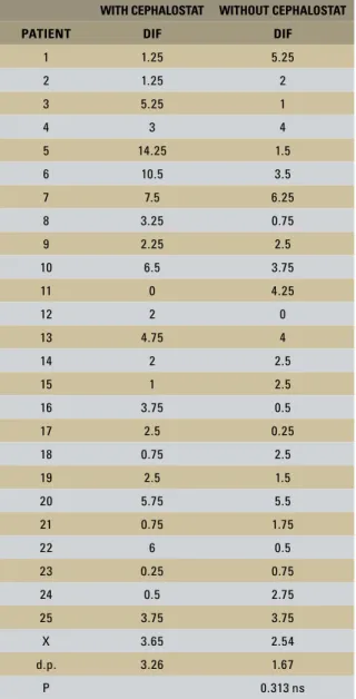
Table 1 displays the results of the measure-ments made with and without the cephalostat and with a 15-day interval between the photo sessions. Wilcoxon’s test revealed no statistically significant differences between the angle values in T1 and T2 when the photos taken with the cephalostat (p = 0.484) or without the cepha-lostat (p = 0.425). Table 2 displays the absolute angular differences between T1 and T2 for the photos taken with and without the cephalostat. The Mann-Whitney test revealed no statistical-ly significant differences between the absolute angles on the images obtained with and without the cephalostat (p = 0.313). The Kruskal-Wallis test revealed no statistically significant differ-ences between T1 and T2 values with and with-out the cephalostat (comp = 0.998).
DISCUSSION
The importance of the reproducibility of the NHP is justified by the fact that facial analysis is essential to orthodontic diagnoses.1
According to Moorrees,19 little attention has
been given in orthodontic publications regard-ing the appropriate facial orientation durregard-ing photographic recording. In some cases, pa-tients with malocclusion Class II are docu-mented before treatment with the head tilted down and after treatment with the head tilted up in order to highlight the correction of man-dible retrognathia.
The Frankfurt horizontal plane (parallel to soil) is the most commonly used reference for positioning patients during photographic or radiographic records, as recommended by
Burstone.5 However, according to Viazis28 and
subsequently confirmed by Arnett and Berg-man,1 people do not assume this posture in their
day-to-day life. Viazis29 states that a variation of
as much as 4° could be tolerated in the records based on the NHP. This margin was found in most cases in the present study. Lin and Arild15
WITH CEPHALOSTAT WITHOUT CEPHALOSTAT
PATIENT T1 (degrees) T2 (degrees) T1 (degrees) T2 (degrees)
1 24 22.75 21 26.25
2 20 18.75 19.25 21.25
3 21.5 16.25 18.5 17.5
4 11.5 8.5 9 5
5 21.25 7 17 18.5
6 11 21.5 17.5 21
7 21.75 14.25 21.75 15.5
8 26.5 23.25 22.75 22
9 21 23.25 23.25 20.75
10 15.75 22.25 15.25 19
11 19 19 19.25 23.5
12 17.5 19.5 21 21
13 20.5 25.25 22.25 26.25
14 18.25 20.25 18 20.5
15 22 23 25.5 23
16 20.75 17 17.5 18
17 30.5 33 32.5 32.25
18 18.25 19 14.25 16.75
19 30 32.5 30 28.5
20 18.5 12.75 18.25 12.75
21 15.75 16.5 19.75 18
22 15 21 14 13.5
23 28 27.75 22.75 23.5
24 13 12.5 20.5 17.75
25 28.25 32 31 27.25
X 20.38 20.35 20.47 20.37
d.p. 5.15 6.45 5.12 5.42
p 0.808 ns 0.898 ns
TABLE 1 - Comparisons (Wilcoxon test) between T1 and T2 angular mea-surements (Ls-Pog’ .LV) from photographs taken with and without the aid of a cephalostat.
found a large variation in the NHP of children between six and nine years of age assessed longi-tudinally. Few studies have evaluated children in orthodontic treatment.9 Angular variations tend
to decrease in adult patients.21
Arnett and Bergman2 prefer the NHP in
fa-cial analysis, stating that it does not exhibit the
same variation as intercranial points. This lesser variation has been confirmed by a number of au-thors.6,7,9,15,17 Thus, the NHP has been a preferred
reference position for assessing facial morphology, as it is the position individuals naturally assume. Arnett et al3 detailed the analysis of soft tissues
based on the philosophy of “Facial Keys”, which reinforces the need for the use of the NHP.
Although the NHP is recognized in the lit-erature as a reliable position, different methods for obtaining this position are cited.7,8,18,20,28,29
There are few studies on profile photographs, which hinders the discussion of what method is best for obtaining the NHP. The use of the mirror promotes better reproducibility regard-ing this position when compared with images obtained without the aid of a mirror.8,14 There
are divergences regarding the need for the use of a cephalostat, especially when recording the NHP in children. Cooke and Wei8 analyzed the
use of the ear rods of the cephalostat and found no significant influence regarding the obtain-ment of the NHP.
The present study found that the NHP is a method with good reproducibility, corroborating previous studies.7,8,21 It was also confirmed that
the NHP can be safely used in profile photogra-phy involving children. As there was no increase in accuracy when obtaining the NHP with the assistance of the cephalostat, this device seems unnecessary. Moreover, the children reported discomfort caused by the ear rods.
In 1971, Solow and Tallgren27 found a
stan-dard deviation of 2.48° in the natural position when using the self-balance method, while the standard deviation with the aid of the mirror was 1.43°. A mirror was used in the present study and the standard deviation was 1.67° without the aid of the cephalostat and 3.26° with the cephalostat.
Another situation that caused discomfort in the patients was the presence of the string rep-resenting the true vertical line (VL), which was TABLE 2 - Comparison (Mann-Whitney test) between T1 and T2 angular
variation from photographs taken with and without the aid of a cepha-lostat.
ns = not statistically significant.
WITH CEPHALOSTAT WITHOUT CEPHALOSTAT
PATIENT DIF DIF
1 1.25 5.25
2 1.25 2
3 5.25 1
4 3 4
5 14.25 1.5
6 10.5 3.5
7 7.5 6.25
8 3.25 0.75
9 2.25 2.5
10 6.5 3.75
11 0 4.25
12 2 0
13 4.75 4
14 2 2.5
15 1 2.5
16 3.75 0.5
17 2.5 0.25
18 0.75 2.5
19 2.5 1.5
20 5.75 5.5
21 0.75 1.75
22 6 0.5
23 0.25 0.75
24 0.5 2.75
25 3.75 3.75
X 3.65 2.54
d.p. 3.26 1.67
FIGURE 4 - Photographs taken from the same patient positioned in a cephalostat (A – first photograph; B – after 15 days). Obvious postural variation was observed.
A B
located in front of the patient. This suggests that it would be better to position the line simulating the VL alongside rather than in front of the patient in order to avoid this possible nuisance when photo-graphing children. Another alternative would be to attach the string to the light box.
While there were cases in which the NHP did not vary with the use of cephalostat (Fig 3), there were also cases in which the mean values were different between the two photo sessions (initial and after 15 days) (Fig 4). A similar situation occurred when the photographs were taken without the use of the cephalostat, with some cases exhibiting no significant differences between photo sessions (Fig 5) and others ex-hibiting considerable variation (Fig 6). In most cases, however, the measures exhibited little variability in the photographs with and without the cephalostat (Fig 7).
Foster et al,10 Ludström and Ludström17 and
Rino Neto et al25 all report that slight
differ-ences in the recording of the natural head posi-tion is a smaller problem than the variaposi-tion in intracranial reference lines. However, clinicians should assess whether the photography was real-ly carried out with the patient in the NHP prior to performing the facial analysis. According to Reche et al,23 the analysis of the facial profile
using standardized photographs is valid and reli-able for orthodontic documentation.
Based on the protocol of the present study, the researcher did not interfere in the position-ing of the patient, as the intention of the study was to observe the reproducibility of the NHP in children. However, it should be noted that, in ev-eryday practice, there is a need for trained tech-nicians to record the photographs in the NHP. The cases in Figures 4B and 6B clearly show that
FIGURE 3 - Photographs taken from the same patient positioned in a cephalostat (A – first photograph; B – after 15 days). No angle variation was observed.
FIGURE 5 - Photographs taken from the same patient without the aid of a cephalostat (A – first photograph; B – after 15 days). No postural variation was observed.
A B
FIGURE 6 - Photographs taken from the same patient without the aid of a cephalostat (A – first photograph; B – after 15 days). Obvious postural variation was observed.
A B
FIGURE 7 - Photographs taken from the same patient at the same day (T1). No angle variation was observed (A – with cephalostat; B – without cephalostat).
A B
the patients were in incorrect positions, requiring a new supervised photograph record. Thus, there should be technicians trained in obtaining photos in the NHP at centers for orthodontic exams, as suggested by Rino Neto et al.25 Thus, the proposal
made by Lundström et al16 regarding the natural
head position (NHP) is confirmed, as previously suggested by Moorrees and Kean.20 This position
is also reinforced by Halazonetis,13 who stresses
CONCLUSION
Although positional variations have suggested that patients in this age group should receive addi-tional orientation on NHP photographs, there were
no significant differences in the NHP within a 15-day interval with or without the aid of a cephalo-stat in the present study. Thus, the NHP proved to be a method with good reproducibility in children.
Submitted: January 2007
Revised and accepted: September 2009
1. Arnett GW, Bergman RT. Facial keys to orthodontic diagnosis and treatment planning. Part I. Am J Orthod Dentofacial Orthop. 1993 Apr;103(4):299-312.
2. Arnett GW, Bergman RT. Facial keys to orthodontic diagnosis and treatment planning. Part II. Am J Orthod Dentofacial Orthop. 1993 May;103(5):395-411.
3. Arnett GW, Jelic JS, Kim J, Cummings DR, Beress A, Worley CM Jr, Chung B, Bergman R. Soft tissue cephalometric analysis: diagnosis and treatment planning of dentofacial deformity. Am J Orthod Dentofacial Orthop. 1999 Sep;116(3):239-53.
4. Bister D, Edler RJ, Tom BD, Prevost AT. Natural head posture – considerations of reproducibility. Eur J Orthod. 2002 Oct;24(5):457-70.
5. Burstone CJ. The integumental profile. Am J Orthod Dentofacial Orthop. 1958 Jan;44(1):1-25.
6. Chen CM, Lai S, Tseng YC, Lee KT. Simple technique to achieve a natural head position for cephalography. Br J Oral Maxillofac Surg. 2008 Dec;46(8):677-8.
7. Cooke MS. Five-year reproducibility of natural head posture: a longitudinal study. Am J Orthod Dentofacial Orthop. 1990 Jun;97(6):489-94.
8. Cooke MS, Wei SH. A summary five factor cephalometric analysis based on natural head posture and the true horizontal. Am J Orthod Dentofacial Orthop. 1988 Mar;93(3):213-23. 9. Ferrario VF, Sforza C, Germanò D, Dalloca LL, Miani A Jr. Head posture and cephalometric analyses: an integrated photographic/radiographic technique. Am J Orthod Dentofacial Orthop. 1994 Sep;106(3):257-64.
10. Foster TD, Howat AP, Naish PJ. Variation in cephalometric reference lines. Br J Orthod. 1981 Oct;8(4):183-7.
REFERENCES
11. Freire-Maia BAV, Pereira MFSM, Paiva JB, Rino Neto J. Avaliação cefalométrica radiográfica da posição craniofacial de pacientes orientados em posição natural da cabeça pré e pós-expansão rápida da maxila. Rev Dental Press Ortod Ortop Facial. 2005 mar/abr;10(2):96-109.
12. Goldreich HN et al. Considerações sobre os erros em cefalometria. Rev Dent Press Ortod Ortop Maxilar. 1998 jan/ fev;3(1):81-90.
13. Halazonetis DJ. Estimated natural head position and facial morphology. Am J Orthod Dentofacial Orthop. 2002 Apr;121(4):364-8.
14. Jiang J, Xu T, Lin J. The relationship between estimated and registered natural head position. Angle Orthod. 2007 Nov;77(6):1019-24.
15. Lin XP, Arild S. Longitudinal study of the stability and reproducibility of natural head position in adolescents with different facial types over time. Shanghai Kou Qiang Yi Xue. 2005 Jun;14(3):238-42.
16. Lundström A, Forsberg CM, Westergren H, Lundström F. A comparison between estimated and registered natural head posture. Eur J Orthod. 1991 Feb;13(1):59-64.
17. Lundström A, Lundström F. The Frankfort horizontal as a basis for cephalometric analysis. Am J Orthod Dentofacial Orthop. 1995 May;107(5):537-40.
18. Marcotte MR. Head posture and dentofacial proportions. Angle Orthod. 1981 Jul;51(3):208-13.
19. Moorrees CF. A. Natural head position - a revival. Am J Orthod Dentofacial Orthop. 1994 May;105(5):512-3.
21. Nouri M, Mir M, Akbarzadeh A, Marami A. Three-year reproducibility of natural head position: a longitudinal study. J Dent. 2006;3(4):178-86.
22. Peng L, Cooke MS. Fifteen-year reproducibility of natural head posture: a longitudinal study. Am J Orthod Dentofacial Orthop. 1999 Jul;116(1):82-5.
23. Reche R, Colombo Vl, Verona J, Moresca CA, Moro A. Análise do perfil facial em fotografias padronizadas. Rev Dental Press Ortod Ortop Facial. 2002 jan/fev;7(1):37-45.
24. Rino Neto J, Paiva JB, Freire-Maia BAV, Miasiro Jr H, Attizzani MF, Crivello Jr. O. Avaliação da reprodutibilidade da posição natural da cabeça: estudo radiográfico. Ortodontia. 2002 out/ dez;35(4):55-68.
25. Rino Neto J, Freire-Maia BAV, Paiva JB. Método de registro da posição natural da cabeça para obtenção da radiografia cefalométrica lateral – considerações e importância do método no diagnóstico ortodôntico-cirúrgico. Rev Dental Press Ortod Ortop Facial. 2003 maio/jun;8(3):61-71.
26. Siersbaek-Nielsen S, Solow B. Intra- and interexaminer variability in head posture recorded by dental auxiliaries. Am J Orthod. 1982 Jul;82(1):50-7.
27. Solow B, Tallgren A. A natural head position in standing subjects. Acta Odontol Scand. 1971 Nov;29(5):591-607. 28. Viazis AD. A cephalometric analysis based on natural head
position. J Clin Orthod. 1991 Mar;25(3):172-81.
29. Viazis AD. Posição natural da cabeça. In: Viazis AD. Atlas de Ortodontia: princípios e aplicações clínicas. São Paulo: Ed. Santos; 1996. p. 41-3.
Contact address
Adriana Likes Pereira Rua Brasil 1667