CEREBELLAR LIPONEUROCYTOMA
A newly recognized clinico-pathological entity
Nádia Montagna
1, Daniel Moreira
1, Luiz Carlos Vaz
2, Marcelo Reis
2ABSTRACT - The term “cerebellar liponeurocytoma”, recently adopted by the World Health Organization Working Group (WHO), replaced many other different terms used up to now to give name to this rare tumor. To our knowledge, less than 20 cases have been related up to now under different names like as ”lipomatous medulloblastoma, lipidized medulloblastoma, neurolipocytoma, medullocytoma and lipomatous glioneurocytoma”. The new nomenclature eliminates the word “medulloblastoma”, reinforces its benign caracter, and includes it in the category of glioneuronal tumors. We describe an adictional case of this distinct clinico-pathological entity removed from the right cerebellar hemisphere of a 53-year-old woman. With the present case report, we hope to contribute to the knowledge on the diagnostic and prognostic implications derived from the finding of mature adipose-like tissue within a medulloblastomatous tumour.
KEY WORDS: liponeurocytoma, medulloblastoma, adipose tissue, mixed tumour.
Liponeurocitoma cerebelar: uma nova entidade clínico-patológica
RESUMO - O termo “liponeurocitoma cerebelar” recentemente adotado pela Organização Mundial de Saúde (Classificação de Tumores Cerebrais - versão 2000), surgiu em substituição a vários outros utilizados até então, para denominar esta rara neoplasia. De nosso conhecimento há na literatura menos de 20 casos relatados sob termos diferentes tais como “meduloblastoma lipomatoso, meduloblastoma lipidizado, neurolipocitoma, medulocitoma e glioneurocitoma lipomatoso”. A nova nomenclatura elimina a palavra “meduloblastoma”, enfatiza seu caráter benigno e o coloca na categoria dos tumores glioneuronais. Descrevemos mais um caso desta rara entidade clínico-patológica, ressecada do hemisfério cerebelar direito em uma mulher de 53 anos. Com este caso esperamos contribuir para o melhor conhecimento sobre o diagnóstico, prognóstico e possibilidades terapêuticas advindas da presença de tecido adiposo em tumor meduloblastomatoso de adultos.
PALAVRAS-CHAVE: liponeurocitoma, meduloblastoma, tecido adiposo, tumor misto.
1Department of Pathology, Rio de Janeiro State University, Rio de Janeiro RJ, Brazil; 2Hospital Geral da Posse, Health Ministery, Brazil.
Received 27 February 2002. Accepted 10 May 2002.
Dra. Nadia Montagna - Department of Pathology, Medical School, Rio de Janeiro State University - Avenida Professor Manoel de Abreu 48/3o andar - 20550-170 Rio de Janeiro RJ - Brazil. E-mail: mmontag@matrix.com.br
Medulloblastoma is the most common primitive ne-uroectodermal tumor of the central nervous system. It occurs usually in childhood in the first decade of life1, being also found in adults2-4. According to World Health Organization (WHO) it is considered a grade IV neoplasm5. Its heterogeneity as well as its potential to differentiate has been largely related in the literature, happening in 40% of cases6. Cartilaginous, rhabdomyoblastic and neu-ro-glial variants have been reported7-9. The uncommon finding of adipose tissue in medulloblastomatous neo-plasms has been related rarely and correlates with bet-ter prognosis10-15. This kind of tumor was recently inclu-ded in the WHO working group for classification of central nervous system neoplasms under the name “Cerebellar Liponeurocytoma”5. The term adopted
omits the word ”medulloblastoma” clearly pointing to a better prognosis.
We describe here such a particular posterior fossa neoplasm in a 53-year-old woman, especially com-posed of mature adipose tissue closely admixed with undifferentiated small blue cell areas consistent with medulloblastoma.
CASE
surgical exploration with subtotal resection of the lesion. Because of a residual tumor the patient received postope-rative cranial radiotherapy (5600 rads).
The surgically removed tumor fragments were received at the laboratory fixed in 10% formallin. They were proces-sed for paraffin sectioning and stained with hematoxylin-eosin (H&E), periodic acid-Schiff (PAS), Gomori, Fontana-Masson and reticulin.
No fresh tumor tissue was available to make oil red O stain to lipid droplets or ultrastructural examination. Im-munohistochemical staining using DAKO monoclonal anti-bodies following avidin-biotin techniques included: glial fibrillary acidic protein (GFAP-1:200 dilution), vimentin (Vim-1:200 dilution), neuron-specific enolase (NSE-1:400 dilution), synaptophysin (Syn-1:300 dilution) and Ki 67 - 1:25 dilution).
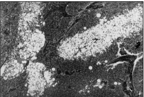
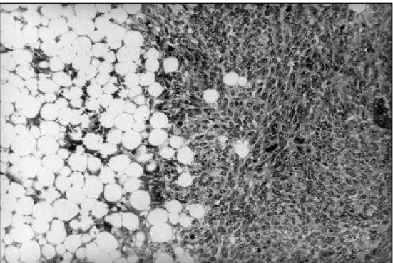
Pathologic findings - Microscopically we observed a neo-plasm consisting of two distinct cellular elements: a predo-minant poorly differentiated component of small cells, in-termixed with another element indistinguishable from matu-re fat cells at optic microscopy examination (Fig 1). No dis-tinct separation was observed between these two elements (Fig 2). The small cell component disclosed scanty cyto-plasm and round or oval nuclei arranged in a fibrillary background (Fig 3). The lipomatous component was com-posed of grouped vacuolated cells indistinguishable from mature fat cells (Fig 4). Other tumor areas contained smal-ler vacuoles recalling an apparent transition between the two populations. In the lipomatous component there was no cellular immaturity or nuclear atypia. Only few mitosis were found in poorly differentiated areas. Recent hemor-rhage as well as necrotic areas were also observed in both
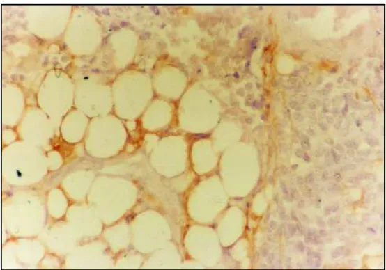
components. The non-tumoral cerebellar tissue circumja-cent to tumor was devoid of fat cells, disclosing only hypo-xic cellular alterations. The lipomatous cells showed a peri-pheral ring shape immunopositivity to GFAP (Fig 5), VIM and NSE. Medulloblastomatous areas were immunopositive to NSE and SYN. MIB 1 antibody immunolabelling index was positive in less than 5% of the cells.
DISCUSSION
Medulloblastoma, a malignant cerebellar tumor of childhood, is heterogeneous in regard to tissue pat-tern. Its potential of differentiation has been largely reported in the literature, including neuronal, glial and mesenchymal elements6-8. Authors have attempted to correlate some histological characteristics with prog-nosis and this has lead, sometimes, to different conclu-sions4. We report a case that represents a rare example of cerebellar tumor in adult, exhibiting an extraordinary morphology characterized by an uncommon mixture of mature adipose tissue with medulloblastomatous areas. The presence of adipose cells in neuroglial neo-plasms has been exceptionally related in the literature not being restricted to cerebellum neither to medul-loblastomas16. It has also been described in spinal cord neoplasms17 and in supratentorial ependymomas18. The origin of these adipocytic elements is still disputed and it hasn’t yet been completely elucidated. Some authors consider that the presence of such cells sug-gests that the neoplasm has evolved in dysgenetic ar-eas where distinct cellular elements co-exist intimately9.
Some others have found ultrastructural evidences that there is a progressive accumulation of lipid vacuoles in the cell’s cytoplasm, probably due to some dege-nerative or metabolic change. So, those cells would become similar to adipocytes at optic microscopy. Ana-lyzed under immunohistochemistry their neuroglial ori-gin manifest themselves18. This explains the immuno-reactivity in the remaining cytoplasm ring related by
some authors14,15,18. What mechanism is in action in these tumors is still under consideration.
Apart from histopathological considerations, it sho-uld be pointed out that this variant, characterized by adult onset, shows a much more benign evolution. All these observations strongly suggest it is a clinico-pathological entity distinct from its conventional cor-respondent.
Fig 2. Higher power of the neoplasm demonstrating both cellular components merging one into another (HE; x 200).
Fig 5. Immunostaining to GFAP showing positivity in a cytoplasmic ring of vacuolated cells (HE; x 400). Fig 4 . High power of the lipomatous component shows large and small vacuolated fat cells (HE; x 400).
For the last years, many different terms have been proposed to designate these tumors, such as lipo-matous medulloblastoma15, lipidized medulloblas-toma14, neurolipocytoma10 and medullocytoma13. Most of these terms clearly point to a more benign lesion than classical medulloblastoma. In accordance to a recent publication of the WHO working group on “Classification of Tumors of the Nervous System”,
they were included in the category of mixed glio neu-ronal tumors. The term “Cerebellar Liponeurocyto-ma” has been proposed and should be used from now on5. Bearing in mind that this kind of lesion is not restricted to cerebellum16, maybe the word “ce-rebellar” should have been omitted.
consensus about the use of complementary radio-therapy19. In the majority of cases that had evidence of residual tumor, patients were submitted to radio-therapy19,20. Despite the presumed good prognosis, in one single case tumor behaved in a more aggres-sive way21. In our case, despite the presence of large necrotic areas, factor known to be associated with a poor prognosis, the patient is doing well almost four years after surgery and postoperative radiotherapy. We really believe the ideal post operative conduct is still to be determined.
The present report represents another step to de-termine whether the presence of lipomatous cells in medulloblastomatous tumors does, in fact, have a prognostic value, distinguishing them from conven-tional medulloblastomas.
REFERENCES
1. Becker LE, Hinton D. Primitive neuroectodermal tumors of the central nervous system. Hum Pathol 1983;14:538-550.
2. Kopelson G, Linggood RM, Kleinman GM. Medulloblastoma in adults: improved survival with supervoltage radiation therapy. Cancer 1982; 9:1334-1337.
3. Russel DS, Rubinstein LJ (eds). Pathology of tumors of the nervous system. 6.Ed. London: Edward Arnold, 1998:460-470.
4. Caput AJ, Mccullough DC, Manz HJ, Patterson K, Hammock MK. A review of the factors influencing the prognosis of medulloblastoma. J Neurosurg 1987;66:80-87.
5. Kleihues P, Cavenee WK. Pathology & Genetics: tumours of the nervous system. Lyon: IARC Press, 2000:110-111.
6. Burger PC, Grahmann FC, Bliestle A, Kleihues P. Differentiation in the medulloblastoma: a histological and immunohistochemical study. Acta Neuropathol (Berl) 1987;73:115-123.
7. Anwer UE, Smith TW, De Girolami U, Wilkinson HA. Medulloblasto-ma with cartilaginous differentiation. Arch Pathol Lab Med 1989;113:94-98. 8. Dickson DW, Hart MN, Menezes A, Cancilla, PA. Medulloblastoma with glial and rhabdomyoblastic differentiation: myoglobin and glial fibrillary acidic protein immunohistochemical and ultrastructural study. J Neuropathol Exp Neurol 1983;42:639-647.
9. Bechtel JT, Patton JM, Takei Y. Mixed mesenchymal and neuroectodermal tumor of the cerebellum. Acta Neuropathol (Berl) 1978;41:261-263. 10. Ellisson DW, Zygmunt SC, Weller RO. Neurocytoma/lipoma
(neuroli-pocytoma) of the cerebellum. Neuropathol Appl Neurobiol 1993;19:95-98. 11. Budka H, Chimelli, L. Lipomatous medulloblastoma in adults: a new tumor
type with possible favorable prognosis. Hum Pathol 1994;25:730-731. 12. Chimelli L, Hahn MD, Budka, H. Lipomatous differentiation in a
medulloblastoma. Acta Neuropathol (Berl) 1991;81:471-473. 13. Giancaspero F, Cenachi G, Roncaroli F, et al. Medullocytoma (lipidized
medulloblastoma): a cerebelar neoplasm of adults with favorable prog-nosis . J Neuropathol Exp Neurol 1995;54:423.
14. Davis DG, Wilson D, Schmitz M, Markesbery WR. Lipidized medullo-blastoma in adults. Hum Pathol 1993;24:990-995.
15. Soylemezoglu F, Soffer D, Onol B, Schwecheimer K, Kleihues P. Li-pomatous medulloblastoma in adults: a distinct clinicopathological entity. Am J Surg Pathol 1996;20:413-418.
16. Selassie L, Rigotti R, Kepes JJ, Towfighi J. Adipose tissue and smooth muscle in a primitive neuroectodermal tumor of cerebrum. Acta Neuropathol (Berl) 1994;87:217-222.
17. Roda JM, Gutierrez-Molina M. Multiple intraspinal low-grade astro-cytomas mixed with lipoma (astrolipoma): case report. J Neurosurg 1995; 82:891-894.
18. Ruchoux MM, Kepes JJ, Dhellemmes P, Hamon M, et al. Lipomatous differentiation in ependymomas: a report of three cases and compari-son with similar changes reported in other central nervous system neo-plasms of neuroectodermal origin. Am J Surg Pathol 1998;22:338-346. 19. Jackson TR, Regine WF, Wilson D, Davis DG. Cerebellar liponeurocytoma:
case report and review of the literature. J Neurosurg 2001;95:700-703. 20. Taddei Gl, Buccoliero AM, Caldarella A, et al. Cerebellar