C
a s eR
e p o Rt1 8 9 Arq Bras Oftalmol. 2016;79(3):189-91 http://dx.doi.org/10.5935/0004-2749.20160054
INTRODUCTION
Toxoplasmosis is the leading cause of infectious posterior uveitis
worldwide, accounting for 80% of the cases in some regions(1). Although
toxoplasmic retinochoroiditis usually has a self-limiting course, it can lead to an irreversible visual loss, particularly when the macula and optic nerve head are involved. Vascular involvement typically consists of diffuse or segmental sheathing produced by antigen-antibody com-plex deposition in the vessel wall, as well as by localized mononuclear cell infiltrates(1). Retinal vein occlusion may occur, but artery obstruc-tion is rare(2-4). We herein report an atypical presentation of toxoplasmic retinochoroiditis characterized by the occurrence of combined branch retinal vein and artery occlusion.
CASE REPORT
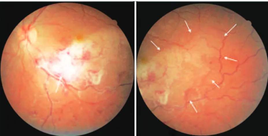
A 22-year-old healthy man complained of low visual acuity and pain in his left eye [oculus sinister (OS)] for five days. His past ocular and family history were unremarkable. Best-corrected visual acuity (BCVA) was 20/20 in the right eye [oculus dexter (OD)] and 20/400 in the left eye (OS). His anterior segment examination was normal OD and revealed 2+ anterior chamber reaction OS. Intraocular pressure by applanation tonometry was 10 mmHg in both eyes at 08:00. Fun-duscopy showed a normal OD and a white, poorly demarcated, and slightly elevated retinal lesion at the proximal portion of the infero-temporal vascular arcade OS. There were retinal hemorrhages in the inferotemporal region extending to the periphery with venous dila-tion and increased tortuosity, as well as ischemic retinal whitening along the inferotemporal vascular arcade, 2+ vitreous cells, and diffu-se periphlebitis. The striking macular appearance was reminiscent of viral retinitis, given its necrotic and hemorrhagic features (Figure 1).
Blood tests revealed normal leukocyte and erythrocyte counts and erythrocyte sedimentation rates. Serological tests for human immunodeficiency virus (HIV) and syphilis (venereal disease research laboratory and fluorescent treponemal antibody absorption tests) were negative. Chest X-ray films were normal. Toxoplasma globulin (Ig) G antibodies were positive in an enzyme-linked immuno-sorbent assay (132 IU/mL), whereas anti-toxoplasma IgM antibodies were negative.
Two days after initial presentation, the patient was started on pyrimethamine at 50 mg daily, sulfadiazine at 2 g daily, folinic acid at 15 mg three times a week, prednisone at 60 mg daily, in a tapering schedule, as well as topical prednisolone acetate 1% and tropicamide 1%. Five days later, BCVA was maintained, and the ophthalmologic examination did not reveal any changes. Three weeks later, BCVA was 20/150 OS. His anterior segment examination was unchanged OD and showed 1+ anterior chamber reaction OS. Intraocular pressure was 16 mmHg OD and 14 mmHg OS. Funduscopy was unchanged OD and showed a well-demarcated, whitish inferotemporal retino-choroidal lesion. There was sheathing of the inferotemporal retinal artery, mild juxtafoveal inferior retinal whitening, a macular star, and 1+ vitreous cells (Figure 2, left). Six weeks after the beginning of treatment, BCVA was 20/30 OS. There was no anterior chamber or vi-treous cells. The inferotemporal retinochoroidal lesion showed signs of scarring (Figure 2, middle). Oral medication was discontinued, and the prednisolone acetate 1% drops were tapered.
Three months after the initial presentation, BCVA was 20/30 OS. His anterior segment examination revealed no changes. The intrao-cular pressure was 16 mmHg OD and 15 mmHg OS. Funduscopy showed an inferotemporal retinochoroidal scar and partial resolution
Combined branch retinal vein and artery occlusion in toxoplasmosis
Oclusão combinada de ramo arterial e venoso retinianos em toxoplasmose
Fabio bom aggio1, Fernando Joséde novelli1, evandro luis rosa1, mário Junqueira nobrega1
Submitted for publication: April 1, 2015 Accepted for publication: June 18, 2015
1 Hospital de Olhos Sadalla Amin Ghanem, Joinville, SC, Brazil.
Funding: No specific financial support was available for this study.
Disclosure of potential conflicts of interest: None of the authors have any potential conflict of interest to disclose.
Corresponding author: Fabio Bom Aggio. Rua Camboriú, 35 - Joinville - SC - 89216-222 - Brazil - E-mail: [email protected]
ABSTRACT
A 22-year-old man complained of low visual acuity and pain in his left eye for five days. His ophthalmological examination revealed 2+ anterior chamber reaction and a white, poorly defined retinal lesion at the proximal portion of the infero-temporal vascular arcade. There were retinal hemorrhages in the inferoinfero-temporal region extending to the retinal periphery. In addition, venous dilation, increased tortuosity, and ischemic retinal whitening along the inferotemporal vascular arcade were also observed. A proper systemic work-up was performed, and the patient was diagnosed with ocular toxoplasmosis. He was treated with an anti-toxoplasma medication, and his condition slowly improved. Inferior macular inner and middle retinal atrophy could be observed on optical coherence tomography as a sequela of ischemic injury. To our knowledge, this is the first report of combined retinal branch vein and artery occlusion in toxoplasmosis resulting in a striking and unusual macular appearance.
Keywords: Ocular toxoplasmosis; Uveitis; Retinal artery occlusion; Retinal vessels; Visual acuity; Optical coherence tomography; Vision disorders
RESUMO
Um paciente do sexo masculino, com 22 anos de idade, queixou-se de redução da acuidade visual no olho esquerdo por 5 dias. O exame oftalmológico mostrou reação de câmara anterior 2+ e uma lesão retiniana esbranquiçada, pouco definida, na porção proximal da arcada vascular temporal inferior. Foram observadas hemorragias retinia-nas na região temporal inferior estendendo-se à periferia, assim como ingurgitamento venoso, aumento da tortuosidade e palidez isquêmica da retina no mesmo quadrante. Exames laboratoriais corroboraram o diagnóstico de toxoplasmose ocular. O paciente melhorou lentamente após tratamento apropriado. Foi evidenciada atrofia da retina macular inferior interna e média à tomografia de coerência óptica, como sequela da isquemia retiniana. Para nosso conhecimento, este é o primeiro relato de oclusão retiniana combinada de ramo arterial e venoso em toxoplasmose ocular, levando a um aspecto fundoscópico atípico e peculiar.
Co m b i n e db r a n C hr e t i n a lv e i n a n da rt e ryo C C l u s i o ni nt o x o p l a s m o s i s
1 9 0 Arq Bras Oftalmol. 2016;79(3):189-91
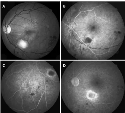
of the macular star (Figure 2, right). Fluorescein angiography revealed focal hypofluorescence with surrounding early hyperfluorescence corresponding to the inferotemporal scar and blocked hypofluo-rescence corresponding to the retinal hemorrhages. No areas of non-perfusion were detected (Figure 3). Optical coherence tomogra-phy showed signs of partial posterior hyaloid detachment, inferior macular inner and middle retinal atrophy, hyperreflective intraretinal dots superior to the fovea corresponding to the hard exudates, as well as a mild foveal discontinuity of the outer retina and pigment epithelium layer (Figure 4).
DISCUSSION
The diagnosis of ocular toxoplasmosis is usually straightforward; however, challenging cases may occur. In the case presented here, vascular occlusion of the inferotemporal retinal artery and vein led to unusual fundus findings characterized by ischemic retinal white-ning and numerous scattered retinal hemorrhages obscuring the boundaries of the focal inflammatory lesion. The appearance of the fundus resembled viral retinitis. Considering the high prevalence of
toxoplasmosis in Brazil(5), the health status of the patient, and
absen-ce of peripheral retinitis, toxoplasmosis was strongly suspected. An anti-toxoplasma medication was introduced, and the patient was closely followed.
Ocular toxoplasmosis begins in the superficial retina. As retinitis progresses, the involvement of full-thickness retina, adjacent choroid, vitreous, and even sclera may occur. The initial lesion with ill-defined margins due to retinal edema tends to become more defined with treatment. After a variable time period, pigmentation mainly occurs
in the lesion margins(1). These findings were observed in our case.
Vascular changes in toxoplasmosis typically involve veins, but arteries may also be affected, particularly in the form of Kyrieleis
ar-teritis(1), characterized by nodular periarterial plaques that were
obser ved in our case (Figure 2). Although classically described in toxoplasmosis, Kyrieleis arteritis may occur in syphilis, rickettsial reti-nal vasculitis, acute retireti-nal necrosis, and other diseases. Its presence should raise suspicion of an infectious cause of retinal vasculitis(1).
Branch artery occlusion may result from direct artery compression by retinochoroiditis or by arteriolar contraction associated with increa-sed viscosity of blood and inhibition of coagulation due to heparin release from mast cells as a response to acute inflammatory stimuli. It may also be a consequence of perivasculitis, which may cause
thi-ckening of the vessel wall, disruption of blood flow, and thrombosis(6).
Yu et al.(7) identified the following three patterns of retinal capillary
ischemia using optical coherence tomography in arterial occlusive disease in the acute phase: (1) thickening and hyperreflectivity of the inner retinal layers owing to ischemia of the superficial capillary plexus, (2) hyperreflective band at the level of the inner nuclear layer
represen-Figure 1. Initial presentation: Fundus photography of the left eye showing a white, poorly demarcated, and slightly elevated retinal lesion at the proximal portion of the inferotemporal vascular arcade (arrow, left). There were retinal hemorrhages in the inferotemporal region extending to the periphery with venous dilation and increased tortuosity, as well as ischemic retinal whitening along the inferotemporal vascular arcade (arrows, right), 2+ vitreous cells, and difuse periphlebitis.
Ag g i o FB, e tA l.
1 9 1
Arq Bras Oftalmol. 2016;79(3):189-91
Figure 3. Fluorescein angiography (FA) frames of the left eye three months after the beginning of treatment. (A) Red-free fundus photography. (B) Early FA frame disclosing hypoluorescence corresponding to the inferotemporal lesion surrounded by early hyperluorescence. (C) Interme-diate FA frame of the inferior region. No areas of non-perfusion are observed. (D) Late FA frame showing no leakage at the macular region. Blocked hypoluorescence is seen corresponding to the scattered retinal hemorrhages.
A
C
B
D
Figure 4. Vertical cross-sectional optical coherence tomography image of the left eye showing signs of partial posterior hyaloid detachment, inferior macular inner and middle retinal atrophy, hyperrelective intraretinal dots superior to the fovea, as well as mild foveal discontinuity of the outer retina and pigment epithelium layer.
ting ischemia of the intermediate and deep capillary plexuses, and (3) diffuse thickening and hyperreflectivity of the inner and middle retinal layers representing both superficial and deep capillary ischemia. In the chronic phase, thinning and atrophy of the affected retinal layers were seen. In our case, optical coherence tomography in the chronic phase revealed inner and middle inferior retinal macular atrophy, consistent with both superficial and deep capillary ischemia.
Atypical forms of ocular toxoplasmosis include punctate outer re -tinal toxoplasmosis, neuroretinitis, neuritis, pseudomultifocal retino-choroiditis, multifocal retinoretino-choroiditis, peripheral lesions, and anterior uveitis(1). Atypical presentations have been reported in patients with HIV
infection, iatrogenic immunosuppression, or advanced age(8). Moshfeghi
et al. reported findings on 25 eyes with widespread retino choroiditis
mimicking acute retinal necrosis(8). In their cases, lesions were atypical, large, multifocal, bilateral, or associated with diffuse retinal involvement or panophthalmitis. The diagnosis of toxoplasmosis was confirmed by the response to anti-toxoplasmosis medications, intraocular diagnostic studies such as polymerase chain reaction, intraocular antibody analysis,
ocular fluid culture, or histopathological examination identifying
Toxo-plasma gondii in necrotic retinal tissue.
In conclusion, we report an uncommon presentation of toxoplas-mosis, characterized by combined branch artery and vein occlusion, resulting in ischemic retinal whitening and numerous scattered reti-nal hemorrhages in the inferotemporal region along with a poorly de-fined proximal whitish retinal lesion. The possibility of toxoplasmosis in cases presenting with these features must be considered because prompt treatment may prevent a poor visual outcome.
REFERENCES
1. Oréfice F, Vasconcelos-Santos DV, Cordeiro CA, Oréfice JL, Costa RA. Toxoplasmosis. In: Foster CS, Vitale AT. Diagnosis and treatment of uveitis. New Delhi, India: Jaypee - Highlights; 2013. p.543-68.
2. Williamson TH, Meyer PA. Branch retinal artery occlusion in toxoplasma retinochoroi-ditis. Br J Ophthalmol. 1991;75(4):253.
3. Chiang E, Goldstein DA, Shapiro MJ, Mets MB. Branch retinal artery occlusion caused by toxoplasmosis in an adolescent. Case Rep Ophthalmol. 2012;3(3):333-8. 4. Arai H, Sakai T, Okano K, Aoyagi R, Imai A, Takase H, et al. Presumed toxoplasmic
central retinal artery occlusion and multifocal retinitis with perivascular sheathing. Clin Ophthalmol. 2014;8:789-92.
5. Dubey JP, Lago EG, Gennari SM, Jones JL. Toxoplasmosis in humans and animals in Brazil: high prevalence, high burden of the disease, and epidemiology. Parasitology. 2012;139(11):1375-424.
6. Kahloun R, Mbarek S, Khairallah-Ksiaa I, Jelliti B, Yahia SB, Khairallah M. Branch retinal artery occlusion associated with posterior uveitis. J Ophthalmic Inflamm Infect. 2013;3(1):16. 7. Yu S, Pang CE, Gong Y, Freund KB, Yannuzzi LA, Rahimy E, et al. The spectrum of
super-ficial and deep capillary ischemia in retinal artery occlusion. Am J Ophthalmol. 2015; 159(1):53-63.