273
J Bras Patol Med Lab, v. 53, n. 4, p. 273-275, August 2017 CaSE rEPort
Lymphoepithelial cyst on the tongue:
case report at unusual location
Cisto linfoepitelial em língua: relato de caso em localização incomum
Rafaella B. Leite; Mara Luana B. Severo; Patrícia T. Oliveira; Ana Miryan C. Medeiros; Carlos Augusto G. Barboza; Ericka Janine D. Silveira
Universidade Federal do Rio Grande do Norte (UFRN), Rio Grande do Norte, Brazil.
First submission on 26/04/17; last submission on 27/06/17; accepted for publication on 10/07/17; published on 20/08/17
aBStraCt
Oral lymphoepithelial cyst (OLEC) is an uncommon lesion that develops in oral lymphoid tissue. The aim of the present study was to report a clinical case of OLEC in the tongue. A 22-year-old patient presented a nodular lesion, yellowish, with a softened consistency, measuring 0.5 cm in the ventral surface of the tongue. Under the clinical hypotheses of mucocele and OLEC, excisional biopsy was performed. The histopathological examination revealed a cystic lesion covered by a parakeratinized stratiied squamous epithelium, which presented in its ibrous capsule a prominent lymphoid tissue. Based on the deinitive diagnosis, surgical excision of the lesion was performed.
Key words: oral pathology; non-odontogenic cysts; treatment result.
introDuCtion
Oral lymphoepithelial cyst (OLEC) is a rare and benign lesion of the oral cavity, corresponding to 0.09% to 0.18% of lesions affecting this region(1). It is a lesion that develops within the oral
lymphoid tissue, microscopically similar to the branchial cleft cyst (cervical lymphoepithelial cyst), but much smaller in size(1, 2). It
usually occurs in adults with mean age around the fourth decade of life; it is a lesion rarely found in children. The loor of the mouth is reported as the site of higher incidence (70.7%), followed by lateral border (10.7%) and ventral surface of the tongue (7.3%)(3).
Typically, OLEC appears as a small submucosal mass, generally smaller than 1 cm in diameter; in rare cases, the lesion will be greater than 1.5 cm. The cyst may be irm or soft to palpation, and the covering mucosa is soft and non-ulcerated. The lesion is white or yellow, asymptomatic and often contains keratin in its light with cheesy or creamy appearance. The cyst is asymptomatic, although occasionally some patients complain of swelling or drainage. Pain is rare, but it can occur after trauma(1, 3).
Histopathologically, OLEC is characterized by the presence of a cyst covered by a parakeratinized stratiied squamous epithelium
and a capsule of dense ibrous connective tissue, permeated by a moderate inlammatory mononuclear iniltrate through moderate vascularization(3-5).
The treatment of OLEC consists of conservative surgical removal, and relapses are rare, leading to a good prognosis(6). Due
to its low clinical morbidity and nonspeciic symptoms, diagnosis is still a challenge(1).
The present work aims to report a clinical case of OLEC in the ventral surface of the tongue, as well as to carry out the discussion on important clinical and pathological aspects and treatment of this condition.
CaSE rEPort
A male patient, 22-year-old, leucoderma, presented at the Dental clinics of the Department of Dentistry of the Universidade Federal do Rio Grande do Norte (UFRN), Natal (RN), Brazil, complaining of painless swelling in the tongue with evolution of three months. Past medical history was noncontributory, and the patient did not have human immunodeiciency virus (HIV).
274
Lymphoepithelial cyst on the tongue: case report at unusual location
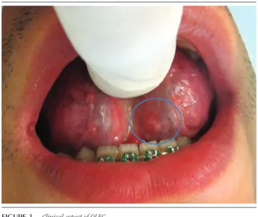
Extraoral examination of gland and neck lymph nodes showed signs of normality; the intraoral physical examination showed presence of a nodular lesion, with exophytic growth, sessile implantation, yellowish coloration and softened consistency, measuring approximately 0.5 cm in the ventral surface of the
tongue (Figure 1). From the clinical hypotheses of mucocele or
OLEC, an excisional biopsy was performed under local anesthesia. The minor salivary glands associated with the lesion were removed. Histopathological examination revealed the presence of a cystic lesion covered by parakeratinized stratiied squamous epithelium surrounded by a capsule of ibrovascular connective tissue. The presence of lymphoid tissue in the cystic capsule was evidenced, besides germinative centers, and the diagnosis of OLEC was issued
(Figure 2). After nine months of follow-up, no evidence of relapse
was found.
DiSCuSSion
OLEC is a rare lesion in the mouth. There is variation in the literature regarding its most common location. Yang et al. (2012)(3)
observed that the loor of the mouth is the anatomical site of higher incidence (70.7%) of OLEC, followed by the lateral border (10.7%) and the ventral surface (7.3%) of the tongue. However, Uchoa-Vasconcelos et al. (2014)(1) found that the tongue, followed by the
loor of the mouth, are the most frequent location. The occurrence of OLEC in the ventral surface of the tongue is rare, as there are only 91 cases described in the literature(7, 8). The present case reported is
the 92nd case of OLEC in the ventral surface of the tongue.
OLEC mainly occurs in adults around the fourth decade of life with female predilection 60%-80%(1, 3), which differed from the
present case, since it occurred in a male patient.
Yang et al. (2012)(3) performed a clinical analysis of 120 cases
of OLECs and found a predilection for the female gender, with a ratio between men and women of 1:2 and ages ranging from 2 to 75 years, with mean of 44.1 years. Meanwhile, Ahamed et al. (2014)(9)
identiied that such lesion was more frequent between the second and third decades of life, corroborating the present case reported.
The etiopathogenesis of OLEC is uncertain. Some authors have hypothesized that the ectopic foci of the embryonic epithelium are trapped in the lymphoid tissue and may proliferate to form a cyst(10). However, other authors have suggested that OLECs are the
result of obstruction of normal oral tonsil crypts(2, 5, 10). It is also
possible that the traumatic implantation of epithelial cells into deeper tissue may lead to the formation of OLEC(2).
Other studies were carried out in an attempt to clarify a possible association of OLEC with HIV infection as part of diffuse iniltrative lymphocytosis syndrome, with an occurrence of 3%-10% of HIV-positive patients(9). This hypothesis is due to the increased
incidence of OLEC in HIV-positive patients, thus suggesting that it is one of the clinical manifestations of this infection(11). However,
the relationship between HIV infection and OLECs has not been fully elucidated yet.
Clinically, OLEC has features similar to other nodular lesions affecting the oral cavity. The evolution, growth rate and symptoms are not speciic, therefore it is always dificult distinguishing this lesion and other benign lesions, such as mucocele, mucous retention cyst, lipoma, ibroma, sialolithiasis and dermoid cyst(2, 3, 10).
Due to its low clinical morbidity and nonspeciic symptoms, the diagnosis of OLEC remains a challenge(3). Microscopic indings are
critical to conclude the diagnosis. Microscopic indings demonstrate a cystic cavity covered by stratiied squamous epithelium, adjacent to
figurE 1 − Clinical aspect of OLEC
Nodular lesion, in the ventral surface of the tongue, yellowish, slow and submucosal growing, measuring approximately 0.5 cm.
OLEC: oral lymphoepithelial cyst.
figurE 2 − Histopathological aspect of OLEC
A and B) presence of a pathological cavity covered by parakeratinized stratified squamous epithelium surrounded by a connective fibrovascular tissue capsule (100×); C) presence of lymphoid tissue in the cystic capsule, and germinal centers (400×).
OLEC: oral lymphoepithelial cyst.
275
Rafaella B. Leite; Mara Luana B. Severo; Patrícia T. Oliveira; Ana Miryan C. Medeiros; Carlos Augusto G. Barboza; Ericka Janine D. Silveira
it exhibit lymphocytes masses with lymphoid follicles, and underlying a ibrous connective tissue capsule(2). These characteristics are
compatible with the lesion presented in this report.
OLEC treatment includes a conservative approach to the lesion. Decompression may be performed by aspirating the intralesional luid, thereby reducing cystic osmotic pressure. Subsequently, deinitive treatment is performed by complete enucleation of the lesion associated with excision of the involved gland(3). This
treatment was similar to that adopted in the case reported here, in which was performed the complete enucleation of the lesion associated with the excision of the accessory glands involved.
rEfErEnCES
1. Uchoa-Vasconcelos AC, Oliveira DJF, Roman-Martelli SJ, Etges A, Neutzling-Gomes AP, Chaves-Tarquínio SB. Demographic proile of oral nonodontogenic cysts in a Brazilian population. Med Oral Patol Oral Cir Bucal. 2014; 19(4): 308-12.
2. Khelemsky R, Mandel L. Lymphoepithelial cyst of mouth loor. J Oral Maxillofac Surg. 2010; 68: 3055-7.
3. Yang X, Ow A, Zhang CP, et al. Clinical analysis of 120 cases of intraoral lymphoepithelial cyst. Oral Surg Oral Med Oral Pathol Oral Radiol. 2012; 113: 448-52.
4. Flaitz CM, Davis SE. Oral and maxillofacial case of the month: oral lymphoepithelial cyst. Tex Dent J. 2004; 121(7): 624-31.
5. Castro LGJ, Ferreira GM, Mendonça EF, Castro LA. A rare occurrence of lymphoepithelial cyst in the palatine tonsil: a case report and discussion of the etiopathogenesis. Int J Clin Exp Pathol. 2015; 8(4): 4264-8. 6. Hung T, Jacob A, Shahab R. Idiopathic lymphoepithelial cyst of the pharynx masquerading as peritonsillar abscess. J Laryngol Otol. 2001; 115(8): 666-7.
After nine months of follow-up, no signs of lesion recurrence were found, supporting the literature indings, which showed that most patients are completely recovered by excision with no lesion recurrence rates(1).
Based on the literature and in the reported case, OLEC is typically present as small asymptomatic nodules located in the oral cavity, which emphasizes the importance of a detailed clinical examination for small lesions that are often neglected. Conservative surgical excision is the treatment for OLEC, and in cases of larger lesions, marsupialization produces excellent
results(12).
rESuMo
O cisto linfoepitelial oral (CLEO) é uma lesão incomum que se desenvolve no tecido linfoide oral. O objetivo do presente trabalho foi relatar um caso clínico de CLEO na língua. Paciente de 22 anos de idade exibia uma lesão nodular, de coloração amarelada e consistência amolecida, medindo 0,5 cm na região ventral de língua. Sob as hipóteses clínicas de mucocele e CLEO, foi realizada biópsia excisional. O exame histopatológico revelou lesão cística revestida por epitélio escamoso estratificado paraceratinizado, que apresentava em sua cápsula fibrosa tecido linfoide proeminente. Com base no diagnóstico definitivo, foi realizada a excisão cirúrgica da lesão.
Unitermos: patologia bucal; cistos não odontogênicos; resultado de tratamento.
7. Juliasse REL, Resende MCA, Maia PA, Nonaka WFC, Galvão CH, Pinto PL. Cisto linfoepitelial oral: relato de quatro casos e revisão de 119 casos apresentados na literatura. J Bras Patol Med Lab. 2010; 46(2): 129-34.
8. Costa GWF, Pereira AMK, Viana AST, Cavalcante BR, Nogueira SA. Ocorrência simultânea rara de cistolinfoepitelial e carcinoma epidermoide em cavidade oral. Braz J Otorhinolaryngol. 2011; 77: 270.
9. Ahamed AS, Kannan VS, Velaven K, Sathyanarayanan GR, Roshni J, Elavarasi E. Lymphoepithelial cyst of the submandibular gland. J Pharm Bioallied Sci. 2014; 6(1): 185-7.
10. Stramandinoli-Zanicotti RT, de Castro Ávila LF, de Azevedo Izidoro AC, Izidoro FA, Schussel JL. Lymphoepithelial cysts of oral mucosa: two cases in different regions. Bull Tokyo Dent Coll. 2012; 53: 17-22.
11. Shivhare P, Shankarnarayan L, Jambunath U, Basavaraju SM. Benign lymphoepithelial cysts of parotid and submandibular glands in a HIV-positive patient. J Oral Maxillofac Pathol. 2015; 19(1): 107. 12. Choi CJ, Choi SW, Cho JG, Woo JS. Bilateral lymphoepithelialcysts of the thyroid gland. Thyroid. 2010; 20: 111-3.
CorrESPonDing author
Rafaella B. Leite