RBCCV 44205-1404 DOI: 10.5935/1678-9741.20120073
1 . Biologist - UNIMONTES-MG, Brazil. Technical adviser. Author 2 . Biomedical - Methodist University, São Paulo, SP, Brazil. 3 . Biomedical - Methodist University, Physiologist - ABC University,
São Paulo, SP, Brazil
Work carried out at Heart Hospital, São Paulo, SP, Brazil.
Edison Emidio dos Reis
1, Livia Dutra de Menezes
2, Caio César Lanaro Justo
3Microembolia gasosa em operação cardíaca com uso de circulação extracorpórea: emprego de shunt
venoarterial como método preventivo
Gaseous microemboli in cardiac surgery with
cardiopulmonary bypass: the use of veno-arterial
shunt as a preventive method
Correspondence address: Edison Emidio dos Reis
Rua Desembargador Eliseu Guilherme, 147 – Paraíso – São Paulo, SP, Brazil – Zip Code: 04004-030
E-mail: [email protected]
Article received on March 5th, 2012
Article accepted on August 23rd, 2012
Abstract
Neurological complications are an important cause of morbidity in the postoperative period of cardiac surgery and its incidence reaches up to 75% of patients. An important cause of these events is the formation of microbubbles in the bloodstream during cardiopulmonary bypass. Integrative review was carried out on gaseous microemboli in cardiopulmonary bypass. This study analyzed studies with different methodological approaches, but that address the issue. The result suggests the denitrogenation of blood by hyperoxia dissolved microbubbles in the blood and venoarterial shunt can balance the respiratory parameters changed with hyperoxia.
Descriptors: Extracorporeal circulation. Embolism, air. Cognition disorders. Cardiac surgical procedures.
Resumo
As complicações neurológicas representam importante causa de morbidade no período pós-operatório de cirurgia cardíaca e sua incidência alcança até 75% dos pacientes. Uma importante causa desses eventos é a formação de microbolhas na corrente sanguínea durante a circulação extracorpórea. Realizou-se revisão integrativa sobre microembolia gasosa na circulação extracorpórea. Esse trabalho analisou estudos com abordagens metodológicas diferentes, mas que contemplam o tema. O resultado sugere que a desnitrogenação do sangue causada pela hiperoxia dissolve microbolhas formadas no sangue e o shunt venoarterial pode equilibrar os parâmetros respiratórios alterados pela hiperoxia.
Cerebral embolism may be the primary cause of encephalic injure or of the aggravation of pre-existing damage [10]. Gaseous microemboli stand out as the principal cause of neurological disturbances, in addition to inducing inflammatory response by activation of completion, and according to Barak [12] and Rodriguez [13], it can also affect clotting.
Gaseous microemboli can be caused by a number of different sources during cardiopulmonary bypass such as the circuit and oxygenators themselves, in addition to the surgical and perfusion techniques used [14]. Gaseous microemboli can be suddenly displaced from areas where they are and released into the circulation [14]. The process of cooling and reheating the blood modifies the physical properties of the dissolved gases (their solubility) and this predisposes the formation of microbubbles in the bloodstream [10,15]. Perfusionist interventions to administer drugs and collect blood samples produce gaseous microbubbles [2,11,16], as well as vacuum assisted drainage [17,18].
Here we address the application of the veno-arterial shunt, despite having been devised in order to decrease the inflammatory response caused by the passage of blood in the oxygenator, especially in the region of the membrane, it can be an alternative to controlling the production of gaseous microemboli during the operation. Is this system capable of preventing or minimizing these gaseous embolic events? That is what will be discussed below.
METHODS
This study raised the hypothesis that hyperoxia can prevent the formation of microbubbles using a veno-arterial shunt in the membrane oxygenators for CPB. Thus, we proposed an integrative review [19] on the subject of gaseous microemboli during CPB. This type of work allows us to analyze studies with different methodological approaches that address the issue. The selected studies in this type of work lead to the building of a body of knowledge necessary for technical-scientific improvement [20]. The bibliographic research was conducted between December 2010 and September 2011 in the following databases:
The Journal of Extracorporeal Technology, www.ctsnet.org, http://www.scielo.br/,
www.anesthesiology.com, http://www.anesthesia -analgesia.org/, and www.perfline.com, http:// circ.ahajournals.org/. Publications from the last 20 years including books of relevance to the study, original, experimental and revision articles were also consulted .The keywords used were: shunt, gaseous microemboli, hyperoxia, microbubbles, neurologic injure after cardiopulmonary by-pass, cerebral air embolism, nitrogen microemboli and cognitive dysfunction after cardiac surgery.
Abbreviations, acronyms & symbols
Atm atmosphere
C a O 2 Oxygen content of arterial blood C c O 2 Capillary Oxygen Content FEO2 Fraction of expired oxygen
Hb hemoglobin
N2 Nitrogen
PAO2 Alveolar oxygen tension. P H 2 O Water vapor pressure PN2 Arterial Nitrogen Pressure
Pox.O2 Oxygen saturation of the hemoglobin of arterial blood
P V C Polyvinyl chloride
Q s / Q t Shunt as percent of total blood flow S a O 2 Oxygen saturation of the hemoglobin of
arterial blood
S v O 2 mixed venous oxygen saturation V O 2 oxygen demand
INTRODUCTION
Technology has led to a reduction of morbimortality in cardiac surgery [1-2]. However, neurological complications related to it still represent an important cause of morbidity in the postoperative period [3,4]. Cardiopulmonary bypass (CPB) was introduced into cardiac surgery 60 years ago, and since then, it has been reported that certain patients develop some type of neurological repercussion [1,2,5].
From then on, neurological aftereffects and CPB itself have become the focus of much research [6]. It is believed that the incidence of these complications, in differing degrees, affects up to 75% of patients [5-8]. Fortunately, the great majority are sub-clinical. But clinically, significant cases of cerebral air embolization are largely sub-diagnosed, sub-treated, and sub-notified [3,9,10].
After the reading the analysis of 169 selected articles and 3 books on specific chapters relating to the content, we selected 48 articles that addressed the topic and were related to our objective. The articles were selected, accessed and printed electronically from the site of the database and through the acquisition of journals.
LITERATURE REVIEW
Transcranial Doppler
The use of transcranial Doppler (TCD) made it possible to detect the occurrence of microbubbles in the circulation [12]. The TCD allows continuous measurement of blood flow velocity in cerebral arteries and is able to pinpoint echogenic signals of solid and gaseous origin [5,12,13].
The detection of gaseous microemboli (GME) by application of TCD has been described by several authors among different groups of patients for its accuracy. Newer models have the ability to qualify and quantify the occurrence of microbubbles as the EDAC ® quantifier (Luna Technologies, USA) [21-23] and Gampt BC200 (GAMPT mbH, Germany) [16], which, according to the authors, has sufficient sensitivity to identify microbubbles with a diameter of 10ìm and count up to 1000 microbubbles per second in streams with 0.2 to 6 L/min [21-23].
The physiology of blood gases
CPB is an artificial cardiopulmonary bypass in which the flow of blood, responsible for transporting oxygen is produced by a peristaltic or centrifugal pump, being that the peristaltic generates pulsatile flow hydrodynamically (not physiologically) and the centrifuge generates a continuous flow, where the gaseous exchanges are performed by a membrane oxygenator which mimics the function of the pulmonary dynamics of exchange, namely by diffusion. The diffusion of a gas depends on the pressure gradient between the means of the gas exchange. Thus,
the higher the gas pressure the greater its diffusion between these means through the membranes [1].
Air is a gas mixture consisting of nitrogen, oxygen and carbonic gas and other gases to a lesser extent, as shown in Table 1. The pressure exerted by each gas component of air is defined as partial pressure [1].The general characteristics of the diffusion of gases allow us to quantify how quickly a gas can diffuse, that is called diffusion coefficient. Oxygen, by its diffusion characteristics in living organisms, has a diffusion coefficient of 1. The diffusion of other gases is measured in relation to oxygen [24]. Table 2 lists the diffusion coefficient of some gaseous components of air.
The concentration of a gas in a solution depends on the solubility coefficient; the gases which are dissolved in water in larger amounts have a greater solubility coefficient. A change in that solubility is an important factor for determining the risk of air microemboli [1,9,15,24,25]. Oxygen and carbon dioxide have high solubility in blood while nitrogen has low solubility and can, therefore, remain in the blood in the form of gas for 48 hours [9].
According to Boyle’s and Graham’s laws, the speed and solubility of a gas in a liquid is directly proportional to the temperature and the average pressure applied to it and inversely proportional to the square root of its molecular weight, i.e., the greater the molecular weight of the gaseous solute, the lower the diffusion rate, as well as the gas solubility [12, 25,26].The N2 has the highest partial pressure, both in the air and in the blood, and has little coefficient of solubility, characteristics that make it the main gas inside the bubbles formed in the blood.
Decompression sickness
Decompression sickness is caused by nitrogen bubbles that expand blood or tissue and can cause gaseous microemboli. It occurs in divers returning from greater depths of immersion without appropriate decompression due to changes of pressure and solubility of nitrogen (N2) in blood and tissues, causing air embolism. Severe cases are treated with hyperbaric oxygen therapy (HBOT) [27,28]. Table 1. Atmospheric air composition
Atmospheric gases Nitrogen Oxygen Carbonic gas Water vapor Total Concentration 78.62% 20.84% 0.04% 0.50% 100% Partial pressure(P) 597 mmHg 159 mmHg 0.3 mmHg 3.4 mmHg 760 mmHg
Table 2. Diffusion coefficient of the gases
Gas Oxygen Carbonic gas Carbon Monoxide Nitrogen Helium Coefficient 1 20.3 0.81 0.53 0.95
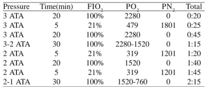
Hyperbaric Oxygen Therapy has many applications, including treatment of decompression sickness, carbon monoxide poisoning and arterial embolism. The treatment is performed in an enclosed chamber where the patient is subjected to pressures ranging up to 3.0 atmospheres absolute pressure (ATA). At these pressures, the concentration of O2 in the body is increased by up to 1900%, and PaO2 higher than 2000 mmHg in sessions lasting 20 to 30
minutes and as a consequence the PaN2 is reduced to
minimum values, as shown in Table 3 for Treatment of Decompression Sickness type 1 from the United States Navy. Murphy et al. [29], between 1970 and 1984, treated 16 patients for embolism, of which 2 cases were for embolism during a CPB. Guy et al. [30] succeeded in treating a patient with HBOT who had suffered large gaseous emboli during a cardiac surgery. Ziser et al. [31] used HBOT to treat cerebral arterial embolism in 17 patients undergoing CPB.
Grist [28], based on the use of HBOT suggested pure O2 in CPB in order to treat and prevent N2 microemboli. The analogy made by Grist between the hyperbaric chamber and the oxygenation chamber of the membrane oxygenator shows a resemblance in general characteristics of closed systems where there is the possibility of using pure oxygen and reduce PaN2. The low pressure of the nitrogen in the blood promotes the tendency of the solubilization of this gas in the formed microbubbles.
Hyperoxia
The oxygenation of blood during infusion corresponds
to a PO2 above 100mmHg and below 200mmHg, called
normoxia. Its maintenance occurs according to the offer of greater or lesser fraction of inspired oxygen at (FIO2) in the gas line connected to the oxygenator during CPB. Hyperoxia is defined as a high concentration of oxygen in the blood, with this condition being produced by the oversupply of oxygen.
Hyperoxia can produce reperfusion injury, excessive production of free radicals [32,33] and even exposure to the toxic effects of oxygen [34]. The use of hyperoxia in CPB is considered unnecessary by most perfusionists. Toraman et al. [35] showed that between 35 and 45% FI02 during CPB is sufficient to allow oxygen extraction safely.
Veno-arterial shunt
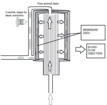
Moraes [38] evaluated the use of a diversion of veno-arterial blood. This deviation was done by a bypass connecting the venous line before entering the oxygenator to an arterial line, which allowed the diversion of part of the circulation, so that a fraction of blood flow was not passed through the oxygenation chamber and returned to the normal circuit system after the oxygenator. This author aimed to reduce the volume of blood in contact with the membrane oxygenator, as shown in Figure 1. The objective was to
demonstrate that the shunt could minimize the inflammatory response induced by cpb. The concept was based on the physiology of fetal circulation, where blood circulating in a fetus is at its greatest volume, a mixture of oxygenated and non-oxygenated blood.
In fetal circulation the only oxygenated blood reaches the fetus from the placenta exclusively by umbilical vein to the point of confluence with the inferior vena cava through the
ductus venosus. The mixing of oxygenated and non-oxygenated
blood with SaO2 = 62% in the thoracic aorta is sufficient for the fetus because their metabolic needs are reduced [36]. Moraes [37] published a comparative where he noted that there was less bleeding and a lesser need for transfusions in the group using veno-arterial shunts. This concept is applied in the manufacture of oxyshunt ® oxygenators (Instrumental Zammi, Duque de Caxias, Rio de Janeiro, Brazil).
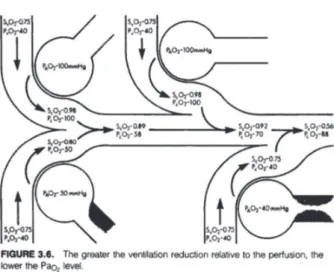
By observing this system based on the physiology of pulmonary shunts, which also occurs between arterial and mixed venous blood, we notice an important relationship. The pulmonary shunt is defined as a disturbance in the pulmonary ventilation-perfusion relation, where a fraction of inadequately ventilated pulmonary blood [38,39] follows without sustaining gas exchange, as illustrated in Figure 1. This non ventilated blood removes some oxygen from the oxygenated blood, which results in a slight reduction in arterial PO (PaO2), hemoglobin oxygen saturation (SaO2) and, consequently in arterial oxygen content (CaO2) (Figures 2 and 3).
In a normal, healthy individual, the physiological shunt represents 2-4% of the heart debt [1], the larger the fraction of the shunt, the lower the arterial oxygen content (CaO2)
The pulmonary shunt fraction is measured by the classical shunt equation [38,39].
Qs/Qt = (CcO2 – CaO2) / (CcO2-CvO2) Where:
Qs: Shunt Flow Qt: Total blood flow
Qs/Qt: % of blood flow that is diverted to the shunt CcO2: Content of the O2 capillary
CaO2: Arterial content of O2 CvO2: Venous content of O2 Calculations
CcO2= (Hb . 1.34 %ScO2) + (0.003 . PaO2) CaO2= (Hb . 1.34 %SaO2) + (0.003 . PaO2) CvO2= (Hb . 1.34 %SvO2) + (0.003 . PaO2)
CcO2, CaO2 and CvO2 are given in ml of O2 by 100 ml of blood (ml/100ml).
Applying the equation, considering:
PaO2= 100 mmHg SaO2= 100%
PvO2= 40 mmHg
SvO2= 75%
Hb= 10g/dL
FIO2= 100% (pure oxygen)
Atmospheric Pressure =760 mmHg, at sea level. Replacing the CcO2 with Cox.O2 (blood content in the blood after passing through the membrane) and ScO2 with
Sox.O2 (oxygen saturation after passing through the
membrane).
When applying this equation using parameters taken from a cardiopulmonary bypass with an oxygenator, with veno-arterial shunt in use, this relationship is as follows:
We have:
Qs/Qt = (Cox. O2– CaO2) / (Cox.O2– CvO2), the result is given as a % of total blood flow.
Qs/Qt = [(Hb.1.34.%Sox.O2)+(0.003.760 mmHg)] –
[(Hb.1.34. %SaO2) + (0,003.PaO2)] / [(Hb. 1.34.%Sox.O2) + (0.003.760 mmHg)] – [(Hb.1.34.%SvO2) + (0.003.PvO2)] =
Qs/Qt = [(10 g/dL.1.34.1) + (0.003.760 mmHg)] - [(10g/ dL.1.34.1) + (0.003.100 mmHg)] / [(10 d/L.1.34.1) + (0,003.760 mmHg)] - [(10d/L.1.34.0.75) + (0.003.40 mmHg)] =
Qs/Qt = [15.5] – [13.7] / [15.5] – [10.17] = Qs/Qt = 1,8 / 5,33 =
Qs/Qt= 0.33 or 33%. Admitting Qt = 5 L/min,
Qs = 0.33 x 5 L/min = 1.65 L/min. Or that is, for Qt = 5 L/ min, um Qs = 1.65
L / min will be sufficient to control hyperoxia generated with FIO2 = 100%.
In other words 33% of the perfusion flow passing through the shunt permits an adequate perfusion with the hyperoxia duly controlled.
The idea then occurred to evaluate the prospect of the
shunt to control the pure O2 induced hyperoxia, as this
could prevent the formation of microbubbles in CPB and this relationship seems to elucidate the question of microbubbles of nitrogen.
We compared the functional anatomy of the ventilatory unit shown in Figure 4 with the anatomy of the Oxyshunt ® Zammi oxygenator (Figure 5).
Fig. 2 - Adapted from Essentials of oxygenation, T. Ahrens, 1993
The dynamics of blood in the oxygenator follow the principles of hemodynamics, the passage of blood by the membrane area produces high strength, and the tendency of the flow segment is to follow by the segment of least resistance (due to the shunt deviation), which makes necessary a constraint mechanism to control the flow of blood running through the shunt. The deviation causes a smaller portion of blood flow to traverse the membrane so there is a tendency of a lower incidence of hemolysis.
The control of venous blood flow (shunt) is made by a clamp mounted on the shunt line, this clamp has an adjustable diameter control line and it is driven by pulse oximetry in the arterial and venous lines, where the targets are 95 and 75% of arterial and venal saturation respectively with a 100% FIO2 oxygen gas/arterial blood flow of 1:1
Thus, the removal of CO2 by the membrane behaves in
the same manner as in oxygenators that utilize a gas mixer. An “online” Oximetry is necessary due to the risk of hypoxia produced by an inadvertently kept larger fraction of shunt and by the contrary, hyperoxia.
Both the veno-arterial shunt and the pulmonary shunt permit the reduction of blood passing through the exchange membrane. This diverted blood does not receive oxygen, and as it returns to the arterial line, it mixes with the blood that passed through the membrane. At the moment of the
mixture there is a gradient of PaO2 that is balanced by
diffusion in a short segment of arterial line, thus hyperoxia does not occur by the use of pure oxygen avoiding the possible occurrence of reperfusion injury and the harmful effects of excess O2.
Table 4. Partial pressure of Nitrogen
Partial pressure PO2
PCO2 PN2 PH2O Total
Air 158 0.3 596 5.7 760
Arterial blood 100
40 573
47 760
Venous blood 40 46 573 47 760
Tissue 40 46 573 47 706 Fig. 4 - Dynamics of the blood as it travels the pulmonary capillary.
Adapted from Essentials of oxygenation, T. Ahrens, 1993
Nitrogenation
Nitrogen gas is responsible for arterial embolization, this occurs because of its low solubility coupled with its high concentration in blood as well as in tissue (Table 4).
The partial pressure of nitrogen in the blood is 573 mmHg, according toTable 4.
The use of pure oxygen produces denitrogenation of
blood, reducing PaN2 and hence the content of dissolved
nitrogen in the body, Tovar et al. [9], and increases the window of oxygen, or that is, with lowered PaN2, the blood has a lower content of dissolved nitrogen, which allows oxygen to fill this empty space formerly occupied by nitrogen.
With the low pressure of N2, there is greater tendency for solubilization of the N2 microbubbles in blood and this still prevents the microbubbles which may be formed in the vascular system. With this concept Berry & Myles [40] produced alveolar denitrogenation with ventilation to a FIO2 at 100%, and recorded FEO2 (Fraction expiratory O2) of 96% after 3 minutes exposure, this experiment shows that this process is rapid and probably happens in the same condition in the membrane oxygenator.
Table 5. Relationship between diameter, volume and surface area of the N2 microbubble
Bubble diameter 1 cm = 10 mm 0.1 cm = 1 mm 0.01 cm 0.001 cm
Bubble volume 0.5 ml 0.5 µl 5x10⁴ µl
5x10⁷ µl
Volume of bubble x Number of bubbles
0.5 ml 0.5 ml 0.5 ml 0.5 ml Number
of 1 1000 1000000 1000000000
Bubble surface 3.14 cm2
3.14 cm2
31.4 µm2
314 µm
Surface x Number of bubbles 3.14 cm2
31.4 cm2
314 cm2
3140cm2
Fig. 7 - The flowchart shows how the shunt venoarterial could control hyperoxia used to produce denitrogenation of blood Fig. 6 - The flowchart shows a sequence of events resulting in the
formation of cavitation microbubbles that culminates with the final outcome embolism, the same sequence of events can occur during CPB
Georgiads et al. [41] subjected 185 patients who were mechanical valve carriers to ventilation with FIO2 to 100% while he monitored microembolic events produced by cavitation valves with TCD. This study showed a significant decrease in the production of microbubbles compared with room air ventilation, suggesting that the principal source of gas embolism in those patients consisted of N2 bubbles. Rodriguez & Belway [42] developed a flow chart reproduced here, which relates the mechanisms involved in the events that follow up to the cerebral embolism produced by cavitation (Figure 6).
Among the mechanisms mentioned are, complement activation, adhesion of plasma proteins and platelet destruction leading to thrombus formation and vascular injury and embolism from the formation of nitrogen bubbles in the blood.
In the description of Tovan et al.[9], who sees a proportional relationship between volume, area and diameter of the Nitrogen microbubbles formed (Table 5), where gas
bubbles enter the bloodstream in dynamic equilibrium with the dissolved gas in plasma, it is noted that these will be enlarged or reduced according to the partial pressure of the gas in solution [11].
However, using the veno-arterial shunt with the flow fraction described above will cause the denitrogenation produced to break this balance and the microbubbles tend strongly to dissolve in the blood.
For the situation of microbubbles formed which passed through the veno-arterial shunt (e.g., bubbles formed in the venous reservoir), when they encounter the arterial blood which passed through the membrane (denitrogenized), there is a strong tendency to dissolve.
Similarly, possible O2 microbubbles formed in the
REFERENCES
1. Souza MHL, Elias DO. Fundamentos da circulação extracorpórea. 2ª ed. São Paulo: Centro Editorial Alfa Rio; 2006.
2. Barbosa NF, Cardinelli DM, Ercole FF. Determinantes de complicações Neurológicas no uso da circulação extracorpórea. Arq Bras Cardiol. 2010;95(6):e151-7.
3. Weitkemper HH, Oppermann B, Spilker A, Knobl HJ, Körfer R. Gaseous microemboli and the influence of microporous membrane oxygenators. J Extra Corpor Technol. 2005;37(3):256-64.
4. Fearn SJ, Pole R, Burgess M, Ray SG, Hooper TL, McCollum CN. Cerebral embolisation during modern cardiopulmonary bypass. Eur J Cardiothorac Surg. 2001;20(6):1163-7.
5. Groom RC, Quinn RD, Lennon P, Donegan DJ, Braxton JH, Kramer RS, et al; Northern New England Cardiovascular Disease Study Group. Detection and elimination of microemboli related to cardiopulmonary bypass. Circ Cardiovasc Qual Outcomes. 2009;2(3):191-8.
6. Deverall PB, Padayachee TS, Parsons S, Theobold R, Battistessa SA. Ultrasound detection of micro-emboli in the middle cerebral artery during cardiopulmonary bypass surgery. Eur J Cardiothorac Surg. 1988;2(4):256-60.
7. Liu YH, Wang DX, Li LH, Wu XM, Shan GJ, Su Y, et al. The effects of cardiopulmonar bypass on the number of cerebral microemboli and the incidence of cognitive dysfunction after coronary bypass graft surgery. Anesth Analg. 2009;109(4):1013-22.
RESULTS
The denitrogenation promoted by hyperoxia prevents the formation of microbubbles. The veno-arterial shunt as described by Moraes obeys the physiology of pulmonary shunts and can control hyperoxia used to produce denitrogenation of blood during CPB according to the flowchart in Figure 7.
DISCUSSION
Since the first oxygenator developed by Gibbon to current models, many problems inherent to the CPB were identified, problems related to effectiveness, such as adequate oxygenation, removal of CO2 [42], and others related to safety such as inflammatory activation trauma and blood microemboli. This evolution of new knowledge minimized the deleterious effects of CPB and with the addition of accessories such as arterial filters and bubble detectors; it has reduced the morbimortality in cardiac surgery procedures with CPB [38,43-45]. However, the incidence, still frequent of neurological complications remains a challenge because, despite all the advances, neurologic complications still occur and are causes of serious damage [4]. Many studies have been conducted with the accuracy of the TCD, which has made it possible to quantify and qualify the occurrence of microbubbles in both the CPB circuit as well as in cerebral arteries and to measure the resulting GME.
It is known that microbubbles have nitrogen gas as
their main component. The removal of N2 from blood is
well documented; the denitrogenation decreases the
tension of N2 pressure, so the bubbles thus formed are
dissolved. This occurrence of denitrogenation implies the
use of pure O2 in oxygenation which would lead to
hyperoxia and its toxic effects, but that can be avoided in cardiopulmonary bypass procedures with the use of veno-arterial shunts in the oxygenator.
Weitkemper et al. [3] stated that “gaseous microemboli are still an unsolvable problem of the CPB circuit.” However, if we consider the information presented here, we believe that this paradigm dissolves with the content presented here. The veno-arterial shunt that at the time was developed with the goal of reducing inflammatory activation in CPB [38] and was not associated with denitrogenation by the use of pure O2, which appears to be its main advantage and benefit to the patient. And, moreover, it has proven to be a simple and practical method, provided it is properly controlled, the benefit of which has great importance for those who require the use of CPB in heart and organ management operations, with greater cost savings, and mainly eliminating the complications of air microemboli during cardiopulmonary bypass, as well as an evolution in practice. This work sheds light on this possibility and we
suggest that randomized controlled trials are effected to confirm the benefits of the possibility of damage control and vascular complement activation cited by Rodriguez et al. [13].
CONCLUSION
8. Mathew JP, Mackensen GB, Phillips-Bute B, Stafford-Smith M, Podgoreanu MV, Grocott HP, et al. Effects of extreme hemodilution during cardiac surgery on cognitive function in the elderly. Anesthesiology. 2007;107(4):577-84.
9. Tovar EA, Del Campo C, Borsari A, Webb RP, Dell JR, Weinstein PB. Postoperative management of cerebral air embolism: gas physiology for surgeons. Ann Thorac Surg. 1995;60(4):1138-42.
10. Lelis RGB, Auler Jr JOC. Lesão neurológica em cirurgia cardíaca: aspectos fisiopatológicos. Rev Bras Anestesiol. 2004;54(4):607-17.
11. Borger MA, Peniston CM, Weisel RD, Vasiliou M, Green RE, Feindel CM. Neuropsychologic impairment after coronary bypass surgery: effect of gaseous microemboli during perfusionist interventions. J Thorac Cardiovasc Surg. 2001;121(4):743-9.
12. Barak M, Katz Y. Microbubbles: pathophysiology and clinical implications. Chest. 2005;128(4):2918-93.
13. Rodriguez RA, Rubens F, Belway D, Nathan HJ. Residual air in the venous cannula increases cerebral embolization at the onset of cardiopulmonary bypass. Eur J Cardiothorac Surg. 2006;29(2):175-80.
14. Souza MHL, Elias, DO. Microembolias na CEC. Boletim Informativo Tecnologia Extracorpórea, 2008;06,07:2.
15. Nollert G, Nagashima M, Bucerius J, Shin’oka T, Jonas RA. Oxygenation strategy and neurologic damage after deep hypothermic circulatory arrest. I. Gaseous microemboli. J Thorac Cardiovasc Surg. 1999;117(6):1166-71.
16. Hammon JW Jr, Stump DA, Kon ND, Cordell AR, Hudspeth AS, Oaks TE, et al. Risk factors and solutions for the development of neurobehavioral changes after coronary artery bypass grafting. Ann Thorac Surg. 1997;63(6):1613-8.
17. Taylor RL, Borger MA, Weisel RD, Fedorko L, Feindel CM. Cerebral microemboli during cardiopulmonary bypass: increased emboli during perfusionist interventions. Ann Thorac Surg.1999;68(1):89-93.
18. Norman MJ, Sistino JJ, Acsell JR. The effectiveness of low-prime cardiopulmonary bypass circuits at removing gaseous emboli. J Extra Corpor Technol. 2004;36(4):336-42.
19. Mendes KDS, Silveira RCCP, Galvão CM. Revisão integrativa: método de pesquisa para a incorporação de evidências na saúde e na enfermagem. Texto contexto Enferm. 2008;17(4):758-64.
20. Souza MT, Silva MD, Carvalho R. Revisão integrativa: o que é e como fazer. Einstein. 2010;8(1 Pt 1):102-6.
21. Lynch JE, Wells C, Akers T, Frantz P, Garrett D, Scott ML, et al. Monitoring microemboli during cardiopulmonar bypass
with EDAC quantifier. J Extra Corpor Technol. 2010;42(3):212-8.
22. Urbanek S, Tiedtke HJ. Improved methods for measurement of gaseous microbubbles during extracorporeal circulation. Perfusion. 2002;17(6):429-34.
23. Sleep J, Syhre I, Evans E. Blood temperature management and gaseous microemboli creation: an in-vitro analysis. J Extra Corpor Technol. 2010;42(3):219-22.
24. Blauth CI. Macroemboli and microemboli during cardiopulmonary bypass. Ann Thorac Surg. 1995;59(5):1300-3.
25. Gill MC, Dando H, John D. In the air handling capability of the quadrox D pump dependent within an ECMO circuit? An in vitro study. J Extra Corpor Technol. 2010;42(3):203-11.
26. Dye TE. Macroscopic bubbles from dissolved nitrogen during CPB. Ann Thorac Surg. 1997;64(5):1527.
27. Guyton AC. Tratado de fisiologia médica. 8ª Ed. Rio de Janeiro:Guanabara Koogan;1992. p.415-8.
28. Grist G. Gaseous microemboli and hyperoxia. J Extra Corpor Technol. 2006;38(4):367-9.
29. Murphy BP, Harford FJ, Cramer FS. Cerebral air embolism resulting from invasive medical procedures. Treatment with hyperbaric oxygen. Ann Surg. 1985;201(2):242-5.
30. Guy TS, Kelly MP, Cason B, Tseng E. Retrograde cerebral perfusion and delayed hyperbaric oxygen for massive air embolism during cardiac surgery. Interact Cardiovasc Thorac Surg. 2009;8(3):382-3.
31. Ziser A, Adir Y, Lavon H, Shupak A. Hiperbaric oxygen therapy for massive arterial air embolism during operations. J Thorac Cardiovasc Surg. 1999;117(4):818-21.
32. Brown DM, Holt DW, Edwards JT, Burnett RJ 3rd. Normoxia vs. hyperoxia: impact of oxygen tension strategies on outcomes for patients receiving cardiopulmonary bypass for routine cardiac surgical repair. J Extra Corpor Technol. 2006;38(3):241-8.
33. Caputo M, Mokhtari A, Rogers CA, Panayiotou N, Chen Q, Ghorbel MT, et al. The effects of normoxic versus hyperoxic cardiopulmonary bypass on oxidative stress and inflammatory response in cyanotic pediatric patients undergoing open cardiac surgery: a randomized controlled trial. J Thorac Cardiovasc Surg. 2009;138(1):206-14.
34. Joachimsson PO, Sjöberg F, Forsman M, Johansson M, Ahn HC, Rutberg H. Adverse effects of hyperoxemia during cardiopulmonary bypass. J Thorac Cardiovasc Surg. 1996;112(3):812-9.
cardiopulmonary bypass. Asian Cardiovasc Thorac Ann. 2007;15(4):303-6.
36. Moraes DJ, Moraes MCJ, Dias JRJ, Martins P, Moraes ZCJ, Souza CG, et al. Uso de oxigênio puro e shunt veno-arterial nos oxigenadores de membrana. Rev Bras Cir Cardiovasc. 1997;12(1):77-82.
37. Moraes MCJ, Moraes DJ, Bastos ES, Murad H. Circulação extracorpórea com desvio veno-arterial e baixa pressão parcial de oxigênio. Rev Bras Cir Cardiovasc. 2001;16(3):251-61.
38. Guyton A C. Tratado de fisiologia médica. 8ª ed. Rio de Janeiro:Guanabara Koogan;1992. p.381.
39. Ahrens T, Basham KAR. Essentials of oxygenation: implication for clinical practice. 1ª ed. Boston:Jones & Bartlett Publishers;1993. p.21-31.
40. Berry CB, Myles PS. Preoxygenation in heathy volunteers: a graph of oxygen “washin” using end-tidal oxygraphy. Br J Anaesth.1994;72(1):116-8.
41. Georgiadis D, Wenzel A, Lehmann D, Lindner A, Zerkowski HR, Zierz S, et al. Influence of oxygen ventilation on Doppler microemboli signals in pacients with artificial heart valves. Stroke. 1997;28(11):2189-94.
42. Rodriguez RA, Belway D. Comparison of two different extracorporeal circuits on cerebral embolization during cardiopulmonary bypass in children. Perfusion. 2006;21(5):247-53.
43. Perthel M, El-Ayoubi L, Bendisch A, Laas J, Gerigk M. Clinical advantages of using mini-bypass systems in terms of blood product use, postoperative bleeding and air entrainment: an in vivo clinical perspective. Eur J Cardiothorac Surg. 2007;31(6):1070-5.
44. Barbut D, Lo YW, Gold JP, Trifiletti RR, Yao FS, Hager DN, et al. Impact of embolization during coronary artery bypass grafting on outcome and length of stay. Ann Thorac Surg. 1997;63(4):998-1002.