The use of biomarkers in clinical osteoporosis
HEBERT WILSON SANTOS CABRAL1*, BRUNA FERREIRA GALONE ANDOLPHI2, BRUNNA VILA COUTINHO FERREIRA2,
DANIELLE CRISTINA FILGUEIRA ALVES2, RENATO LÍRIO MORELATO3, ANTÔNIO CHAMBO FILHO4, LIZÂNIA SPINASSÉ BORGES5
1Post-Doctoral degree in Neuroscience – Adjunct Professor of Medicine and Permanent Professor for the Masters Program in Public Policies and Local Development at Escola Superior de Ciências da Santa Casa de
Misericórdia de Vitória (Emescam), Vitória, ES, Brazil
2MD at Emescam, Vitória, ES, Brazil
3PhD in Physiological Sciences – Head of the Geriatric Service and Adjunct Professor at Emescam, Vitória, ES, Brazil 4PhD in Medicine – Full Professor of Gynecology and Obstetrics, Emescam, Vitória, ES, Brazil
5PhD, degree in Sciences. Post-Doctoral research at Université Paris Sud 11, Paris, France
S
UMMARYStudy conducted at Escola Superior de Ciências da Santa Casa de Misericórdia
de Vitória (Emescam), Vitória, ES, Brazil
Article received: 12/18/2014
Accepted for publication: 3/23/2015
*Correspondence:
Address: Av. Nossa Senhora da Penha, 2190, Bela Vista
Vitória, ES – Brazil Postal code: 29027-502
http://dx.doi.org/10.1590/1806-9282.62.04.368
Osteoporosis is a disease of ascending character in the world population; in this context, bone biomarkers are being increasingly studied in order to aid in the diagnosis and monitoring of these patients. The main objective of this study was a literature review of articles whose main theme was the use of biomarkers for bone formation and degradation, and to evaluate their possible applicabili-ty in clinical practice. Literature review was performed through articles indexed and published in the last five years in the PubMed database. The findings of this study showed that most of the previously selected articles were published in the last two years, and the most cited markers were bone resorption, C-terminal col-lagen telopeptide (CTX), showing the highest correlation with the dynamics of bone, and the biomarker of bone formation, bone-specific alkaline phosphatase (BAP), which is increased in the event of fracture or may suggest another bone disease. There was an increase in published articles, associating different bone biomarkers and their clinical applicability, especially for treatment control. Our findings suggest that in recent years there has been significant increase in pub-lications evaluating the use of bone turnover biomarkers for bone formation and resorption and their possible clinical applicability, especially in the moni-toring of treatment. Still, we believe that further studies need to be conducted to confirm these findings, given the advantages that bone biomarkers can deliv-er in the clinical management of the disease.
Keywords: osteoporosis, bone remodeling, biological markers.
I
NTRODUCTIONOsteoporosis (OP) is a systemic disease characterized by decreased bone mass and microstructural deterioration of bone tissue, with consequent increase of fragility and susceptibility to fracture.1 Most of the fractures caused
by osteoporosis have considerable impact on economi-cally active individuals, and there is a substantial risk of increased mortality and mobility in the elderly popula-tion, interfering directly in the quality of life.2 Ethnic,
genetic, anthropometric, socio-cultural and economic difference and other life habits contribute to explaining the differences in the incidence and prevalence of osteo-porosis.3,6
According to data from the World Health Organiza-tion (WHO), OP affects more than 75 million people in the United States (USA), Europe and Japan, accounting for more than 8.9 million fractures annually around the world, with more than 4.5 million occurring in the Americas and Europe.4 In Brazil, osteoporosis is estimated to affect 10
million people, with a prevalence of 11 to 23.8% for all types of bone fracture.3 According to the Ministry of Health, in
the year 2007 around BRL 51.8 million was spent on hos-pitalizations, and each year spending arising from the treat-ment of fractures increases, especially in elderly people.5
effective in preventing fractures, thus being approved for the treatment of OP, are bisphosphonates (alendronate, risedronate and ibandronate), categorized as bone antire-sorptive medication and remaining the most popular and widely used pharmacological treatment for osteoporosis. However, when these drugs are contraindicated or cause serious side effects, new treatments and alternative phar-macological forms are available. Currently, there has been a great difficulty in assessing response to treatment, since Bone Densitometry (DXA), considered the gold standard in both the diagnosis and monitoring of these patients, is a diagnostic method not often available in basic health ser-vices.7 The latest data from the literature has been
suggest-ing an alternative to the use of DXA in the clinical moni-toring of OP patients: biomarkers of bone resorption and formation. Bone remodeling can be easily measured using a variety of biochemical markers, which are largely divid-ed into two categories: markers of bone resorption, which reflect the activity of osteoclasts, are mostly the products of type 1 collagen degradation; and markers of bone for-mation, reflecting the activity of osteoblasts and are the byproducts of collagen synthesis (matrix proteins or os-teoblastic enzymes). The main markers of bone formation in the blood are: a) osteocalcin (bone Gla-protein) – OC, b) total alkaline phosphatase (ALP), c) procollagen type 1 car-boxy-terminal propeptide, and d) procollagen 1 amino-ter-minal propeptide (P1NP). Bone resorption and bone for-mation processes are coupled, so in some situations these markers will reflect on the changes in bone remodeling.2
Bone resorption markers in the blood are: a) tartrate-resis-tant acid phosphatase (TRACP), b) procollagen type 1 c-terminal portion (S-CTX). Bone resorption markers found in urine include: a) Pyridinoline and deoxypyridinoline (U-PYD and DPD-U), b) hydroxyproline (U-HYP), c) amino-terminal portion of procollagen I (U-NTX), d) carboxy-ter-minal portion of procollagen I (U-CTX).26
The dosage of these biomarkers would be another use-ful tool in monitoring therapy for this pathology. While DXA would be evidence of a positive response to treatment with increased bone mineral density (BMD), 1 to 2 years after the increase of formation biomarkers, a reduction of those related to bone resorption could be detected 3 to 6 months after the drug intervention. Recent studies have shown limitations on the use of biomarkers of bone re-sorption, suggesting that more studies need to be conduct-ed for their application in routine clinical practice.8
M
ETHODThe present study is a review of the literature using arti-cles published over the last five years and stored in a
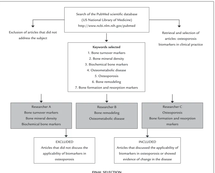
sci-entific database. The main objective was to determine the clinical applicability of bone biomarkers over the years and nowadays, as described in the flowchart (Figure 1).
Selection and searching of articles by keywords
In order to define the descriptors to be used in the re-search, searches were carried out on the PubMed web-site (US Library of Medicine) using the keywords: “bio-marker osteoporosis”, and the following filters: review papers, completed text in descending order based on date of publication. According to our search the most cited descriptors were: a) bone mineral density osteopo-rosis, b) biochemical bone markers, c) bone turnover, d) osteometabolic diseases, e) osteoporosis, f) bone forma-tion and resorpforma-tion markers.
The search was distributed to three researchers, in such a way that the findings relating to the first three mentioned descriptors were evaluated by Researcher A, the two following descriptors by Researcher B and the last two by Researcher C (Figure 1). The exclusion crite-ria were defined as: a) review articles that only cited the biomarkers, but did not discuss their applicability in the clinical practice, b) articles whose main theme did not in-clude biomarkers in osteoporosis. Thus, the inclusion cri-teria were: a) the articles that discussed the clinical use of biomarkers in osteoporosis and presented evidence in the occurrence of clinical conditions associated with the dis-ease. Our last search of the database was performed in December 2014.
Data collection
Data collection was carried out over a period of 12 months, during which the information extracted from articles previously selected was: a) names of the biomarkers an-alyzed in publications, b) classification of biomarkers as to their role in the bone metabolism phase, c) date of publication of the article, d) number of authors, e) sam-ple size and period of study. After the selection of arti-cles published over the last five years, they were strati-fied and divided into two groups: publications made over the past two and a half years and articles published more than two and a half years ago. The number of authors was also grouped into two categories: up to three au-thors and more than three auau-thors. We noted that the number of participants was described in variable and continuous manner. Study duration in the articles was divided into four groups: up to 1 year; 2 to 5 years; 5 to 10 years and more than 10 years.
Identiication and classiication of biomarkers
Bone markers were listed according to their description in the articles as per the corresponding classification. The classification regarding type of biomarkers evaluated in the clinical trials was divided into the following groups: i) Formation biomarkers: These are represented by enzymes or products that participate in the genesis of the bone ma-trix and function as bone formation markers if dosed;9 ii)
Degradation biomarkers: classified as proteins or their fragments released during formation of the bone degra-dation process, which when dosed have the function of bone resorption markers.10 We noted that both markers
were cited in the same article and that other types of bio-markers used were not appropriate for classification in our study, that is, were not considered as bone formation or degradation biomarkers. Biomarkers classified as asso-ciated in scientific publications address one or more
bio-markers, named as “others” in this study, and were added to the dosage of bone resorption formation markers.
Data analysis
For data analysis, we used descriptive statistics in order to summarize data into: i) Number, ii) simple frequen-cies, iii) mean and standard deviations, and iv) construc-tion of tables. The data was processed using chi-squared test to compare simple frequencies and Student’s t test to compare averages of the parameters. The Statistical Package for Social Science for Windows (SPSS) 17 soft-ware was used in all statistical analyses, adopt p<0.05 and a confidence level of 95%.
R
ESULTSFollowing the methodology described in the present study, in our results we examined 7,996 articles relating to the
Search of the PubMed scientiic database (US National Library of Medicine) http://www.ncbi.nlm.nih.gov/pubmed
Exclusion of articles that did not address the subject
Retrieval and selection of articles: osteoporosis biomarkers in clinical practice Keywords selected
1. Bone turnover markers 2. Bone mineral density 3. Biochemical bone markers
4. Osteometabolic disease 5. Osteoporosis 6. Bone remodeling
7. Bone formation and resorption markers
Researcher A Bone turnover markers
Bone mineral density Biochemical bone markers
Researcher B Bone remodeling Osteometabolic disease
Researcher C Osteoporosis
Bone formation and resorption markers
EXCLUDED
Articles that did not discuss the applicability of biomarkers in
osteoporosis
FINAL SELECTION
INCLUDED
Articles that discussed the applicability of biomarkers in osteoporosis or showed
evidence of change in the disease
descriptors “osteoporosis”, 2,729 “bone formation” and 152 “resorption markers”, 7 “osteometabolic diseases”, 1,788 “bone remodelling”, 1,753 “bone turnover
mark-ers’’, 172 “biochemical bone markmark-ers’’, 1,395 “bone min-eral density’’. 66 articles were considered eligible for this study and were categorized according to the type of bio-marker: Bone formation and bone degradation, both of which were cited in the references used. We also noted other biomarkers, but they did not apply to the classifi-cation used in our target of study. Our findings show that the data from the literature classifies some biomarkers with the associated term, covering formation and degra-dation biomarkers along with non-classifiable ones, as can be observed in Table 1. According to this table, we see that more than 50% of the articles discuss formation and degradation biomarkers in conjunction. On the other hand, analyses of non-classifiable biomarkers were ob-served at a rate of 7.6%, when analyzed in isolation, and 6.1% in association with formation and degradation mark-ers. The chi-squared statistical tests showed significance at a p-value<0.05 but the number of biomarkers defined as “others” and “associated” was low, and this result should be evaluated carefully. Nevertheless, this demonstrates greater significance compared with the data observed in the clinical trials, which address bone formation and deg-radation biomarkers concomitantly.
TABLE 1 Number and percentage of selected articles that
discuss the subject of osteoporosis biomarkers.
Type of marker Frequency Percentage (%)
Formationa 9 13.6
Degradationb 13 19.7
Bothc 35 53.0
Otherd 5 7.6
Associatede 4 6.1
Total 100
aSubstances present in the genesis of bone matrix; bsubstances released during the process of
bone degradation; cformation and degradation biomarkers cited in the same article; d
bioma-rkers that are not categorized as formation or degradation; ecitations of formation and
degra-dation biomarkers along with non-classiiable ones (others).
Main types of biomarkers cited in the selected articles and their clinical applicability
In the texts retrieved, we found 13 different types of bio-markers: a) BAP (bone alkaline phosphatase), b) OPG (os-teoprotegerin), c) RANKL (nuclear factor Kappa E), d) P1CP (type 1 procollagen carboxy-terminal propeptide), e) P1NP (amino-terminal propeptide) f) OC (Osteocalcin), g) TRACP (tartrate-resistant acid phosphatase), h) TNF--alpha (tumor necrosis factorTNF--alpha), i) PYD (pyridinoline),
j) DPD (deoxypyridinoline), k) CTX (carboxy-terminal telopeptide of type 1 collagen), l) NTX (amino-terminal telopeptide of type 1 collagen), m) ALP (serum alkaline phosphatase). These are shown in Chart 1, which repre-sents the total frequency of citations of the biomarkers and their applicability in a more generalized form. Note that the biomarkers cited the most were P1NP (procolla-gen 1 amino-terminal propeptide), BAP of bone forma-tion and CTX of bone degradaforma-tion. It is important to note that a considerable number of each biomarker has con-firmed clinical applicability; for example, we can mention the degradation biomarker CTX, observed in 35 citations with applicability confirmed by 25 of them. In relation to formation biomarkers, P1NP was cited in 25 clinical tri-als demonstrating clinical application in 18 citations.
Evolution of the percentage of papers published in the last ive years
Evaluating all of the selected works, we found that ap-proximately 57.8% were published in the last five years. The period observed was from April 2011 to March 2012, where there was a large increase in the number of publi-cations in scientific databases. However, a reduction of publications was noted in the periods between April 2010 and March 2011. This data is displayed in Chart 2.
As for types of biomarkers, the increased variability studied is well-known, given that in studies published between April 2008 and March 2009, there are just two sub-types of articles, that is, those addressing formation biomarkers only, and those covering formation and deg-radation biomarkers together, whereas in the publica-tions in the period between April 2012 and April 2013, five subtypes were analyzed, that is, formation biomark-ers, degradation biomarkbiomark-ers, both, others and associat-ed biomarkers.
Average number of participants studied in the selected articles
distri-CHART 1 Types of bone biomarkers and their medical applicability.
BAP: bone alkaline phosphatase; OPG: osteoprotegerin; RANKL: receptor activator of nuclear factor kappa-E; P1CP: carboxy-terminal propeptide of type 1 procollagen; P1NP: amino-terminal pro-peptide; OC: osteocalcin; TRACP: tartrate-resistant acid phosphatase; TNF-alpha: tumor necrosis factor-alpha; PYD: pyridinoline; DPD: deoxypyridinoline; CTX: carboxy-terminal telopeptide of type 1 collagen; NTX: amino-terminal telopeptide of type 1 collagen; ALP: serum alkaline phosphatase.
CHART 2 Analysis of the number of studies grouped by type of biomarker, published in the period.
Formation: substances present in the genesis of bone matrix; degradation: substances released during the process of bone degradation; both: formation and degradation biomarkers cited in the same article; other: citations of biomarkers that are not categorized as formation or degradation; associated: citations of formation and degradation biomarkers along with non-classiiable ones (others).
Number of citations Applicability – yes BAP
26
14
8 8
3
2 2 2
25
18 23
14
6 5
1 1 1 1 4
2 35
25
21
13
3 2
OPG RANKL P1CP P1NP OC TRAP alfa-TNF PYD DPD CTX NTX FAL 40
35
30
25
20
15
10
5
0
April/08-March/09 April/09-March/10 April/10-March/11 April/11-March/12 April/12-March/13
Formation Degradation Both Other Associated 14
12
10
8
6
4
2
butions do not assume normality, showing that employ-ing Student’s t-test as method was not the best choice, and suggesting that the results between the comparisons should be made using nonparametric tests in this kind of analysis.
Duration of patient monitoring in the selected articles
During the period of study, patients remained similar, meaning that 31.8% of the studies lasted from two to five years. It is important to note that 30.3% of the arti-cles did not report the assessment period precisely, rep-resenting a selection bias in this evaluation. Regarding patient monitoring, we did not find long-term studies, with those conducted for more than 6 years represent-ing only 9.1% of the sample. We performed some multi-ple comparisons through chi-square test, showing sig-nificance (p<0.05) between some of the items observed, but since some sub-items were represented by zero and numbers less than five, the chi-squared test cannot be considered valid.
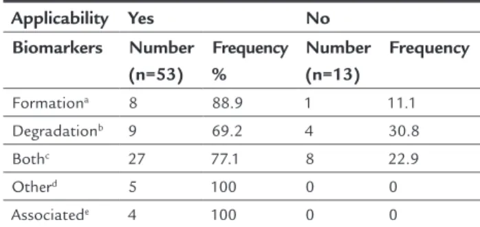
Clinical applicability of the use of biomarkers in the selected articles
Of the articles selected and indexed, approximately 80.3% showed any clinical applicability of the biomarkers with statistical significance (p<0.001). During our analysis, subdividing the items in the 66 articles, the comparisons were limited by chi-squared test for the reasons mentioned above. Observing Table 2 again, we can see that the bio-markers allocated as “others” and “associated” and cited in smaller numbers obtained 100% clinical applicability, while in the group defined as “both”, present in a great-er numbgreat-er of publications, thgreat-ere was 77.1% applicability, both following an overall trend in the study. When con-ducting an individual analysis of the biomarkers, we not-ed that P1CP (carboxy-terminal propeptide of type 1 pro-collagen) presented 100% applicability, but was only cited in two references. BAP has a higher frequency in the cita-tions, with a clinical applicability confirmed in 54% of the publications analyzed. By analyzing PYD (Pyridinoline), a degradation biomarker, applicability was confirmed de-spite the fact that this marker was reviewed only in one article, while CTX, cited the most in the references, and showed 71% of possible use. OPG (Osteoprotegerin), cit-ed most often within the group of non-classifiable bio-markers, presented 100% applicability for clinical use. The results with TNF-α and RANKL (receptor activator of nu-clear factor kappa-E) should be analyzed and interpret-ed with a larger sample size for confirmation of their ac-tual application.
TABLE 2 Applicability in the articles according to the type
of biomarker.
Applicability Yes No Biomarkers Number
(n=53)
Frequency %
Number (n=13)
Frequency
Formationa 8 88.9 1 11.1
Degradationb 9 69.2 4 30.8
Bothc 27 77.1 8 22.9
Otherd 5 100 0 0
Associatede 4 100 0 0
aSubstances present in the genesis of bone matrix; bsubstances released during the process of
bone degradation; cformation and degradation biomarkers cited in the same article; d
bioma-rkers that are not categorized as formation or degradation; ecitations of formation and
degra-dation biomarkers along with non-classiiable ones (others).
D
ISCUSSIONBone turnover biomarkers have contributed to a better understanding of the physiology of bones as well as the pathogenesis of metabolic bone diseases. In recent years, several technologies have been developed in order to de-termine markers in osteoporosis. Our study looked at 66 articles over the last five years. During this period we observed a predominance of references that addressed both formation and degradation marker, with around 53% of citations mentioning both. This fact may be at-tributed to prior knowledge involving bone formation and degradation that are bound and dependent. It is be-lieved that these processes need to be analyzed in con-junction in order to evaluate bone metabolism in a more suitable manner.11 The most relevant findings of the
se-lected publications are based on the application of bio-markers with the purpose of monitoring the treatment of osteoporosis.12 The findings in the study by Grey et al.
suggest that bone metabolism markers were low in 40% of patients undergoing treatment with zoledronate com-pared to a placebo group.12 Teriparatide, an
osteoana-bolic drug for the treatment of osteoporosis, was also used often. Studies conducted recently suggest that this drug may be effective to speed up the healing of frac-tures, as well as to stimulate the process in patients with delayed healing.13 According to Jakob et al. this
corrob-orates previous observations when concluding that wom-en after mwom-enopause diagnosed with severe osteoporosis benefited from the use of teriparatide even with the in-terruption of therapy 18 months beforehand.14
findings, as a document recently released by the Bone Marker Standards Working Group, in conjunction with the International Osteoporosis Foundation (IOF) and the International Federation of Clinical Chemistry and Laboratory Medicine (IFCC), suggested the use of bone formation and resorption markers in clinical trials, such as P1NP (formation marker) and CTX (resorption mark-er).15 In view of the difficulty of establishing an ideal
mark-er, the reference standards were chosen based on criteria such as adequate characterization and definition of each marker in relation to specificity to the bone, the results observed in clinical trials, biological variability, ease for analyzing the data, the availability of the method in anal-ysis laboratories, and the means of measuring the biolog-ical sample (serum or urine).16
In the last five years, around 57.6% of the articles were published in the period from 2011 to 2014, i.e. over the last 3.5 years. Our observational data suggest a gradual increase in the number of publications about this sub-ject, justified by increasing advances in research on isola-tion and characterizaisola-tion of cells and studies on extracel-lular components of the bone matrix, resulting in the development and implementation of new technologies for serum or urinary measurement of new biochemical markers of bone metabolism.17 Advances in molecular
bi-ology are providing a better understanding of the patho-physiology mechanisms of osteoporosis. One of the most significant contributions in this area was the identifica-tion of a system known as OPG (osteoprotegerin)/RANKL/ RANK (receptor activator of nuclear factor kappa B li-gand), discovered in the mid-1990s. This system is respon-sible for skeletal health, and recent studies indicate that RANKL\RANK signaling also play an important role in other tissues.18
The data in this study related to the average number of patients studied showed that 477 participants were in-cluded in our study. The standard deviation was 1,357.6, and this can be explained by a great variability in the num-ber of patients investigated in the selected studies, from 8 to 9,846 participants. We observed a lack of standard-ization in the concepts used and methodological differ-ences may justify this variation. In the studies that eval-uated the therapeutic response, differences found in relation to the sample size are considered extensive.19 As
for duration of patient monitoring, we did not observe major differences between the studies, and those lasting two to five years (31.8%) presented a discreet predomi-nance over the others.
In 2007, a study conducted by Vieira et al. suggested that the effectiveness of therapeutic monitoring in the
treatment of osteoporosis, carried out using the dosage of bone biomarkers, can be measured from one to three months after the beginning of the follow-up, as observed in the present study. We believe that although not stan-dardized, the treatment time might serve as the basis for other studies given that, when bone densitometry is used, changes are observed only after one to two years. These findings can be explained through the use of qualitative (x-rays) or quantitative (bone densitometry, quantitative CT) x-ray absorption techniques, allowing the examina-tion of the bone structure through the detecexamina-tion of radi-opaque crystals. However, the metabolic, physiological or pathological phenomena that can affect the bone tis-sue, only significantly affect its radiopaque structure af-ter a considerable period of time. Thus, the use of these techniques is limited for a more dynamic and short-term study on bone metabolism. Therefore, there is an inter-est in defining methodologies that can quantify substanc-es that could reprsubstanc-esent the metabolic procsubstanc-esssubstanc-es under-way in the tissue.17 Studies evaluating the outcome of
clinical applicability showed that 80.3% of the studies concluded that biomarkers show statistical significance with p<0.001. The data evaluated in this study revealed the potential of bone biomarkers in clinical practice, with the most frequent indicators being: i) Control of thera-peutic efficacy, ii) predicting the response in relation to adherence to treatment, iii) prediction of the risk of frac-ture. There are also further possibilities discussed in oth-er articles, such as forecast bone loss and selection of pa-tients for treatment.20 Evaluation of the control of
therapeutic efficacy is believed to be the main clinical ap-plication.
According to Kerschan-Schindl et al. there was a pos-itive correlation between early changes of biomarkers among patients under treatment and subsequent struc-tural changes, when 1,637 post-menopausal women us-ing teriparatide or a placebo were assessed. An increase in BAL and P1NP markers was observed in the first month in conjunction with histomorphometric indexes for ¼ computed microtomography (CT) after 22 months of treatment.21,22 When evaluating adherence to treatment,
with the use of antiresorptive medication (bisphospho-nates), and a 30 to 40% reduction when catabolic agents (raloxifene) are administered. Thus, the absence of these reductions would indicate low adherence to treatment by the patient or incorrect drug administration.23 When we
evaluated the prediction of risk of BMD fractures (corre-lated positively with bone strength), measurement using DEXA remains the best choice. Other relevant use of these biomarkers is the ability to identify women who will pres-ent a high rate of loss of bone mass during the years fol-lowing menopause, in order to initiate a prevention strat-egy for osteoporosis. In summary, the markers of bone renewal with other demographic variables could predict around 30 to 40% of the variations of bone loss in wom-en with untreated mwom-enopause.24 Currently the choice of
treatment based on bone markers is not being applied in routine clinical practice.25 One of the limitations of this
study is the small number of samples analyzed in this work, and the reduced number of national publications analyzed.
C
ONCLUSIONIn recent years there has been great progress in trying to understand the use of biomarkers and their applications in osteoporosis, due to major advances in science. If we use the physiology of the bone remodeling process, the ideal markers would be those able to form the diagnosis of osteoporosis and differentiate patients classified as slow or fast losers, adding sensitivity and specificity to the bone density measurement in the fracture risk assess-ment, leading to more appropriate treatassess-ment, the iden-tification of potential patients that would benefit from antiresorptive measures (fast losers or high bone turn-over) or increased bone formation measures (slow losers or low bone turnover), and serving as markers for thera-peutic response in order to monitor the patient’s adher-ence to treatment. Despite new studies and tests being developed quickly and leading in this direction, up to now a series of restrictions and considerations have lim-ited the use of biomarkers in clinical practice. However, the markers of bone remodeling have brought great ad-vances to the knowledge of the pathophysiology of bone tissue, although different serum and urine concentra-tions due to characteristics that are not only biological but also analytical make it difficult to interpret the re-sults in daily clinical practice. The articles analyzed indi-cate the possible applicability of these biomarkers to mon-itor patients, especially under treatment. Our analysis also allows us to conclude that there is no defined con-sensus as yet on the use of biomarkers. We believe that
further research will be conducted in order to define the use of biomarkers in clinical practice, based on their po-tential use.
R
ESUMOO uso dos biomarcadores na clínica da osteoporose
A osteoporose é uma doença de caráter ascendente na po-pulação mundial. Nesse contexto, os biomarcadores ós-seos vêm sendo cada vez mais estudados com o propósito de auxiliar no diagnóstico e acompanhamento desses pa-cientes. Os principais objetivos deste estudo incluem rea-lizar uma revisão da literatura dos artigos cujo principal tema estudado foi a utilização dos biomarcadores de for-mação e degradação óssea, e avaliar uma possível aplica-bilidade desses biomarcadores na prática clínica. A revisão da literatura foi realizada com artigos indexados e publi-cados nos últimos cinco anos, utilizando a base de dados PubMed. Os achados deste trabalho mostraram que a maio-ria dos artigos previamente selecionados foram publica-dos nos últimos dois anos, e osmarcadores mais citados foram o de reabsorção óssea, o C-telopeptídeo do coláge-no (CTX), que mostra maior correlação com a dinâmica do osso, e o biomarcador de formação óssea, a fosfatase alcalina específica do osso (BAP), cujos valores aumenta-dos estão relacionaaumenta-dos à vigência de fratura ou sugerem uma outra doença óssea. Foi observado um aumento dos artigos publicados associando os diferentes biomarcado-res ósseos e uma possível aplicabilidade clínica, principal-mente no controle do tratamento. As nossas conclusões sugerem que nos últimos anos houve aumento significa-tivo das publicações avaliando o uso dos biomarcadores de remodelação óssea de formação e reabsorção e uma pos-sível aplicabilidade clínica, principalmente na monitori-zação do tratamento. No entanto, acreditamos que novos estudos precisam ser conduzidos a fim de confirmar esses achados, tendo em vista as vantagens que os biomarcado-res ósseos apbiomarcado-resentam no manejo clínico da doença.
Palavras-chave: osteoporose, remodelação óssea, marca-dores biológicos.
R
EFERENCES1. Garnero P. Bone markers in osteoporosis. Curr Osteoporos Rep. 2009; 7(3):84-90.
2. Eastell R, Hannon RA. Biomarkers of bone health and osteoporosis risk. Proc Nutr Soc. 2008; 67(2):157-62.
4. Pinheiro MM, Eis SR. Epidemiologia de fraturas pela osteoporose no Brasil: o que temos e o que precisamos. Arq Bras Endocrinol Metab. 2010; 54(2):164-70.
5. Brasil. Ministério da Saúde. Quedas de idosos. SUS gasta quase R$ 81 milhões com fraturas em idosos em 2009. [cited 2014 Nov]. Available from: http:// www.singlecare.com.br/news/sus-gasta-quase-r$-81-milh%C3%B5es-com-fraturas-em-idosos-em-2009-saiba-mais!/.
6. Costa-Paiva L, Horowitz AP, Santos AO, Fonsechi-Carvasan GA, Pinto-Neto AM. Prevalência de osteoporose em mulheres na pós-menopausa e associação com fatores clínicos e reprodutivos. Rev Bras Ginecol Obstet. 2003; 25(7):507-12.
7. Grey A, Bolland M, Wong S, Horne A, Gamble G, Reid IR. Low-dose zoledronate in osteopenic postmenopausal women: a randomized controlled trial. J Clin Endocrinol Metab. 2012; 97(1)286-92.
8. Lee J, Vasikaran S. Current recommendations for laboratory testing and use of bone turnover markers in management of osteoporosis. Ann Lab Med. 2012; 32(2):105-12.
9. Watts NB. Clinical utility of biochemical markers of bone remodeling. Clin Chem. 1999; 45(8 Pt 2):1359-68.
10. Lenora J, Gerdhem P, Obrant KJ, Ivaska KK. Bone turnover markers are correlated with quantitative ultrasound of the calcaneus: 5-year longitudinal data. Osteoporos Int. 2009; 20(7):1225-32.
11. Heaney RP. Pathophysiology of osteoporosis. Endocrinol Metab Clin North Am. 1998; 27(2):255-65.
12. Grey A, Bolland M, Wong S, Horne A, G Gamble, Reid IR. Low-dose zoledronate in osteopenic postmenopausal women: a randomized controlled trial. J Clin Endocrinol Metab. 2012; 97(1):286-92 .
13. Borges JLC, Freitas A, Bilezikian JP. Accelerated fracture healing with teriparatide. Arq Bras Endocrinol Metab. 2013; 57(2):153-6.
14. Jakob F, Oertel H, Langdahl B, Ljunggren O, Barrett A, Karras D, et al. Effects of teriparatide in postmenopausal women with osteoporosis pre-treated with bisphosphonates: 36-month results from the European Forsteo Observational Study. Eur J Endocrinol. 2012; 166(1):87-97.
15. Vasikaran S, Eastell R, Bruyère O, Foldes AJ, Garnero P, Griesmacher A, et al.; IOF-IFCC Bone Marker Standards Working Group. Markers of bone turnover for the prediction of fracture risk and monitoring of osteoporosis
treatment: a need for international reference standards. Osteoporos Int. 2011; 22(2):391-420.
16. Vasikaran SD, Cooper C, Kanis JA. Recommendations for bone marker standards in osteoporosis: what, why and where to now? Ann Clin Biochem. 2011; 48(Pt2):91-2.
17. Vieira JGH. Diagnóstico laboratorial e monitoramento das doenças osteometabólicas. J Bras Patol Med Lab. 2007; 43(2):75-82.
18. Boyce BF, Xing L. Biology of RANK, RANKL, and osteoprotegerin. Arthritis Res Ther. 2007; 9(Suppl 1):S1.
19. Kuno R, Roquetti MH, Gouveia N. [Concepts and determination of reference values for human biomonitoring of environmental contaminants]. Rev Panam Salud Publica. 2010; 27(1):74-9.
20. Vasikaran SD, Glendenning P, Morris HA. The role of biochemical markers of bone turnover in osteoporosis management in clinical practice. Clin Biochem Rev. 2006; 27(3):119-21.
21. Kerschan-Schindl K, Mikosch P, Obermayer-Pietsch B, Gasser RW, Dimai HP, Fahrleitner-Pammer A, et al. Current controversies in clinical management of postmenopausal osteoporosis. Exp Clin Endocrinol Diabetes. 2014; 122(8):437-44.
22. Schafer AL, Vittinghoff E, Ramachandran R, Mahmoudi N, Bauer DC. Laboratory reproducibility of biochemical markers of bone turnover in clinical practice. Osteoporos Int. 2010; 21(3):439-45.
23. Garnero P, Shih WJ, Gineyts E, Karpf DB, Delmas PD. Comparison of new biochemical markers of bone turnover in late postmenopausal osteoporotic women in response to alendronate treatment. J Clin Endocrinol Metab. 1994; 79(6):1693-700.
24. Vasikaran SD, Cooper C, Kanis, JA. Recommendations for bone marker standards in osteoporosis: what, why and where to now? Ann Clin Biochem. 2011; 48(Pt 2):91-2.
25. Chesnut CH 3rd, Bell NH, Clark GS, Drinkwater BL, English SC, Johnson CC Jr, et al. Hormone replacement therapy in postmenopausal women: urinary n-telopeptide of type I collagen monitors therapeutic effect and predicts response of bone mineral density. Am J Med. 1997; 102(1):29-37. 26. Romero Barco CM, Manrique Arija S, Rodríguez Pérez M. Biochemical