In Vitro Evaluation of Portuguese Propolis and
Floral Sources for Antiprotozoal, Antibacterial
and Antifungal Activity
Soraia I. Falcão,1,2Nuno Vale,3Paul Cos,4Paula Gomes,3Cristina Freire,2Louis Maes4 and Miguel Vilas-Boas1*
1CIMO - Escola Superior Agrária, Instituto Politécnico de Bragança, Campus de Sta. Apolónia, Apartado 1172, 5301-855 Bragança, Portugal 2REQUIMTE - Departamento de Química e Bioquímica, Faculdade de Ciências da Universidade do Porto, Rua do Campo Alegre, 687, 4169-007 Porto, Portugal
3CIQ-UP, Departamento de Química e Bioquímica, Faculdade de Ciências da Universidade do Porto, Rua do Campo Alegre, 687, 4169-007 Porto, Portugal
4Laboratory of Microbiology, Parasitology and Hygiene (LMPH), University of Antwerp, Universiteitsplein 1, B-2610 Antwerp, Belgium
Propolis is a beehive product with a very complex chemical composition, used since ancient times in several ther-apeutic treatments. As a contribution to the improvement of drugs against several tropical diseases caused by protozoa, we screened Portuguese propolis and its potentialfloral sources Populus x Canadensis andCistus ladaniferagainstPlasmodium falciparum,Leishmania infantum,Trypanosoma bruceiandTrypanosoma cruzi. The toxicity against MRC-5fibroblast cells was evaluated to assess selectivity. Thein vitroassays were performed following the recommendations of WHO Special Programme for Research and Training in Tropical Diseases (TDR) and revealed moderate activity, with the propolis extracts presenting the relatively highest inhibitory effect againstT. brucei. Additionally, the antimicrobial activity againstStaphylococcus aureus, Candida albicans, Trichophyton rubrum and Aspergillus fumigatus was also verified with the better results observed against T. rubrum. The quality of the extracts was controlled by evaluating the phenolic content and antioxidant activity. The observed biological activity variations are associated with the variable chemical composition of the propolis and the potentialfloral sources under study. Copyright © 2013 John Wiley & Sons, Ltd.
Keywords:propolis; antiprotozoal activity; antimicrobial activity; phenolic compounds; antioxidant activity.
INTRODUCTION
Propolis is a resinous bee product collected by honey-bees (Apis melifera L.) from parts of plants, buds and exudates on the vegetation around the hive and enriched with wax, salivary and enzymatic secretions (Marcucci, 1995). This natural resinous substance plays an important role as construction and defence material against infections in the hive (Bankova et al., 2000) and preserves some of the medicinal properties of certain plants. For instance,Cistusspecies secrete large amounts of a strong aromatic resin on the surface of leaves and stems, rich in polyphenols and used since ancient times to treat diarrhea, dysentery and menstruation problems (Robles et al., 2003). The Populus bud exudates have long been used in popular medicine for treating wounds and ulcers. Their antiseptic, anti-inflammatory and anti-microbial properties have been documented (Scaysbrook et al., 1992; Zhanget al., 2006).
During the last decades, propolis has become the subject of increased scientific interest for its wide variety of pharmacological and biological properties, such as antibacterial (Sforcin et al., 2000), antifungal (Millet-Clerc et al., 1987), antiviral (Amoros et al., 1992),
antiprotozoal (Monzote et al., 2012), antioxidant, hepatoprotective, antitumor and anti-inflammatory (Banskota et al., 2001) activities. Approximately half of the propolis content corresponds to phenolic com-pounds, while beeswax, volatiles and pollen account for, respectively, 30%, 10% and 5% (Burdock, 1998). The chemical composition of propolis is highly variable and complex, depending strongly on the plant sources available at the site of collection and thus on the geographic and climatic characteristics of the apiary location. In regions of temperate climate, bees obtain resins from the buds ofPopulusspp., and derived prop-olis is mainly composed by flavonoids, phenolic acids and their esters. In tropical areas, species of Baccharis spp. in Brazil and Clusia spp. in Cuba and Venezuela are the main sources of propolis, with prenylated p-coumaric acids and polyisoprenylated benzophenones as the main compounds inBaccharisandClusia-derived propolis, respectively (Bankovaet al., 2000). Despite the compositional differences between propolis types, their biological properties are very similar, e.g. all possess antimicrobial activity.
Propolis from North-eastern Portugal was recently characterized, providing identification of 37 phenolic compounds, with pinocembrin, chrysin and pinobanksin-3-O-acetate being the most abundant (Falcão et al., 2010). The phenolic compounds show strong antioxidant activity and decrease erythrocyte membrane fragility in hereditary spherocytosis (Moreiraet al., 2011). Prop-olis extracts also strongly suppress the proliferation of
* Correspondence to: Miguel Vilas-Boas, CIMO - Escola Superior Agrária, Instituto Politécnico de Bragança, Campus de Sta. Apolónia, Apartado 1172, 5301-855 Bragança, Portugal.
E-mail: mvboas@ipb.pt
Published online 30 May 2013 in Wiley Online Library (wileyonlinelibrary.com)DOI: 10.1002/ptr.5013
primary renal cancer cells in a concentration-dependent manner (Valenteet al., 2011) and are able to exert moder-ate neuroprotection through the inhibition of caspase-3 activation (Cardoso et al., 2011). Portuguese propolis extracts were shown to have antimicrobial activity againstStaphylococcus aureus,Pseudomonas aeruginosa, Escherichia coliandCandida albicans(Silvaet al., 2012), with the Gram-negative bacteria being more sensitive to the bee glue. To the best of our knowledge, there are yet no studies on the activity of Portuguese propolis against pathogenic protozoa.
In this study, Portuguese propolis phenolic extracts and their potential plant sources were screened in vitro for their activity against the pathogenic protozoaPlasmodium falciparum, Leishmania infantum, Trypanosoma cruzi andTrypanosoma brucei. To assess selectivity of action, cytotoxicity against MRC-5fibroblasts and antibacterial and antifungal activities were evaluated in parallel. The quality of the extracts was assessed by evaluating their phenolic content and antioxidant activity.
MATERIALS AND METHODS
Chemicals and reagents. Standard compounds such as galangin, pinocembrin, caffeic acid, chloroquine, melarsoprol, benznidazole, miltefosine, tamoxifen, as well as other chemicals, such as aluminium chloride, potassium ferricyanide, ferric chloride, trichloroacetic acid, 2,2-diphenyl-1-picrylhydrazyl (DPPH) and dimethyl sulfoxide (DMSO) were all purchased from Sigma Chemical Co (St. Louis, MO, USA). Analytical grade reagents like so-dium carbonate, potassium hydroxide, Folin–Ciocalteau reagent, acetic acid, sulphuric acid, formic acid, ethanol and methanol were obtained from Panreac (Barcelona, Spain). 2,4-Dinitrophenylhydrazine (DNP), ampicillin and miconazole were from Fluka Chemical Co (St. Louis, MO, USA). Water was treated in a Milli-Q water purifi ca-tion system (TGI Pure Water Systems, USA).
Samples origin and preparation. The study was per-formed on propolis and plant samples present in the hive neighborhood that were reported (Bankova et al., 2000) as propolis floral sources. Two different propolis samples were collected from beekeepers after the honey harvesting season by scratching the hive walls and frames, followed by the removal of debris of wood and bees. Sample A1 was collected in North-eastern Portugal (Bragança county), while sample A2 was col-lected from the centre of Portugal (Leiria county). For thefloral sources of the bee glue, the buds exudates and surface material present on the leaves and stems of Populus x Canadensis Moenchen (an hybrid species of Populus) male (PM) and female specimens (PF) and Cistus ladanifer L. (C) were collected during spring. All samples were stored at 20C until analysis. The voucher specimens are deposited at the herbarium of Escola Superior Agrária of Instituto Politécnico de Bragança with the reference number BRESA 5174, BRESA 5355 and BRESA 5356 for C, PF and PM, respectively.
The extraction was made according to the work previ-ously described (Falcãoet al., 2010). Prior to the extrac-tion, 1 g of powdered propolis sample was homogenized
and mixed with 10 mL of 80% of ethanol/water and kept at 70C for 1 h. The resulting mixtures werefiltered, and the residues were re-extracted in the same conditions. After the second extraction, thefiltrates were combined, concentrated and freeze-dried. For biological studies, stock solutions of the extracts were prepared in 13% DMSO/water (propolis) or 100% DMSO (floral sources) at 20 mg/mL.
Total phenolic content.The total phenolic content was determined with a modified Folin–Ciocalteu method (Singleton and Rossi, 1965). An ethanolic extract ali-quot (0.5 mL) was mixed with 0.25 mL Folin–Ciocalteu’s reagent. After 3 min, 1 mL of a saturated sodium car-bonate solution was added to the mixture, and the vol-ume adjusted to 5 mL with distilled water. The solution was then heated at 70C for 10 min, cooled in the dark for 30 min, and the optical density was measured at 760 nm (Analytikijena 200–2004 spectrophotometer, Analytik Jena, Jena, Germany). The ethanolic extracts were evaluated at the final concentration of 0.05 mg/L, and the total phenolic content was expressed in milli-grams per gram of caffeic acid:galangin:pinocembrin (1:1:1) equivalents. For each extract, measurements were performed in three independent experiments.
Flavone and flavonol content. The content of flavone andflavonol was determined based on the method previ-ously described (Cveket al., 2007) with minor modifi ca-tions. Briefly, 2 mL of the ethanolic extract was added to 0.2 mL of aluminium chloride solution (2% aluminium chloride in 5% acetic acid/methanol), and the volume was adjusted to 5 mL with 5% acetic acid/methanol. After 30 min at room temperature, the optical density was measured at 415 nm. The ethanolic extracts were eval-uated in triplicate at a final concentration of 0.1 mg/L, and theflavone andflavonol contents were expressed as milligrams per gram of galangin equivalents.
Flavanone and dihydroflavonol content.Flavanones and dihydroflavonols were determined using a previously described method (Popova et al., 2004). Briefly, 1 mL of the test solution and 2 mL of DNP solution (1 g of DNP was dissolved in 2 mL of 96% sulphuric acid and the volume was adjusted to 100 mL with methanol) were heated at 50C for 50 min. After cooling to room temperature, the mixture was diluted to 10 mL with 10% potassium hydroxide in methanol (w/v). An ali-quot (1 mL) of the resulting solution was added to 10 mL of methanol and diluted to 50 mL with methanol. Finally, the optical density was measured at 486 nm. The content inflavanones and dihydroflavonols was eval-uated in triplicate and expressed as milligrams per gram of pinocembrin equivalents.
DPPH free radical-scavenging activity.The antioxidant effect on DPPH radical was measured according to the procedure described previously (Brand-Williams et al., 1995) with some modifications. The reaction was per-formed in a 96-well microplate where an aliquot of propolis extract (0.08 mL) in 80% ethanol containing
different extract concentrations (2.5–40mg/mL) was added to 0.220 mL of DPPH (0.025 g/L in 80% ethanol, daily prepared). After 45 min at room temperature, optical den-sity was measured at 515 nm using an ELX800 Microplate Reader (Bio-Tek Instruments, Inc.). A mixture of pheno-lic compounds (caffeic acid: galangin: pinocembrin; 1:1:1) was used as standard. The percentage of radical inhibi-tion was calculated from the absorbance of the DPPH solution without sample (ADPPH) and of the DPPH solu-tion with sample (Asample), using the following equation: % Inhibition = [(ADPPH-Asample)/ADPPH]100.
This percentage was plotted against the extract con-centration to obtain the amount of antioxidant needed to decrease the initial DPPH concentration by 50% (EC50). The assay was performed in triplicate.
Reducing power. The reducing power of the propolis extracts was determined according to the method of Oyaizu (1986). 2.5 mL of the propolis ethanolic extract (10–200mg/mL) was mixed with 2.5 mL phosphate buffer (0.2 mol/L, pH 6.6) and 2.5 mL of 1% potassium ferricyanide. The mixture was incubated at 50C for 20 min. Then, 2.5 mL of 10% trichloroacetic acid was added to the mixture followed by centrifugation at 3000 rpm (Centurion K2R series) for 10 min. The upper layer of the solution (2.5 mL) was mixed with distilled water (2.5 mL) and FeCl3 (0.5 mL, 0.1%). Finally, the optical density was measured at 700 nm. A mixture of caffeic acid: galangin: pinocembrin (1:1:1) was used as standard. An increase of the optical density for the reac-tion mixture indicates a higher reducing power. The complex with absorbance is the result of the phenolic compound reaction during the reduction of iron. The assay was run in triplicate in independent experiments.
Test plate production for antimicrobial evaluation.The experiments were performed in 96-well plates (Greiner) at four-fold dilutions in a dose-titration range of 64mg/mL to 0.25mg/mL. Dilutions were carried out by a pro-grammable precision robotic station (BIOMEK 2000, Beckman, USA). Each plate also contained medium-controls (blanks: 0% growth), infected untreated con-trols (negative control: 100% growth) and reference controls (positive control). Tests were run in duplicate in two independent experiments.
Biological screening tests. The integrated panel of mi-crobial screens for the present study and the standard screening methodologies were adopted as described before (Coset al., 2006). Extracts with high cytotoxicity and/or non-selective activity against the different proto-zoa were not titrated down to their exact IC50.
Antiplasmodial activity. The chloroquine-susceptible P. falciparumGHA-strain was used. Parasites were cul-tured in human erythrocytes A+at 37C under a low ox-ygen atmosphere (3% O2, 4% CO2 and 93% N2) in a modular incubation chamber (Trager and Jensen, 1976). The culture medium was RPMI-1640 supplemented with 10% human serum. Two hundred microliters of infected human red blood cells suspension (1% parasitemia, 2%
hematocrit) were added to each well of the plates with test compounds and incubated for 72 h. After incubation, test plates were frozen at 20C. Parasite multiplication was measured by the Malstat method (Makler et al., 1993). One hundred microliters of Malstat reagent was transferred in a new plate and mixed with 20 mL of the hemolysed parasite suspension for 15 min at room tem-perature. After addition of 20 mL nitroblue tetrazolium (NBT)/phenazine ethosulphate (PES) solution (1.6 mg of NBT and 0.1 mg of PES) and 2 h incubation in the dark, the optical density was spectrophotometrically read at 655 nm (Biorad 3550-UV microplate reader). Percent-age of growth inhibition was compared to the negative blanks. Chloroquine was used as reference drug.
Antitrypanosomal activity.Trypomastigotes ofT. brucei Squib-427 strain (suramin-sensitive) were cultured at 37C and 5% CO
2in Hirumi-9 medium (Hirumi and Hirumi, 1989), supplemented with 10% fetal calf serum (FCS). Assays were performed by adding 1.5104
trypomastigotes/well. After 72 h incubation, parasite growth was assessedfluorimetrically by adding resazurin (Raz et al., 1997) for 24 h at 37C. Fluorescence was measured using a GENios Tecanfluorimeter (excitation 530 nm, emission 590 nm). Melarsoprol was used as reference drug.
Amastigotes of T. cruzi (Tulahuen CL2 strain, nifurtimox-sensitive) were maintained on MRC-5 cells in minimal essential medium (MEM) supplemented with 20 mM L-glutamine, 16.5 mM sodium hydrogen carbonate and 5% FCS at 37C and 5% CO2. To deter-mine in vitroactivity, 4103 MRC-5 cells and 4104
parasites were added to each well of the test plate with compound. After incubation at 37C for 7 days, parasite growth was assessed by adding b-galactosidase sub-strate chlorophenol redb-D-galactopyranoside (Buckner et al., 1996) for 4 h at 37C. The colour reaction was read at 540 nm, and optical density values were expressed as a percentage of the blank controls. Benznidazole was used as reference drug.
Antileishmanial activity. Amastigotes of L. infantum (MHOM/ET 67), used to infect primary peritoneal mouse macrophages, were collected from the spleen of donor hamsters with an established Leishmania infec-tion of 6 to 8 weeks. After aseptic removal of the spleen, a number of smear impressions were made on a micro-scope slide and stained with Giemsa stain to enumerate the amastigote spleen burden using the Stauber index (total number of amastigotes = weight of spleen (g)
number amastigotes/cell 2 108). To purify the
amastigotes, the spleen was grinded in 10 mL culture medium in a tissue grinder; the splenic cell suspension was transferred in a sterile 15 mL centrifugation tube and centrifuged for 10 min at 300 rpm and 4C to remove most of the cell debris. The supernatant was then transferred to a sterile 15 mL centrifugation tube and centrifuged for 10 min at 2200 rpm and 4C; the super-natant was discarded, and the pellet was re-suspended in 10 mL complete culture medium. After one addi-tional washing cycle and appropriate dilution, this sus-pension was used as infection inoculum for thein vitro macrophage cultures.
To determinein vitroantileishmanial activity, 3104 macrophages were seeded in each well of a 96-well plate. After 48 h outgrowth, 5104 amastigotes/well were added and incubated for 2 h at 37C. Pre-diluted compounds were subsequently added, and the plates were further incubated for 120 h at 37C and 5% CO2. Parasite burdens were determined microscopically after Giemsa staining and expressed as a percentage of the blank controls without propolis sample. Miltefosine was used as reference drug.
Cytotoxicity assay.MRC-5SV2 human foetal lungfi bro-blasts were cultivated in MEM, supplemented with L-glutamine (20 mM), 16.5 mM sodium hydrogen carbonate and 5% FCS at 37C and 5% CO
2. For the assay, 104 MRC-5 cells/well were seeded onto the test plates containing the pre-diluted compounds and incubated at 37C and 5% CO2for 72 h. Cell viability was determined after addition of resazurin. Tamoxifen was used as refer-ence drug.
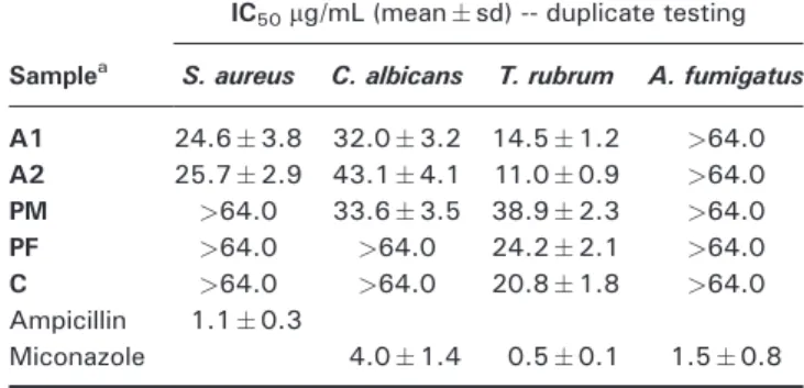
Antibacterial and antifungal assays. These assays have been also performed at the Laboratory of Microbiology, Parasitology and Hygiene (LMPH), Antwerp Univer-sity, Belgium (Cos et al., 2006) against Staphylococcus aureus, Candida albicans, Trichophyton rubrum and Aspergillus fumigatus. IC50 values were determined from five 4-fold dilutions, starting from a maximum concentration of 64mg/mL. Ampicillin was used as refer-ence forS. aureus, while miconazole was used as refer-ence for Candida, Trichophyton and Aspergillus. The impact of toxicity was determined by analyzing the selectivity index (SI), the ratios between the MRC-5SV2 cytotoxic and the antimicrobial IC50 values.
Statistical analysis. For the phenolic composition and antioxidant activity, the assays were carried out in tripli-cate and presented in the figures as mean values with standard deviation. The results were analyzed using one-way analysis of variance followed by Tukey’s HSD test with a = 0.05. The analysis was performed using SPSS v. 18.0 program. The result of this analysis can be found in the figures: samples with the same letter are statistically no different.
For the antiprotozoal, antibacterial and antifungal activity, in each experiment, the 50% of microbial growth (IC50) and human cell growth (CC50) inhibition value was determined from the concentration–response curves, and the results were expressed as the mean
standard deviation of two independent experiments.
RESULTS AND DISCUSSION
Propolis is a bee product with a complex chemical com-position which is largely dependent on its plant origin. Populus bud exudates and Cistus ladanifer L. leaf exudates are documented to be potential resin sources for propolis in Europe (Bankova et al., 2000). In the present study, these plants were abundant in the hive neighborhood, and their phenolic content, antioxidant
and antimicrobial activities were evaluated and com-pared to propolis.
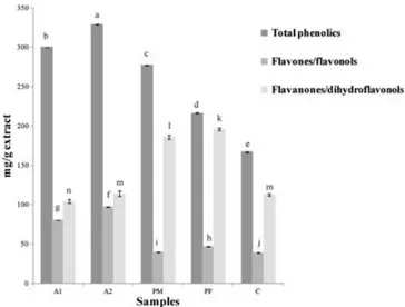
Figure 1 shows the content of total phenolic compounds, flavones/flavonols and flavanones/dihydroflavonols in propolis and in theCistusandPopulusethanolic extracts. All values are statistically different. The highest total phe-nolic content was found in the propolis samples, but there is no great difference between the two samples of dis-tinct geographical origin. The total phenolic content of 329 mg/g for the central propolis (A2) is in line with other studies on Portuguese propolis (Moreira et al., 2008). For the plant extracts, the values ranged from 167 to 278 mg/g with the malePopulussample revealing a higher value thanCistus ladanifer.
For theflavones andflavonols quantified by the alu-minium chloride method, a higher content was also found in propolis compared to the plant extracts. North-eastern and central propolis samples showed values ranging from 81 to 97 mg/g, while the plant ex-tracts contained 39 to 47 mg/g (Fig. 1). The flavanones and dihydroflavonols quantified by the DNP method were present in smaller quantities in the Cistusextract (113 mg/g) compared with propolis andPopulusethanolic extracts (Fig. 1).
Phenolic compounds, due to their hydroxyl groups, are known to act as antioxidants (Rice-Evans et al., 1996). Therefore, the activity of the extracts was evalu-ated with the DPPH free radical scavenging method (Brand-Williamset al., 1995). The EC50values are shown in Fig. 2A with all propolis and plant extracts possessing significant free radical scavenging activity. The extracts of male and female Populus x canadensisbud exudates exhibited the highest activity with EC50 values of 0.014 and 0.015 mg/mL, respectively. These values were close to the standard mixture of pure compounds used in this study (caffeic acid: galangin: pinocembrin, 1:1:1), pointing out the significant activity of these natural extracts. The propolis samples showed an EC50 value around 0.018 mg/mL, while Cistus ladaniferresin presented the lowest scavenging activity (Fig. 2A).
Figure 1. Phenolic composition of propolis and their ethanolic
extracts. A1, North-eastern propolis; A2, Central propolis; PM, Populus x canadensis male; PF,Populus x canadensisfemale; C, Cistus ladanifer. Total phenolics were expressed as caffeic acid: galangin: pinocembrin (1:1:1) equivalents. Flavones/flavonols were expressed as galangin equivalents. Flavanones/dihydroflavonols were expressed as pinocembrin equivalents. In each column, different letters (a–n) mean significant differences between samples (p<0.05)
Fe(III) reduction was used as an indicator of electron-donating activity, which is an important mechanism for phenolic antioxidant action (Yildirim et al., 2001). The reducing power was expressed as caffeic acid: galangin: pinocembrin (1:1:1) equivalents (Fig. 2B). Once again, the best activity was found for the male Populus x canadensis followed by the propolis sample A1 with values of 807 mg/g and 710 mg/g respectively. Sample A2 revealed a very low reduction power, even lower than the plant extracts.
Considering the above, the highest radical scaveng-ing ability or reduction power was not found for the samples with the richer phenolic content, meaning that the observed bioactivity cannot be judged solely on the
basis of the overall phenolic content. In fact, some of the individual phenolic compounds present in the extract can play a more important role in the activity than others. Portuguese propolis phenolic extracts and their potential plant sources were evaluated for in vitro activity against the pathogenic protozoa P. falciparum, L. infantum, T. cruziandT. brucei(Table 1). To assess selectivity of action, cytotoxicity against MRC-5fibroblasts was included.
Activity againstP. falciparumwas found for the prop-olis sample from the central region of Portugal with an IC50 of 8.83mg/mL, while the phenolic extract of male Populus showed the highest SI (SI = 4). Filho et al. (2009) reported an IC50 of 20mg/mL (SI>2.4) for Brazilian green propolis and of 25mg/mL (SI>1.9) for the extract ofBaccharis dracunculifolia, thefloral origin of green propolis. These values are actually in the same range as our results and in fact reveal that our samples A2 and PM show a marginally higher antiplasmodial potential, which may be associated with the richer composition in flavonoids for propolis from Populus (Machado et al., 2007). For an antimalarial ‘hit’, the WHO Special Programme for Research and Training in Tropical Diseases defines an activity criterion to be IC50 <0.2mg/mL with SI >20 (TDR, 2007). Therefore, the relevance of the antimalarial action of our samples requires further research. Propolis toxicity was referred by Marcucci (1995) and Burdock (1998) to be associated with some of the propolis components, namely caffeic acid esters present in the European propolis with origin in the poplar buds. The synergistic effect of all compounds in the entire extract can be responsible for a real loss of activity, while the isolated compounds or fractions of it can indeed reveal a higher activity (Filhoet al., 2009).
ForL. infantum, propolis samples and male and female Populusshowed a similar IC50value of 8.11mg/mL.Cistus ladanifershowed the lowest activity with an IC50value of 32.46mg/mL. Duranet al. (2011) reported antileishmanial activity on two different Turkish propolis samples with IC50 values ranging between 125 and 325mg/mL. Filho et al. (2009) presented IC50 values of 49 and 45mg/mL for a green propolis sample and its plant source B. dracunculifoliaagainstL. donovani.
Our results for the activity againstT. cruzi were very similar between all the samples tested, ranging from 6.16 to 8.59mg/mL (Table 1). Prytzyket al. (2003) and Cunha et al. (2004) tested the activity againstT. cruziin Bulgarian and Brazilian propolis whereby all the extracts showed a lower activity than the Portuguese propolis, with values
Figure 2. Antioxidant activity of propolis and ethanolic plant
extracts (A) against DPPH and (B) reducing power. A1, North-eastern propolis; A2, Central propolis; PM,Populus x canadensis male; PF,Populus x canadensisfemale; C,Cistus ladanifer; S, caffeic acid: galangin: pinocembrin (1:1:1) used as standard. Reducing power was expressed as caffeic acid: galangin: pinocembrin (1:1:1) equivalents. In each column, different letters (a–g) mean significant differences between samples (p<0.05)
Table 1. Antiprotozoal activity againstP. falciparum,L. infantum,T. cruzi,T. bruceiand cytotoxicity in MRC-5fibroblast cells
Samplea
IC50mg/mL (meansd) -- duplicate testing
P. falciparum L. infantum T. cruzi T. brucei MRC-5
A1 30.14.1 8.11.0 6.21.9 1.70.5 12.04.3
A2 8.81.8 8.10.9 7.72.1 3.81.2 9.73.5
PM 10.92.1 8.11.1 7.81.8 5.71.3 38.45.8
PF 28.53.8 8.11.3 7.61.6 5.31.9 33.76.2
C 17.02.5 32.52.7 8.62.0 2.00.4 32.24.5
Chloroquine 0.040.01 - - -
-Miltefosine - 2.40.8 - -
-Benznidazole - - 2.50.6 -
-Melarsoprol - - - 0.0050.001
-Tamoxifen 10.52.5
aSamples: A1: North-eastern propolis; A2: Central propolis; PM:Populus x canadensismale; PF:Populus x canadensisfemale; C:Cistus ladanifer.
ranging between 108.8 and 1065mg/mL for Bulgarian propolis and between 421 and 1437mg/mL for Brazilian propolis. These biological activity variations are likely as-sociated with different chemical compositions presented by the different propolis types. No cytotoxicity tests were performed in that study.
For all antiprotozoal assays, the relatively highest activ-ities were found against T. brucei. Sample A1 was the most active with an IC50 value of 1.70mg/mL (Table 1) which is in the same range as the antitrypanosomal refer-ence drug suramin (Otoguroet al., 2012). Isolation of phe-nolic compounds from propolis can significantly increase the activity:b-phenylethyl caffeate showed a high activity (IC50= 0.013mg/mL; SI = 150), while 2,2-dimethylallyl caffeate exhibited a reduced antitrypanosomal activity (IC50= 12.5mg/mL), demonstrating the potential loss of activity when testing the entire extract (Otoguro et al. (2012).
The activity of the propolis and plant ethanolic extracts was also evaluated against bacteria and fungi (Table 2). The results reveal that the plant extracts do no exhibit relevant antimicrobial activity compared to propolis ethanolic extracts, with exception ofT. rubrum. In gen-eral, both propolis samples revealed a similar antimi-crobial effect, with the highest activity found against T. rubrumand the lowest againstA. fumigatus. Recently, Silvaet al. (2012) verified that propolis from the North and Centre of Portugal was more active againstS. aureus than againstC. albicans, which agrees with our results. In fact, considering that the IC50value forT. rubrumis even lower than forS. aureus, it can be considered a promising result. However, one should also consider the cytotoxic-ity, which was rather high in our study.
In summary, the present study focused on the screening of Portuguese propolis samples and two potential plant sources against pathogenic protozoa, revealing reduced activity and low selectivity. Since propolis has a complex chemical composition, an extract fractionation will be
needed to identify putative active components. Also, these results were evaluated according to criteria set up by the WHO Special Programme for Research and Training in Tropical Diseases, what should be considered by future studies with others propolis samples to enable effective comparisons between scientificfindings.
Acknowledgements
Soraia I. Falcão thanks FCT for the PhD grant SFRH/BD/44855/2008. N. Vale thanks FCT for the Post-Doc grant SFRH/BPD/48345/2008. Thanks also to FCT forfinancial support provided to CIMO (PEst-OE/ AGR/UI0690/2011) and CIQ-UP (PEst-C/QUI/UI0081/2011). Thanks are due to the National Federation of Portuguese Beekeepers, for supplying propolis samples.
Conflict of Interest
The authors report no conflict of interest.
REFERENCES
Amoros M, Sauvager F, Girre L, Cormier M. 1992.In vitroantiviral activity of propolis.Apidologie23: 231–240.
Bankova V, Castro SL, Marcucci MC. 2000. Propolis: recent advances in chemistry and plant origin.Apidologie31: 3–15. Banskota AH, Tezuka Y, Kadota SH. 2001. Recent progress in pharmacological research of propolis. Phytother Res 15: 561–571.
Brand-Williams W, Cuvelier ME, Berset C. 1995. Use of free radical method to evaluate antioxidant activity.LWT28: 25–30. Buckner FS, Verlinde CL, La Flamme AC, Van Voorhis WC. 1996.
Efficient technique for screening drugs for activity against Trypanosoma cruziusing parasites expressing betagalactosidase. Antimicrob Agents Chemother40: 2592–2597.
Burdock GA. 1998. Review of the biological properties and toxicity of bee propolis (propolis). Food Chem Toxicol36: 347–363.
Cardoso SM, Ribeiro M, Ferreira IL, Rego AC. 2011. Northeast Portuguese propolis protects against staurosporine and hydro-gen peroxide-induced neurotoxicity in primary cortical neurons. Food Chem Toxicol49: 2862–2868.
Cos P, Vlietinck AJ, Berghe DV, Maes L. 2006. Anti-infective potential of natural products: How to develop a stronger in vitro‘proof-of-concept’.J Ethnopharmacol106: 290–302. Cunha IBS, Salomão K, Shimizu M,et al. 2004. Antitrypanosomal
activity of Brazilian propolis fromApis mellifera.Chem Pharm
Bull52: 602–604.
Cvek J, Medić-Šarić M, Jasprica I, et al. 2007. Optimiza-tion of an extracOptimiza-tion procedure and chemical characteriza-tion of Croatian propolis tinctures. Phytochem Anal 18: 451–459.
Duran N, Muz M, Culha G, Duran G, Ozer B. 2011. GC-MS analysis and antileishmanial activity of two turkish propolis types. Parasitol Res108: 95–105.
Falcão S, Vilas-Boas M, Estevinho LM, Barros C, Domingues MRM, Cardoso SM. 2010. Phenolic characterization of Northeast Portuguese Propolis: usual and unusual compounds. Anal Bioanal Chem,396: 887–897.
Filho AAS, Resende DO, Fukui MJ, et al. 2009. In vitro antileishmanial, antiplasmodial and cytotoxic activities of phenolics and triterpenoids from Baccharis dracunculifolia D. C. (Asteraceae).Fitoterapia80: 478–482.
Hirumi H, Hirumi K. 1989. Continuous cultivation ofTrypanosoma brucei blood stream forms in a medium containing a low concentration of serum protein without feeder cell layers. J Parasitol75: 985–989.
Machado GMC, Leon LL, De Castro SL. 2007. Activity of Brazilian and Bulgarian propolis against different species ofLeishmania. Mem Inst Oswaldo Cruz102: 73–77.
Makler MT, Ries JM, William JA,et al. 1993. Parasite lactate dehy-drogenase as an assay forPlasmodium falciparumdrug sensi-tivity.Am J Trop Med Hyg48: 739–741.
Marcucci MC. 1995. Propolis: Chemical composition, biological properties and therapeutic activity.Apidologie26: 83–99. Millet-Clerc J, Michael D, Simeray J, Chaumont JP. 1987. Étude
preliminaire des proprietés fongistatiques de la propolis comparées à celles de quelques produits commerciaux.Plant Med Phytother21: 3–7.
Monzote L, Cuesta-Rubio O, Campo Fernandez M, et al. 2012. In vitroantimicrobial assessment of Cuban propolis extracts. Mem Inst Oswaldo Cruz107: 978–984.
Table 2. Antimicrobial activity against Staphylococcus aureus,
Candida albicans,Trichophyton rubrumandAspergillus fumigatus
Samplea
IC50mg/mL (meansd) -- duplicate testing
S. aureus C. albicans T. rubrum A. fumigatus
A1 24.63.8 32.03.2 14.51.2 >64.0
A2 25.72.9 43.14.1 11.00.9 >64.0
PM >64.0 33.63.5 38.92.3 >64.0
PF >64.0 >64.0 24.22.1 >64.0
C >64.0 >64.0 20.81.8 >64.0
Ampicillin 1.10.3
Miconazole 4.01.4 0.50.1 1.50.8
aSamples: A1: North-eastern propolis; A2: Central propolis; PM:
Populus x canadensismale; PF:Populus x canadensisfemale; C: Cistus ladanifer
Moreira L, Dias L, Pereira JA, Estevinho L. 2008. Antioxidant properties, total phenols and polinic analysis of propolis from Portugal.Food Chem Toxicol46: 3482–3485.
Moreira L, Dias T, Dias LG, Rogão M, Da Silva JP, Estevinho LM. 2011. Propolis influence on erythrocyte membrane disorder (hereditary spherocytosis): A first approach.Food Chem Toxicol,49: 520–526. Otoguro K, Iwatsuki M, Ishiyama A, et al. 2012. In vitro antitrypanosomal activity of some phenolic compounds from propolis and lactones from Fijian Kawa (Piper methysticum). J Nat Prod66: 558–561.
Oyaizu M. 1986. Studies on product of browning reaction pre-pared from glucose amine.Jpn J Nutr44: 307–315. Popova M, Bankova V, Butovska D,et al. 2004. Validated methods
for the quantification of biologically active constituents of poplar-type propolis.Phytochem Anal15: 235–240. Prytzyk E, Dantas AP, Salomão K, et al. 2003. Flavonoids and
trypanocidial activity of Bulgarian propolis.J Ethnopharmacol
88: 189–193.
Raz B, Iten M, Grether-Buhler Y, Kaminsky R, Brun R. 1997. The Alamar Blue asssay to determine drug sensitivity of African trypanosomes (T. b. rhodesiense, T. b. gambiense) in vitro. Acta Trop68: 139–147.
Rice-Evans C, Miller NJ, Paganga G. 1996. Structure-antioxidant activity relationships of flavonoids and phenolic acids.Free Radic Biol Med20: 933–956.
Robles C, Bousquet-Mélou A, Garzino S, Bonin G. 2003. Compar-ison of essential oil composition of two varieties of Cistus ladanifer.Biochem Systemat Ecol31: 339–343.
Scaysbrook T, Greenaway W, Whatley FR. 1992. Relation of
“antimicrobial” compounds present in poplar bud exudates to disease resistance by poplars. Z. Naturforsch 47c: 197–200.
Sforcin JM, Fernandes Jr A, Lopes CAM, Bankova V, Funari SRC. 2000. Seasonal effect on Brazilian propolis antibacterial activ-ity.J Ethnopharmacol73: 243–249.
Silva JC, Rodrigues S, Feás X, Estevinho LM. 2012. Antimicrobial activity, phenolic profile and role in the inflammation of propo-lis.Food Chem Toxicol50: 1790–1795.
Singleton VL, Rossi JA. 1965. Colorimetry of total phenolics with phosphomolybdic–phosphotungstic acid reagents.Am J Enol Vitic16: 144–158.
TDR. 2007. Lead discovery for drugs. Business Plan 2008–2013. BL 3.
Trager W, Jensen JB. 1976. Human malaria parasites in continu-ous culture.Science193: 673–675.
Valente MJ, Baltazar AF, Henrique R, Estevinho L, Carvalho M. 2011. Biological activities of Portuguese propolis: Protection against free radical-induced erythrocyte damage and inhibition of human renal cancer cell growth in vitro.Food Chem Toxicol
49: 86–92.
Yildirim A, Mavi A, Kara A. 2001. Determination of antioxidant and antimicrobial activities ofRumex crispusL. extracts.J Agric Food Chem49: 4083–4089.
Zhang X, Hung TM, Phuong PT, et al. 2006. Anti-inflammatory activity of flavonoids from Populus davidiana. Arch Pharm
Res29: 1102–8.