PROCEEDINGS OF VIPIMAGE 2011 - THIRD ECCOMAS THEMATIC CONFERENCE ON COMPUTATIONAL VISION AND MEDICAL IMAGE PROCESSING, OLHAO, ALGARVE, PORTUGAL, 12-14 OCTOBER 2011
Computational Vision and Medical
Image Processing
VipiMAGE 2011
Editors
Joao Manuel R.S. Tavares
&
R.M. Natal Jorge
Faculdade de Engenharia da Universidade do Porta
Porta, Portugal
0
~'~~,;.~~~~""'
Boco Ratan London New York Lei denCRC Press is on imprint of th e
Taylor & Francis Group, an informa business
CRC Press/Balkema is an imprint of the Taylor & Francis Group, mr informa business
© 2012 Taylor & Francis Group, London, UK
Typeset by Vikatan Publishing Solutions (P) Ltd., Chennai, India Printed and bound by CPI Gro up (UK) Ltd, Croydon, CRO 4YY
All rights reserved. No part of this publication or the information contained herein may be reproduced, stored in a retrieval system, or transmitted in any form o r by any means, electronic, mechanical, by photocopying, recording or otherwise, without written prior permission from the publisher.
Although all care is taken to ensure integrity and the quality of this publication and the information herein, no responsibility is assumed by the publishers nor the author for any damage to the property or persons as a result of operation or use of this publication and/or the information contained herein.
Published by: CRC Press/Balkema
P.O. Box 447, 2300 AK Leiden, The Netherlands e-mail: Pub.NL@ taylorandfrancis.com
www.crcpress.com - www.taylorandfrancis.co.uk - www.balkema.nl
ISBN: 978-0-415-68395- 1 (Hbk) ISBN: 978-0-203- 128 18-3 (eBook)
Computational Vision and Medica/Image Processing- Tavares & Natal Jorge (eds) © 2012 Tay/or & Francis Group, London, 978-0-415-68395-1
Table of contents
Preface
Acknowledgements
Invited lectures
Thematic sessions
Scientific committee
Invited lecturers
Towards human-sequence evaluation C. Femande:::, J Gon:::irle::: & X Roca
Finite-context models for image compression
A. J Pinlro
Learning classifier families for object detection and parameter estimation S. Sclaroff, A . Tlumgali, Q. Yuan & V. Ablavsky
Cmllributed papers
Generation of planar radiographs from 3D anatomical models using the GPU
A.S. Cardoso, D. C. Moura & J G. Barbosa
An on-line system for medical and biological image sharing
G. M Pore ides, L.A.P. Ne1•es, L. C. M. de Aquino & G. A. Giraldi
Stapes replacement-different ways to replace its function
F Genii!, C. Gm·be, lvf. Parente, P. Martin.\; RN. Jorge & J Para
Vision-based hand segmentation techniques for human-robot interaction for real-time applications
P. Trigueiros, F Ribeiro & G. Lopes
Database implementation for clinical and computer assisted diagnosis of dermoscopic images
B. S.R Amorim, T. Mendonra. A.R.S. Mm·cal, JS. Marques & J Ro:::eira
The finite element analysis of skull deformation after correction of scaphocephaly
W Wolmlski, M . G:::ik, E. Kawlewska, D. Larysz & P. Larysz
Modeling and simulation of trigonocephaly correction with use of Finite Elements Method
M. G:::ik, W Wolmlski, E. Kawlewska, K Kawlewski & D. Lmysz
Computer-aided diagnosis of dementia using medical imaging processing and artificial neural networks
G. Gavidia, R. Lope::: & E. Soudah
Automated extraction of the Femoral Shaft Axis and its distal entry point from full and reduced 3D models
S. Van Cawer; M. De Beule, A. Van Haver; P. Verdonk & B. Verlregghe
Facial expression recognition using MPEG-4 FAP-based 3D MMM
H. Ujir & M. Spann
V
xi
xiii
XV
xvii
xix
3
9
15
19
23
27
31
37
43
47
51
57
I for olds liVer ·ical ows and >ni-d at cro. :in g. ring
in a
~z ag tical oral ech. 1an- pia-1. & 1idic
Computational Vision and Medica/Image Processing- Tavares & Natal Jorge (eds)
© 2012 Taylor & Francis Group, London, ISBN 978-0-415-68395-1
Flow visualization of trace particles and Red Blood Cells
in a microchannel with a diverging and converging bifurcation
V.
Leble & C. FernandesESTiG, IPB. C Stc1. Apolonia, Braganca, Portugal
R. Dias &
R.
LimaESTiG. IPB. C Sta. Apolonia, Braganca, Portugal CEFT. FE UP, R. D1: Roberto Frias, Porta, Por111gal
T.
Ishikawa & Y. ImaiDepartment of Bioengineering & R obo ti C.\~ Grad. School Engineering. Tohoku Unil'ersity, Aoba, Sendai, Japan
T. Yamaguchi
Department of Biomedical Engineering. Grad. School Engineering. Toho/w Unil'ersity, Aoba, Sendai, Japan
ABSTRACT: This paper aims to investigate the eiTect of both diverging and converging bifurcations on the flow behaviour of Pure Water (PW) and Red Blood Cells (RBCs). A confocal micro-PTV system is used to visualize and measure the flow characteristics of the working fluids. The results show no forma-tion of a Cell-Free Layer (CFL) around the apex of the bifurcaforma-tion.ln contrast, there is a clear formaforma-tion of a triangular CFL just downstream of the confluence apex. As a result, this triangular CFL seems to play an important role on the in vitro blood flow characteristics at this region.
I INTRODUCTION
Blood flow behaviour in both in llii'O and in vitro
environments has been investigated for several
years [1-4]. However, studies performed by Suzuki
et al. [3] and Pries, et al . [4] have found conflicting
results between in vii'O and in 1•itro experiments
with respect to the blood rheological properties.
Potential causes for the observed in vivo/in vitro
discrepancies are the eiTect of the endothelial sur-face layer, the presence of white blood cells and the complex microvascular networks composed by diverging and converging bifurcations [2]. In order to better understand the observed dis-crepancies we need to investigate in more detail the eiTect of both diverging and converging bifur-cations on the rheological properties of blood. Therefore, the aims of the present paper is to visualize and measure the flow characteristics of both trace particles suspended in pure water and
in vitro blood in a diverging and converging bifur-cation. The experimental flow visualizations and measurements will be performed by means of a confocal system combined by image analysis tech-niques from ImageJ.
2 MATERIALS AND METHODS
2.1 Working fluids and microchannel geometl)'
Two working fluids were used in this study: pure water (PW) with fluorescent trace particles of
I J.l.m and Dextran 40 (Dx-40) containing about
14% (14Hct) of human RBCs. The washed RBCs were fluorescently labelled with a lipophilic carbo-cyanine derivative dye, chloromethylbenzamido (CM-Oil, Molecular Probes), using a previously described procedure [5].
The polydimethylsiloxane (PDMS) microchan-nels used in this study were fabricated using a soft lithography technique [6] and consist of a diverg-ing bifurcation and convergdiverg-ing bifurcation (also known as confluence). Fig. I shows the dimensions of both diverging and converging bifurcations used in the present study.
2.2 Experimental set-up
a)
b)
Figure I. Dimensions of the a) diverging and b) con-verging bifurcation used in this study. The channel dimensions arc in J.lm.
laser with an excitation wavelength of 532 nm and a high-speed camera. The PDMS microchannel was placed on the microscope stage with a sur-rounding temperature of about 37"C. By using a syringe pump the flow rate of the working flu-ids could be controlled by adjusting the injection speed. The flow rates were kept constant and approximately the same for both fluids. Hence,
the Reynolds number (Re) used for PW and in
vitro blood was Re ~ 0.04 and Re = 0.008, respec-tively. For the Re used in this study, the flow of PW inside the microchannel can be assumed as a steady, laminar flow of a Newtonian, incompress-ible fluid (Stokes flow). Therefore, change in the
flow rate of PW to achieve the Re of ill vitro blood
will not influence the trajectories of trace particles. Thus, the comparison of trajectories of both fluids is applicable. More detailed information about the experimental set-up, microchannel fabrication and RBC labelling used in this study can be found else-where [I , 5, 6].
2.3 Image mw~ v sis
All the confocal images were recorded aro und the middle of the PDMS microchannel with a resolu-tion of 640 x 480 pixcls, at a rate of lOO frames/s. The recorded images were transferred to the com-puter and then evaluated in the image processing
210
program ImageJ (NIH) [7] by using the manual
tracking MtrackJ plugin [8] and automatic
Parti-cleTracker 2D plugin [9] to track the trace particles
in PW and RBCs in D x 40, respectively.
3 RESULTS AND DISCUSSION
In this section we presen t the flow visualizations results and investigate the effect of both diverging and converging bifurcation on the trace particles in PW (see Figs. 2a and 3a) and on labelled RBCs (see Figs. 2b and 3b).
For the case of trace particles in PW (Figs. 2a and 3a) we observed that the trajectories were almost symmetric and do not present so many fluc-tuations for both geometries. These results are con-sistent with the Stokes flo w regime. In contrast, for the case of labelled RBCs the trajectories a re more asymmetric when compared with PW trajectories. Additionally, we can also observe several fluctua-tions on their trajectories.
From Fig. 3a we also observed that the trace particles tend to flow very close to the inner walls
...
,
a)b)
Figure 2. Trajectories in a d iverging bifurcation of a) fluorescent particles in PW \ind b) labelled RBCs in
.
-
-
-
---
...
,
-
-n)
• - - -
....
- q - - --b)
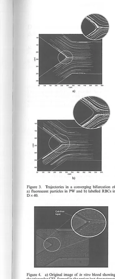
Figure 3. Trajectories in a converging bifurcation of a) nuorescent particles in PW and b) labelled RBCs in D x 40.
Figure 4. a) Original image of iu vitro blood s howing the triangular CFL formed in the region just downstream of the connuence apex.
and as a result they tend to flow in the centre of the microchannel, just downstream of the conflu-ence apex. However, for the case of labelled RBCs we could not measure any trajectory passing in this centre region (see Fig. 3b). This is due to the exist-ence of a cell-free layer (CFL) in both inner walls and a consequent formation of a triangular CFL in the region of the confluence apex (see Fig. 4). As this triangular CFL seems to play an important
role on the in vitro blood flow characteristics, a
detailed quantitative study, to clarify the CFL efTect in the velocity profiles, is currently under way.
ACKOWLEDGEMENTS
The authors acknowledge the financial support
provided by: PTDC/SAU-BEB/ 108728/2008,
PTDC/SAU-BEB/105650/2008 and PTDC/
EME-MFE/099109/2008 from the FCT (Science and Technology Foundation) and COMPETE, Portugal.
REFERENCES
[I] Lima, R., lshikawa, T., lmai, Y., Takeda, M., Wada, S. & Yamaguchi, T. Radial dispersion of red blood cells in blood nowing through glass capillar-ies: role of haematocrit and geometry. Joumal of Biomedrauics 41, 2188- 2196, 2008.
[2] Maeda, N. Erythrocyte rheology in microcircula-tion. Japanese Joumal t!l Plzysiology 46. 1- 14, 1996. [3] Suzuki, Y., Tatcishi , N. , Soutani, M . and Macda, N. Deformation of erythrocytes in microvessels and glass capillaries: effects of erythrocyte dcformabil-ity. Microcirculatimr 3, 49- 57, 1996.
[4] Pries, A. , Secomb, T., et al, Resistance t o blood now in microvcssels in vivo. Circulation R esearch 75, 904-9 15, 1994.
[5] Lima, R ., lshikawa, T., lmai, Y., Takcda, M., Wada, S. and Yamaguchi, T. Measurement of indi-vidual red blood cell motions under h igh hcmatocrit conditions using a confocal micro-PTV system.
Auuals of Biomedical E ngineering, 37, 1546- 59, 2009.
[6] Lima, R. , Femandes, C.S. et al., "Microscalc now dynamics of red blood cells in microchanncls: a n experimental and numericnl analysis", In: Tavares and Jorge (Eds), Computational Vision and Medi-cal Image Processing: Recent Trends, Springer, 19, 297- 309, 20 11.
[7] Abramofl~ M ., Magelhaes, P. and Ram, S. Image Processing with ImageJ. Bioplrot ouics lutemarimral
7 ' 11 ' 36-42, 2004.
[8] Meijering, E. , Smal, L and Danuser, G. "Tracking in Molecular Bioimaging". IEEE Signal Processing Maga::ine. 3, 23, 46- 53, 2006. \ [9] Sbalzarini, I. F. and Koumouts akos, P. Feat tire Point