Introduction
The remarkable movements of chromosomes in mitosis are initiated, controlled and monitored by kinetochores, which are structures that form the interface between the chromosomes and the microtubules of the mitotic spindle. Kinetochores of animal cells can be subdivided into two regions. The inner kinetochore normally forms on highly repetitive DNA sequences and assembles into a specialized form of chromatin that persists throughout the cell cycle. The outer kinetochore is a proteinaceous structure with many dynamic components that assembles and functions only during mitosis. Kinetochore functions include attachment of chromosomes to the spindle microtubules, monitoring those attachments, activating a signalling (checkpoint) pathway to delay cell-cycle progression if defects are detected and helping to power the movements of chromosomes on the spindle. We begin by discussing the molecular organization and assembly pathway of the kinetochore.
Microtubules are metastable polymers of α- and β-tubulin that switch between phases of growth and shrinkage, a phenomenon known as ‘dynamic instability’ (Mitchison and Kirschner, 1984). We discuss below how the highly dynamic nature of microtubule behaviour is integrated with kinetochore function to move and segregate chromosomes. More details about spindle checkpoint function, spindle assembly mechanisms and the dynamics and mechanics of the microtubule plus end are reviewed elsewhere (Musacchio and Hardwick, 2002; Sharp et al., 2000; Compton,
2000; Kapoor and Compton, 2002; Howard and Hyman, 2003). Here we focus on the animal kinetochore and in particular the interface between the outer kinetochore domain and spindle microtubules (for reviews, see Fukagawa, 2004; Amor et al., 2004). [For reviews of budding and fission yeast kinetochores and plant kinetochores, see Yu et al. and others (Yu et al., 2000; Kitagawa and Hieter, 2001; Cheeseman et al., 2002b; Biggins and Walczak, 2003; McAinsh et al., 2003; Cleveland et al., 2003; Westermann et al., 2003; Houben and Schubert, 2003; Hall et al., 2004).]
Animal kinetochore structure
The kinetochore (Fig. 1) is composed of several distinct layers that were first observed by conventional fixation and staining methods for electron microscopy (Brinkley and Stubblefield, 1966; Jokelainen, 1967; Comings and Okada, 1971) (reviewed by Rieder, 1982), and more recently by fast freezing/freeze substitution (McEwen et al., 1998). Innermost is an inner plate, a chromatin structure containing nucleosomes with at least one specialized histone, auxiliary proteins and DNA. The makeup and organization of this DNA remains one of the least understood aspects of the kinetochore in animal cells. The inner plate exists as a discrete heterochromatin domain throughout the cell cycle. Outside this is an outer plate composed primarily, if not solely, of protein (Cooke et al., 1993). This structure forms on the surface of the chromosome The kinetochore is a control module that both powers and
regulates chromosome segregation in mitosis and meiosis. The kinetochore-microtubule interface is remarkably fluid, with the microtubules growing and shrinking at their point of attachment to the kinetochore. Furthermore, the kinetochore itself is highly dynamic, its makeup changing as cells enter mitosis and as it encounters microtubules. Active kinetochores have yet to be isolated or reconstituted, and so the structure remains enigmatic. Nonetheless, recent advances in genetic, bioinformatic and imaging technology mean we are now beginning to understand how kinetochores assemble, bind to microtubules and release them when the connections made are inappropriate, and also how they influence microtubule behaviour. Recent work has begun to elucidate a pathway of kinetochore assembly in animal cells; the work has revealed that many
kinetochore components are highly dynamic and that some cycle between kinetochores and spindle poles along microtubules. Further studies of the kinetochore-microtubule interface are illuminating: (1) the role of the Ndc80 complex and components of the Ran-GTPase system in microtubule attachment, force generation and microtubule-dependent inactivation of kinetochore spindle checkpoint activity; (2) the role of chromosomal passenger proteins in the correction of kinetochore attachment errors; and (3) the function of microtubule plus-end tracking proteins, motor depolymerases and other proteins in kinetochore movement on microtubules and movement coupled to microtubule poleward flux.
Key words: Kinetochore, Microtubule, CENP proteins, Ndc80, Dynein, Centromere, Chromosomal passengers, Aurora B, Ran Summary
The dynamic kinetochore-microtubule interface
Helder Maiato1, Jennifer DeLuca2, E. D. Salmon2,* and William C. Earnshaw3,*
1Laboratory of Cell Regulation, NYSDH – Division of Molecular Medicine, Wadsworth Center, Empire State Plaza, PO Box 509, Albany, NY
12201-0509, USA
2Department of Biology, CB#3280, 607 Fordham Hall, University of North Carolina, Chapel Hill, NC 27599, USA
3Chromosome Structure Group, Wellcome Trust Centre for Cell Biology, Institute of Cell and Molecular Biology, University of Edinburgh, Mayfield
Road, Edinburgh, EH9 3JR, UK
*Authors for correspondence (e-mail: [email protected]; [email protected])
at about the time of nuclear envelope breakdown (Brinkley and Stubblefield, 1966; Ris and Witt, 1981; McEwen et al., 1993). The outer plate of vertebrate kinetochores has about 20 end-on attachment sites for the plus ends of microtubules (termed kinetochore microtubules, kMTs); the outer plate of budding yeast has only one end-on attachment site. The outermost regions of the kinetochore form a fibrous corona that can be visualized by conventional electron microscopy, and usually only in the absence of microtubules. This is composed of a dynamic network of resident and transient components that are involved in the spindle checkpoint and the attachment of microtubules and regulation of their behaviour.
Animal kinetochores form by the assembly of proteins onto a (usually) repetitive DNA sequence, yielding a modular structure that forms a single inner plate as a consequence of chromatin higher-order folding (Zinkowski et al., 1991) (reviewed by Brinkley et al., 1992). Duplicated kinetochores of sister chromatids are first seen to separate from one another during mid-late G2 phase in mammalian cultured cells (Brenner et al., 1981) and at the beginning of prophase in
Caenorhabditis elegans(Moore and Roth, 2001). These pre-kinetochores acquire a mature laminar structure after nuclear envelope breakdown (Moroi et al., 1981; Roos, 1973) in a process that requires components of the inner plate (Tomkiel et al., 1994) (reviewed by Pluta et al., 1995).
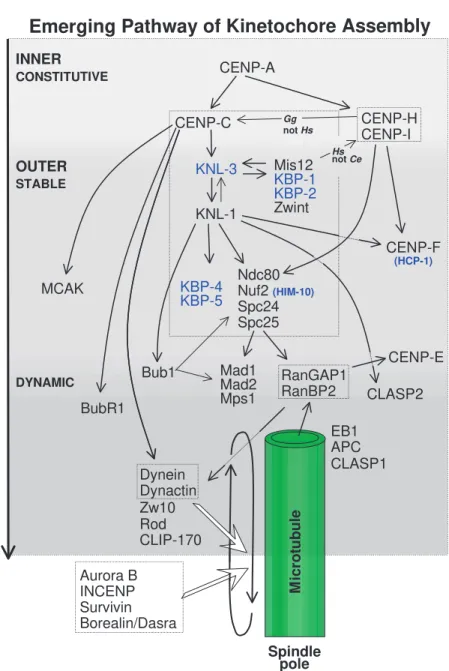
The molecular pathway of kinetochore assembly in higher eukaryotes has been studied in work using gene knockouts in mice and in cultured chicken cells, as well as RNA interference (RNAi) in C. elegans, Drosophilaand human cells. No simple linear pathway can describe the data obtained to date (Fig. 2).
The network of interactions shown in Fig. 2 represents a work in progress; many linkages remain to be discovered and details are likely to change.
The earliest protein known to bind during kinetochore assembly, CENP-A (Cse4 in Saccharomyces cerevisiae), is a specialized isoform of histone H3 (Palmer et al., 1991). CENP-A is required for the recruitment of the inner kinetochore proteins CENP-C, CENP-H and CENP-I/MIS6 (Howman et al., 2000; Oegema et al., 2001; Van Hooser et al., 2001; Fukagawa et al., 2001; Goshima et al., 2003). The relative positions of these proteins in the CENP-A-dependent pathway are not yet clear. CENP-C targeting requires CENP-H in chicken cells, but is independent of CENP-I/MIS6 in human cells (Fig. 2).
In C. elegans, CENP-A and CENP-C direct the assembly of KNL-1 and KNL-3, which colocalize with CENP-C in the inner kinetochore starting during prophase (Desai et al., 2003; Cheeseman et al., 2004). The KNL proteins are required for assembly of multiple components of the outer kinetochore, and the formation of a functional kinetochore-microtubule interface. C. elegans proteins whose targeting depends on KNL-1 include outer plate proteins of the Ndc80 complex (Ndc80/Hec1, Nuf2, Spc24 and Spc25), KNL-binding proteins KBP-1 to KBP-5, MIS12, CENP-F and the checkpoint protein BUB1 (Desai et al., 2003; Cheeseman et al., 2004).
In metazoans, the recruitment of many outer kinetochore proteins is also under the control of the CENP-A-dependent pathway. A homologue of KNL-1, AF15q14, has recently been identified in human cells (Cheeseman et al., 2004), which suggests that the assembly pathway is likely to be conserved.
CENTROMERIC HETEROCHROMATIN
KINETOCHORE
CENTROMERE
MT attachment,
regulation of MT dynamics, & checkpoint signalling INNER PLATE
CENP-A CENP-C CENP-G CENP-H CENP-I/hMis6 CeKNL-1 hMis12
Kinetochore assembly & size determination CENP-B
MCAK INCENP Aurora B Survivin Borealin/Dasra B ICIS
Chromatid pairing, structural support, & MT attachment error correction
INTERZONE
3F3/2 antigens
Tension receptors & checkpoint signalling
FIBROUS CORONA
Ndc80/Hec1 Nuf2 Spc24 Spc25 BUB1 BUBR1 BUB3 MPS1 MAD1 MAD2 Cdc20 CENP-E
RanGAP1 RanBP2 CENP-F Plk PP1 Zw10 Zwint-1 Rod Dynein LIS1 CLASPs CLIP-170 EB1 APC OUTER PLATE
OUTER
KINETOCHORE
INNER
KINETOCHORE
MT PLUS ENDS Microtubule
Interestingly, AF15q14 is a fusion partner for MLL in human leukaemias (Hayette et al., 2000). Other vertebrate and Drosophila kinetochore proteins known to require the CENP-A-dependent pathway for targeting include Polo-like kinase (PLK), ROD, ZW10, ZWINT-1, the microtubule motor dynein, the kinesin motor CENP-E, the spindle checkpoint proteins (MPS1, BUBR1, MAD1 and MAD2) and the non-motor microtubule-associated proteins (MAPs) CLASPs (Wordeman et al., 1996; Blower and Karpen, 2001; Van Hooser et al., 2001; Liu et al., 2003; Desai et al., 2003; Stucke et al., 2004). The spindle checkpoint proteins BUBR1 and BUB1 seem to play a central role as recruiters of the other checkpoint components, but their position in the assembly pathway remains controversial (Sharp-Baker and Chen, 2001; Chen, 2002; Johnson et al., 2004).
Also controversial is the position of MIS12 in the pathway (Goshima et al., 1999). MIS12 binds CENP-A/Cse4 and is required for chromosome biorientation (see below) and the generation of centromere tension in budding yeast (Pinsky et al., 2003). In humans, MIS12 and CENP-A targeting appears to be mutually independent, and MIS12 is required for the subsequent assembly of CENP-I/MIS6 and CENP-H (Goshima et al., 2003). By contrast, C. elegansMIS12 functions downstream of CENP-A (Cheeseman et al., 2004). In human cells, MIS12 is an essential kinetochore component; however, in C. elegans, kinetochores ultimately assemble in the absence of MIS12, although the protein is required for the process to be timely and efficient (Goshima et al., 2003; Cheeseman et al., 2004).
Incorporation of the inner centromeric KinI kinesin MCAK (also known as XKCM1) is dependent on CENP-A and CENP-C but not on KNL-1 in C. elegans, indicating an early bifurcation of the pathway (Desai et al., 2003). Also within the inner centromere before anaphase are the chromosomal passenger proteins, which include the aurora B kinase and its binding partners INCENP, survivin and borealin/dasra B (reviewed by Carmena and Earnshaw, 2003; Gassmann et al., 2004). These inner centromere proteins assemble through a pathway independent of CENP-A (Oegema et al., 2001).
Kinetochore components can be grouped into three classes on the basis of their localization
throughout the cell cycle. Constitutive components, such as CENP-A, CENP-C, CENP-H and CENP-I, are bound to kinetochore-associated chromatin throughout the cell cycle. Other components associate with kinetochores only starting in prophase. Kinetochore proteins can also be grouped by whether their kinetochore concentrations remain constant or vary during mitosis and by whether they turnover slowly (stable) or rapidly (dynamic) at their kinetochore binding sites. Proteins that remain nearly constant in level from prophase through late anaphase include the constitutive components of the inner plate and the stable outer kinetochore components,
such as the Ndc80 complex (Wigge and Kilmartin, 2001; DeLuca et al., 2002), KNL/KBP proteins (Cheeseman et al., 2004), MIS proteins (Cheeseman et al., 2004) and CENP-F (Rattner et al., 1993; Liao et al., 1995). Together with the constitutive components, these proteins appear to form the core kinetochore inner- and outer-plate structures. Cheeseman et al. have recently isolated large multiprotein complexes containing CENP-C, members of the Ndc80 complex, plus KNL, KBP and MIS proteins from both C. elegansand human cultured cells (Cheeseman et al., 2004). This promises to be an exciting breakthrough in characterization of the animal kinetochore.
Mad1 Mad2 Mps1 CENP-A
BubR1
CENP-C
MCAK Ndc80Nuf2(HIM-10) Spc24 Spc25
CLASP2 Mis12
KBP-1 KBP-2
Zwint
CENP-H CENP-I
CENP-F (HCP-1)
INNER
DYNAMIC
OUTER
EB1 APC
Dynein Dynactin Zw10 Rod CLIP-170
Spindle pole
M
ic
ro
tu
b
u
le
Emerging Pathway of Kinetochore Assembly
Bub1
KNL-1
RanGAP1 RanBP2
CENP-E
Aurora B INCENP Survivin Borealin/Dasra
CLASP1
KNL-3
KBP-4 KBP-5
Hs notCe
STABLE
Gg notHs CONSTITUTIVE
Dynamic components that change in concentration at kinetochores during mitosis include the microtubule motors CENP-E and dynein (plus its targeting components ZW10 and ROD), and the spindle checkpoint proteins (e.g. MAD1, MAD2, BUBR1 and Cdc20). These proteins assemble at high concentrations at kinetochores in the absence of microtubules and are reduced in concentration by interactions with spindle microtubules and in particular by kMT formation (Hoffman et al., 2001). By metaphase, CENP-E, BUB3 and BUB1 levels are decreased 3-4-fold relative to those at unattached kinetochores, whereas dynein/dynactin, MAD1, MAD2 and BUBR1 levels fall >10-100-fold (King et al., 2000; Hoffman et al., 2001; Howell et al., 2004; Shah et al., 2004).
The dynamics of protein turnover at kinetochores has been measured by fluorescent recovery after photobleaching (FRAP) of green fluorescent protein (GFP) fusion proteins expressed in cells. CENP-A, CENP-I, CENP-H, Nuf2, MAD1, BUB1 and about 50% of MAD2 are relatively stable components that turn over very slowly over 10 minutes. By contrast, 50% of MAD2 and most of BUB3, BUBR1, Mps1 and Cdc20 are very dynamic components with residence half-lifes of 30 seconds or less (Howell et al., 2000; Kallio et al., 2002a; Howell et al., 2004; Shah et al., 2004). This fast turnover might have a role in the ability of unattached kinetochores to inhibit activation of the anaphase-promoting complex/cyclosome (APC/C) in the cytoplasm (Musacchio and Hardwick, 2002; Cleveland et al., 2003). Importantly, such studies must always be interpreted cautiously and with the caveat that the tagged proteins have typically not been shown to have full biological function.
The above changes in protein concentration and dynamics at kinetochores are partially mediated by microtubules and probably also depend on interactions with the core CENP antigens. Injection of anti-centromere antibodies (ACAs) during G2 phase results in the assembly of kinetochores that look near normal in the presence of colcemid (which blocks spindle assembly) but lack a defined laminar structure if microtubules are present (Bernat et al., 1991). These disrupted kinetochores can still bind to microtubules but appear unable to support chromosome movement. Cytoplasmic dynein associates with kinetochores following the injection of ACA only if microtubule assembly is prevented: in the presence of microtubules, kinetochores of ACA-injected cells lack dynein (Wordeman et al., 1996). Subsequent work has shown that, when ATP is partially depleted in cells, the proteins of the dynamic component, but not members of the core kinetochore structure, are stripped from kinetochores by dynein-mediated transport along microtubules to the spindle poles (Howell et al., 2001). This suggests that there is constant streaming of dynamic components between kinetochores and centrosomes along spindle microtubules. When detectable at kinetochores, MAD1/MAD2 and ROD are seen to cycle continuously between kinetochores and spindle poles in a dynein-dependent manner (Howell et al., 2001; Wojcik et al., 2001; Basto et al., 2004).
Whereas the dynamic outer kinetochore proteins are depleted from the kinetochore when microtubules attach (Hoffman et al., 2001), other components, including EB1, APC and the Ran pathway proteins RanGap1 and RanBP2 (see below), associate with kinetochores only when microtubules are attached (Tirnauer et al., 2002; Kaplan et al., 2001; Fodde
et al., 2001; Joseph et al., 2002; Salina et al., 2003). This might be part of a kinetochore mechanism that recognizes the plus ends of microtubules, ensures they are properly attached and regulates their dynamics while they remain attached (see below).
Initial encounters between kinetochores and microtubules
After nuclear envelope breakdown in animal cells, highly dynamic centrosome-nucleated microtubules continuously probe the cytoplasm with their plus ends to search and capture chromosomes (Kirschner and Mitchison, 1986; Hill, 1985; Holy and Leibler, 1994). Microtubules that encounter a kinetochore become stabilized, whereas those that do not soon depolymerize (Hayden et al., 1990). A single microtubule emanating from the centrosome is sufficient to initiate chromosome alignment, which begins with rapid polewards movement of the captured chromosome involving lateral interactions of the kinetochore with the surface of the microtubules (Rieder et al., 1990). This movement is likely to be mediated by the minus-end-directed motor activity of cytoplasmic dynein (Vaisberg et al., 1993; Sharp et al., 2000; Echeverri et al., 1996), which is highly concentrated at unattached kinetochores (Pfarr et al., 1990; Steuer et al., 1990; Wordeman et al., 1991; Hoffman et al., 2001) (reviewed by Banks and Heald, 2001). Polewards movement slows as chromosomes acquire kMTs and movement becomes governed by changes in the lengths of kMTs (see below).
The high concentrations of dynein at unattached kinetochores correlate with the need for the kinetochore to recruit kMTs. Dynein is released from kinetochores as they acquire their full complement of kMTs (Hoffman et al., 2001; King et al., 2000) and, in mammalian tissue culture cells, is required for inactivation of the spindle checkpoint, but not for chromosome alignment at the spindle equator, normal numbers of kMTs, or anaphase A chromosome segregation (Howell et al., 2001b). There is no evidence for dynein occurring in higher plants or within the nucleus of yeast, but minus-end-directed kinesins might compensate for the lack of dynein function.
CENP-E is a very large kinesin-like protein that is associated with the fibrous corona of mammalian kinetochores from prometaphase through anaphase and is also implicated in the initial encounters with microtubules (Cooke et al., 1997; Yao et al., 1997). Chromosomes lacking CENP-E at their kinetochores often show defects in alignment and a few remain chronically mono-oriented (attached to a single pole) even though most eventually align successfully at a metaphase plate (Schaar et al., 1997; Wood et al., 1997; McEwen et al., 2001; Putkey et al., 2002; Weaver et al., 2003). CENP-E is involved in anchoring kinetochores to shortening microtubules in vitro (Lombillo et al., 1995), and CENP-E-depleted chromosomes have reduced numbers of microtubules bound to their kinetochores (McEwen et al., 2001; Putkey et al., 2002).
initiated from the centromere/kinetochore region, elongate away from the chromosome, and subsequently have their minus ends captured and pulled into the spindle pole/centrosome region by interactions with polar spindle microtubules (Khodjakov et al., 2003). Although we have known for many years that kinetochores (or their proximal centromere regions) have the potential to nucleate microtubules in vitro and in vivo after recovery from microtubule poisons (Telzer et al., 1975; Witt et al., 1980; De Brabander et al., 1981; Mitchison and Kirschner, 1985a), the significance of these results had been questioned. How the kinetochore/centromere region initiates kinetochore fibre formation and how frequently this occurs are important issues to be addressed since this mechanism may contribute significantly not only to initial kMT formation but also to how kinetochores correct attachment errors and regulate movement along kMTs (see below).
The role of the Ndc80 complex in interactions between kinetochores and microtubules
The kinetochore confers unique properties upon its attached microtubules. kMTs are much more resistant to depolymerization induced by cold treatment, high hydrostatic pressure or exposure to calcium (Brinkley and Cartwright, 1975; Salmon et al., 1976; Mitchison et al., 1986) compared with microtubules that have unattached plus ends. Furthermore, kinetochore microtubules turn over much more slowly than astral and spindle microtubules that have free plus ends in vivo (Mitchison et al., 1986), and microsurgical detachment of a chromosome from kinetochore microtubules leads to their rapid depolymerization (Nicklas and Kubai, 1985). In vitro, kinetochores of isolated chromosomes can stabilize the ends of purified microtubules (Mitchison and Kirschner, 1985b), although, under certain circumstances, they can promote microtubule dynamics (Hyman and Mitchison, 1990). How kinetochores stabilize attached microtubules is only now starting to be understood.
Once it became clear that dynein and CENP-E are not essential for kMT formation, the search for other proteins crucial for stable kMT attachment began. Pioneering genetic studies in budding and fission yeast revealed the importance of the Ndc80 protein complex for kMT attachment (Wigge and Kilmartin, 2001; He et al., 2001; Westermann et al., 2003; De Wulf et al., 2003). The budding yeast Ndc80 complex has four components: Ndc80p, Nuf2p, Spc24p and Spc25p. Yeast mutants lacking components of the Ndc80 complex exhibit loss of kinetochore-microtubule attachment without a complete loss of kinetochore structure (Wigge and Kilmartin, 2001; He et al., 2001). By contrast, mutants that completely abolish kinetochore assembly, such as Ndc10 mutants in budding yeast (Goh and Kilmartin, 1993), are deficient not only in microtubule attachment but also in their checkpoint response, presumably because kinetochores serve as a platform for organizing the response. The Ndc80 complex is highly conserved and has been identified in S. pombe, C. elegans,
Xenopus, chickens and humans (Wigge and Kilmartin, 2001; He et al., 2001; Nabetani et al., 2001; Howe et al., 2001; DeLuca et al., 2002; Martin-Lluesma et al., 2002; McCleland et al., 2003). The human homologue of Ndc80, Hec1 (for ‘highly enhanced in cancer cells 1’), has been shown to be
important for chromosome alignment and mitotic progression and to interact with components of the cohesin and condensin complexes (Zheng et al., 1999).
Several recent studies have shown that the Ndc80 complex is crucial for the stable kinetochore-microtubule attachments that are needed to sustain the centromere tensions involved in achieving proper chromosome alignment in higher eukaryotic cells (Howe et al., 2001; DeLuca et al., 2002; Martin-Lluesma et al., 2002; McCleland et al., 2003; Hori et al., 2003; Desai et al., 2003; Bharadwaj et al., 2004; McCleland et al., 2004; Cheeseman et al., 2004). Cells that have impaired Ndc80 complex function (induced by RNAi, gene disruption, or antibody microinjection) have elongated spindles, exhibit loss of tension across sister kinetochores, fail to align their chromosomes (Martin-Lluesma et al., 2002; DeLuca et al., 2002; McCleland et al., 2003; Hori et al., 2003; Desai et al., 2003; Bharadwaj et al., 2004; McCleland et al., 2004) and have few or no kMTs at temperatures low enough to depolymerize non-kMTs selectively (DeLuca et al., 2002; McCleland et al., 2004).
Microinjection of antibodies to Nuf2, Spc24 or Spc25 disrupts or prevents metaphase chromosome alignment, but kinetochores exhibit transient movements along the spindle axis (McCleland et al., 2003; McCleland et al., 2004), as do chromosomes in cultured cells in which levels of Nuf2 or Ndc80/Hec1 are reduced >90% by short interfering (si)RNA (J.D., Y. Dong, P. Hergert, J. Strauss, J. Hickey, E.D.S. and B. McEwen, unpublished). Either transient end-on attachments or lateral microtubule interactions with the kinetochore may produce these transient movements or move chromosomes towards one or the other pole in anaphase (McCleland et al., 2003). In support of this interpretation, electron microscopy studies have found rare microtubule plus-end binding by kinetochores in HeLa cells in which hNuf2 is knocked down by RNAi (J.D. et al., unpublished).
kinetochores and the spindle checkpoint is inactivated (Meraldi et al., 2004).
Disassembly of spindle microtubules by nocodazole results in a substantial recovery of MAD1, MAD2 and dynein at Ndc80/Hec1-depleted kinetochores (DeLuca et al., 2003; Bharadwaj et al., 2004). We hypothesize that interactions with the Ndc80 complex might prevent protein stripping from non-attached kinetochores by dynein-mediated transport along kMTs (Howell et al., 2001; DeLuca et al., 2003; Basto et al., 2004).
The vertebrate Ndc80 complex clearly plays roles in chromosome alignment, kinetochore-microtubule attachment and microtubule-dependent control of MAD1/MAD2 and dynein complexes at kinetochores. Interestingly, there is no evidence that the Ndc80 complex itself directly interacts with microtubules. In yeast, kinetochore-microtubule attachment requires the Dam1-DASH-DDD complex. Some members of this complex bind directly to microtubules, whereas others bind to the Ndc80 complex (Westermann et al., 2003; Courtwright and He, 2002; De Wulf et al., 2003). Thus, the Dam1-DASH-DDD complex could be an essential adaptor between kinetochores and microtubules. However, no animal equivalent of this complex has been identified, and this remains a focus of active investigation.
The role of Ran in kinetochore assembly and function
The small GTPase Ran was first studied as a factor required for nuclear trafficking of proteins (Moore and Blobel, 1994), its function being to differentiate the nuclear interior from the cytoplasm (Gorlich and Kutay, 1999). In the nucleus, Ran-GTP binds to complexes of an importin with import cargo, causing the latter to be released. Nuclear levels of Ran-GTP are high because nuclei contain high levels of a guanine nucleotide exchange factor (GEF), RCC1, that converts GDP to Ran-GTP.
Ran can also have an important role in mitotic spindle assembly (Nachury et al., 2001; Wiese et al., 2001), particularly in cells such as Xenopus oocytes, which lack centrosomes. During interphase, importins bind to and sequester several proteins, including TPX2 and NuMA, which are essential for spindle microtubule assembly and spindle pole formation. In mitosis, Ran-GTP binds to the importins, thereby releasing TPX2 and NuMA to function in spindle assembly. In this case, Ran-GTP is generated in the vicinity of chromosomes by RCC1 bound to the chromatin.
Recent work has revealed that Ran has other essential roles in reassembly of the nuclear envelope, regulation of centriole pairing, and kinetochore assembly and function (Dasso, 2001; Sazer and Dasso, 2000; Clarke and Zhang, 2001; Di Fiore et al., 2004). Thus, Ran acts as a master regulator of cell-cycle-correlated macromolecular assembly processes, presumably by releasing key components that have been sequestered by binding to importins or related molecules.
Interest in the role of the Ran system at kinetochores was ignited when Dasso and coworkers found that, during mitosis, a nuclear pore-associated complex of RanGAP1 (a GTPase-activating protein that stimulates the conversion of Ran-GTP to Ran-GDP) and the Ran-binding protein RanBP2/Nup358 can be detected at kinetochores (Joseph et al., 2002). This appears to be functionally significant because a variety of treatments that raise the levels of Ran-GTP inhibit kinetochore
function, causing the checkpoint components BUB1, BUB3, MAD2 and CENP-E to leave kinetochores (Arnaoutov and Dasso, 2003). Similarly, Salina et al. have found that mitosis is disrupted when levels of RanBP2/Nup358 are lowered by RNAi (Salina et al., 2003). The most common phenotype observed is one in which some chromosomes align at the metaphase plate but others remain near the spindle poles. Importantly, this group also found that the morphology of the kinetochore is abnormal in RanBP2/Nup358-depleted cells and that MAD1, MAD2, ZW10, CENP-E and CENP-F fail to concentrate at kinetochores (Salina et al., 2003). Binding of Ndc80/Hec1, hNuf2, CENP-I, BUB1 and BUBR1 is not affected by RanBP2/Nup358 depletion (Joseph et al., 2004). Thus, RanBP2/Nup358 appears to have an essential role in the behaviour of some but not all kinetochore components.
The targeting of the RanGAP1-RanBP2/Nup358 complex to kinetochores requires kMT formation (Joseph et al., 2002; Salina et al., 2003) and the Ndc80 complex (Joseph et al., 2004). Furthermore, microtubules bound to kinetochores in RanBP2/Nup358-depleted cells appear to be less stable than normal, at least as defined by resistance to lowered temperatures (Joseph et al., 2004). Interestingly, the targeting of a subfraction of a second nuclear-pore-associated complex (containing hNup107 plus eight other nucleoporins) to kinetochores does not require microtubules (Belgareh et al., 2001; Loïodice et al., 2004), prompting the suggestion that this complex is involved in docking of RanGAP1 and RanBP2/Nup358 (Joseph et al., 2004). Why nuclear pore proteins would be associated with the kinetochore is not clear, but it is interesting that, in the primitive dinoflagellate Gyrodinium cohnii, in which the nuclear envelope remains intact at mitosis, chromosomes are connected to spindle microtubules outside the nucleus through modified nuclear pores (Kubai and Ris, 1969; Kubai, 1975).
How can the binding of RanGAP1 and RanBP2/Nup358 be required for binding of MAD1 to kinetochores, particularly if MAD1 binds before RanGAP1 and RanBP2/Nup358, and MAD1 and RanGAP1 cannot be detected on the same kinetochores (Joseph et al., 2004)? One possibility is that RanBP2/Nup358 acts on MAD1 earlier at nuclear pores to render it capable of binding to kinetochores. RanBP2/Nup358 is located in filaments on the outer face of the nuclear pore (Wu et al., 1995), and MAD1 appears to be localized to the inner face of the pore during interphase (Campbell et al., 2001). Another possibility is that RanGAP1 and RanBP2/Nup358 somehow stabilize the kinetochore against the forces exerted by microtubules. When RanBP2/Nup358 is depleted, the force exerted by microtubule-associated motors could then disrupt kinetochore structure and cause components of the outer kinetochore to dissociate. Remember that injection of ACA during G2 phase results in the assembly of kinetochores that appear normal in the presence of colcemid but are disrupted if microtubules are present (Bernat et al., 1991). It will be very interesting to determine whether the various kinetochore components whose binding depends upon RanBP2/Nup358 (this includes dynein) are localized normally following RanBP2/Nup358 depletion in cells that enter mitosis in the absence of microtubules.
sequestration of particular importin target molecules in the vicinity of kinetochores. This could explain recent results suggesting that kinetochores control at least some aspects of spindle formation (Khodjakov et al., 2003). Alternatively, the entire Ran cycle might run in the vicinity of kinetochores. A long-neglected study showed convincingly that RCC1, the Ran GEF, is located at centromeres (Bischoff et al., 1990), where it was termed CENP-D (Kingwell and Rattner, 1987). At kinetochores, this cycle could be part of an uncharacterized switching mechanism that is required for kinetochore stability, microtubule nucleation, or modulation of the dynamic instability of kinetochore-associated microtubules.
Chromosomal passengers and the correction of kinetochore attachment errors in mitosis
During mitosis and meiosis, kinetochores encounter
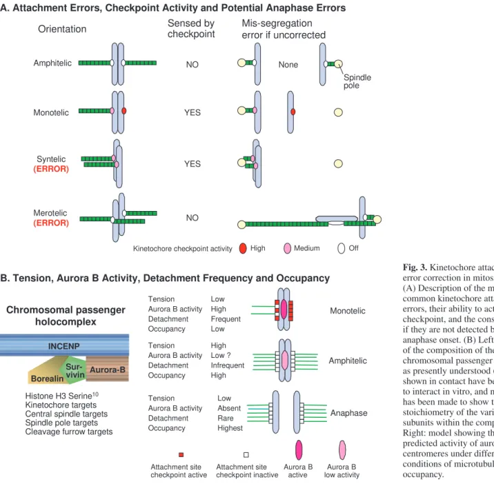
microtubules by chance, and attachment errors are common (e.g. Cimini et al., 2003). After nuclear envelope breakdown, initially chromosomes usually become mono-oriented by one (monotelic attachment) or both (syntelic attachment) sister kinetochores (Fig. 3A). Attachment to both spindle poles (bi-orientation) allows chromosomes to align near the spindle equator and to form a dynamic array referred to as the metaphase plate. For accurate chromosome segregation, the sister kinetochores must achieve amphitelic attachment, where one sister is attached to microtubules solely from one pole whereas the other is attached to microtubules solely from the opposite pole (Fig. 3A). If one or both of the sister kinetochores has microtubule attachments to both poles, this attachment is termed merotelic (Fig. 3A).
The spindle assembly checkpoint detects kinetochores on mono-oriented chromosomes that are either unattached or have syntelic attachment, delaying activation of the APC/C and Amphitelic
Monotelic
Syntelic (ERROR)
Merotelic (ERROR)
NO
YES
YES
NO
A. Attachment Errors, Checkpoint Activity and Potential Anaphase Errors
Orientation Mis-segregation error if uncorrected
None
Sensed by checkpoint
Kinetochore checkpoint activity
Spindle pole
B. Tension, Aurora B Activity, Detachment Frequency and Occupancy
Monotelic
Anaphase Amphitelic
High Low ? Infrequent High
Low Absent Rare Highest Low High Frequent Low Tension
Aurora B activity Detachment Occupancy
Attachment site checkpoint active
Attachment site checkpoint inactive
Aurora B active
Aurora B low activity Tension
Aurora B activity Detachment Occupancy Tension Aurora B activity Detachment Occupancy
Histone H3 Serine10
Kinetochore targets Central spindle targets Spindle pole targets Cleavage furrow targets
Aurora-B INCENP
Sur-vivin Borealin
Chromosomal passenger holocomplex
High Medium Off
therefore the onset of anaphase (reviewed by Musacchio and Hardwick, 2002). The checkpoint is sensitive to the level of kinetochore occupancy by microtubules (Fig. 3B). Whether it can also detect tension exerted by the spindle on kinetochores is still actively debated, in part because the tension generated by bi-orientation stabilizes microtubule attachment (Nicklas and Koch, 1969; Nicklas et al., 2001). However, what is clear is that merotelic attachments of bi-oriented chromosomes, in which kinetochores are attached incorrectly but are under tension, are not detected by the checkpoint (Cimini et al., 2001; Cimini et al., 2002). Nevertheless, most, but not all, merotelic attachments are either corrected before anaphase or are prevented by spindle mechanics from producing inaccurate anaphase chromosome segregation (Cimini et al., 2003).
One key factor in attachment error correction before anaphase appears to be the chromosomal passenger complex (Fig. 1 and Fig. 3B), which consists of the kinase aurora B, its targeting and activation subunit INCENP, and two other subunits whose role is unclear – survivin and borealin/dasra B (Adams et al., 2001a; Gassmann et al., 2004; Sampath et al., 2004). Cells that have chromosome attachment errors accumulate when the function of this complex is disrupted by dominant-negative mutants, RNAi, antibody microinjection, or selective drug targeting (Mackay et al., 1998; Adams et al., 2001b; Kaitna et al., 2002; Honda et al., 2003; Gassmann et al., 2004; Kallio et al., 2002b; Murata-Hori and Wang, 2002; Carvalho et al., 2003; Ditchfield et al., 2003; Hauf et al., 2003; Lampson et al., 2004).
The chromosomal passengers are required for operation of the spindle assembly checkpoint under certain conditions. The checkpoint mechanism functions normally in the absence of survivin or aurora B function if microtubules are completely disassembled by drugs such as nocodazole. By contrast, survivin is required for cells to delay mitotic progression when spindle dynamics are dampened by taxol (Carvalho et al., 2003; Lens et al., 2003), or when formation of monopolar spindles is induced by treatment with the drug monastrol (Lens et al., 2003), both of which reduce centromere tension. Studies using small molecule inhibitors of aurora kinases have yielded similar results (Hauf et al., 2003; Ditchfield et al., 2003), and the budding yeast aurora kinase, Ipl1p, is required for checkpoint signalling specifically when centromere tension is abolished by blocking DNA replication prior to mitotic entry (Biggins and Murray, 2001). The chromosomal passenger complex is required for the stable targeting of checkpoint proteins BUBR1 and MAD2 to kinetochores (Carvalho et al., 2003; Lens et al., 2003; Ditchfield et al., 2003; Murata-Hori and Wang, 2002). Aurora B is also required for the stable targeting of CENP-E, dynein and MCAK to centromeres, but only in the presence of microtubules (Murata-Hori and Wang, 2002; Andrews et al., 2004). This suggests that the chromosomal passenger complex promotes and stabilizes protein recruitment to kinetochores, antagonizing the dynein-driven stripping discussed above.
In contrast to the above results, the checkpoint response induced by loss of microtubules is lost in S. pombeaurora/Ark1 mutants (Petersen and Hagan, 2003) and in human cells expressing dominant-negative aurora B (Murata-Hori and Wang, 2002) or injected with specific antibody (Kallio et al., 2002b). The reason for these differences is not known.
Many studies have shown that aurora B kinase is required to destabilize improper kinetochore microtubule attachments (Fig. 3B) so that chromosomes can achieve an amphitelic orientation and metaphase alignment (reviewed by Adams et al., 2001a; Carmena and Earnshaw, 2003). A clue to the underlying mechanism has emerged from studies in budding yeast, in which Ipl1p mutants (Chan and Botstein, 1993) undergo massive chromosome mis-segregation because they cannot release kinetochore-microtubule attachments normally (Tanaka et al., 2002). To explain this, it has been proposed that Ipl1p is normally located in the inner centromere beneath the kinetochore and that, when sister kinetochores are stretched apart by spindle tension, the kinase can no longer phosphorylate key targets in the kinetochore as the two become physically separated.
Ipl1p phosphorylates several yeast kinetochore proteins, including the constitutive kinetochore component Ndc10p (Biggins et al., 1999), as well as members of the Ndc80 and Dam1-DASH-DDD complexes (Cheeseman et al., 2002a; Kang et al., 2001). Phosphorylation of components of the Ndc80 complex has been shown to destabilize kMT attachment, whereas dephosphorylation produces stabilization (Shang et al., 2003).
Aurora B kinase also appears to influence kMT attachments by a different mechanism. The KinI kinesin MCAK (Wordeman and Mitchison, 1995) (reviewed by Walczak, 2003) is required both for spindle assembly (Walczak et al., 1996; Kline-Smith and Walczak, 2002) and for correction of attachment errors during chromosome alignment (Walczak et al., 2002; Kline-Smith et al., 2004). Phosphorylation of MCAK by aurora B inhibits its ability to promote microtubule disassembly (Andrews et al., 2004; Lan et al., 2004; Ohi et al., 2004). High-resolution light microscopy suggests that aurora B and MCAK largely colocalize in centromeres that are not under tension but that MCAK becomes more closely associated with kinetochores (thereby moving away from the aurora B in the inner centromere) as chromosomes achieve bi-orientation and their centromeres are stretched towards opposite poles (Andrews et al., 2004). Because PP1, the phosphatase that counterbalances aurora B, resides within the kinetochore outer domain (Murnion et al., 2001; Trinkle-Mulcahy et al., 2003), this would be expected to promote the activation of MCAK. The consequences of this observed movement appear to be counter-intuitive. One might imagine that MCAK should be most active during prometaphase, when the largest number of errors is being made in chromosome attachment. Therefore, the colocalization of aurora B and MCAK at this time, which would be expected to result in inactivation of MCAK, is unexpected. However, it could be that the principal function of MCAK is to correct merotelic attachments, and since these persist following the establishment of spindle tension, the spatial segregation of MCAK and the chromosomal passengers might have the expected result.
Another factor that might regulate kMT attachments is ICIS (the ‘inner centromere KinI stimulator’), an activator of MCAK that forms a complex with INCENP and aurora B in
Xenopus eggs (Ohi et al., 2003). How MCAK activity is
Kinetochore movement
Despite the fact that it maintains attachment to microtubules throughout mitosis, the kinetochore is the major site for incorporation of new tubulin subunits into kMTs (Mitchison et al., 1986; Mitchison, 1989; Mitchison and Salmon, 1992; Zhai et al., 1995), and microtubule plus ends captured by the kinetochore can still depolymerize while remaining attached (Koshland et al., 1988; Cassimeris and Salmon, 1991; Coue et al., 1991; Huitorel and Kirschner, 1988; Centonze and Borisy, 1991; Hunt and McIntosh, 1998). One classic model proposes that microtubules insert into sleeves in the kinetochore, attachment being maintained by multiple weak interactions between the polymerized tubulin subunits and the walls of the sleeve (Hill, 1985; Inoue and Salmon, 1995). Such a model leaves the end of the microtubule free to grow and shrink while remaining attached, provided that shrinkage is slow relative to the ability of the microtubule to adjust its position within the sleeve. Other molecular models for dynamic attachment to microtubule plus ends are possible but less developed (Inoue and Salmon,
1995; Mogilner and Oster, 2003; Rogers et al., 2004). Most chromosome movements relative to spindle poles are associated with the lengthening and shortening of kMTs. One of the most intriguing properties of kinetochores is their ability to switch their associated bundle of up to 20 (or more) kMTs from a state of plus-end depolymerization to polymerization. This enables kinetochores in prometaphase mammalian cultured cells (Skibbens et al., 1993) and budding yeast (He et al., 2001; Pearson et al., 2001) to exhibit ‘directional instability’ (Skibbens et al., 1993), switching between persistent phases of polewards and anti-polewards movements that are coupled to alternating states of kMT depolymerization and polymerization, respectively. This kinetochore bi-stability appears to be part of the mechanism for aligning chromosomes at the spindle equator during prometaphase without the loss of mechanical linkage between sister kinetochores and the spindle poles.
It is thought that kinetochore bi-stability is based on the dynamic instability of microtubule plus ends and it is partly controlled by tension at the kinetochore. In mammalian
cultured cells, low tension promotes the switch to kMT depolymerization and high tension promotes switching to kMT polymerization (Rieder and Salmon, 1994; Skibbens et al., 1995; Skibbens and Salmon, 1997). At high tension, kinetochores appear to act like a ‘slip-clutch’ mechanism, switching to polymerization to prevent detachment of depolymerizing ends (Fig. 4A) (Rieder and Salmon, 1994; Maddox et al., 2003). Switching between states of polymerization or depolymerization probably depends on structural changes associated with the dynamic instability of microtubule plus ends (Fig. 4B). Polymerizing ends are typically blunt or slightly flared into open sheets because their protofilaments are straight or slightly curved inside-out as they incorporate tubulin-GTP (Fig. 4B, polymerization state). By contrast, protofilaments are highly curved inside-out at depolymerizing ends that have lost their stabilizing cap of tubulin-GTP (Fig. 4B, depolymerization state). The hydrolysis of GTP bound to tubulin following assembly into the microtubule lattice is thought to provide the energy that drives this inside-out curvature of the tubulin dimer that is seen when
Kinetochore tension regulates microtubule
dynamics Velocity
(µm/min)
Kinetochore tension Polymerization
Depolymerization
A
Kinetochore Bi-Stability
Depolymerisation state -Force generating
Lower Tension
Higher Tension Polymerisation state -Resistive
Centromere tension
Inner plate Flux
EB1
GTP-Tubulin
GDP-Tubulin Kin I Dynein Dynactin CLASPs
Centromere tension
Outer plate Flux
Inner plate
B
NDC80 Complex ?
Outer plate
NDC80 Complex ?
APC MCAK
CENP-E
?
Fig. 4. (A) The kinetochore acts like a slip-clutch mechanism, switching to
polymerization at high tensions to prevent detachment (Maddox et al., 2003). See text for details. (B) Model for the roles played by the cast of characters involved in
tubulin-GDP protofilaments loose their lateral attachments at the microtubule tip (Arnal et al., 2000; Howard and Hyman, 2003). A fundamental unanswered question is how switching between these polymerization and depolymerization states of dynamic instability is controlled by tension or other chemical mechanisms within the kinetochore or gradients within the spindle (Inoue and Salmon, 1995; Kapoor and Compton, 2002; Sprague et al., 2003). Switching probably depends on regulation of depolymerases and +TIP proteins within the kinetochore (Fig. 4B), as discussed in the next section. Another model (Joglekar and Hunt, 2002) proposes that high tension induces the switch from depolymerization to polymerization by causing the loss of all depolymerizing ends from the kinetochore while a sub-population of attached kMTs is still undergoing polymerization and maintaining anchorage within the Hill sleeve (Hill, 1985; Joglekar and Hunt, 2002). This model makes several testable predictions, including the notion that polymerizing and depolymerizing ends coexist within single kMT fibres.
In vertebrate cultured cells, two mechanisms combine to move chromosomes polewards during metaphase oscillations and anaphase A segregation (Fig. 4): ‘Pac-Man’ motility, which is coupled to depolymerization of the plus ends of kMTs within the attachment site at the kinetochore; and poleward microtubule flux, produced by microtubule translocation forces within the spindle and coupled to depolymerization of microtubule minus ends near the spindle poles. Sorting out contributions from Pac-Man and flux-based mechanisms to poleward movement has been made possible by the development of fluorescence photoactivation (Mitchison, 1989) and, more recently, by fluorescence speckle microscopy methods. If one microinjects or transfects cells with fluorescent tubulin subunits at low (<1%) fractions of the endogenous unlabelled tubulin pool, microtubules acquire random distributions of fluorescent subunits. Stochastic clustering during polymerization produces fluorescent speckles of 1-5 fluorophores along the microtubule lattice; these can be imaged relative to fluorescently labelled kinetochores and poles with high resolution and sensitivity using cooled charge-coupled device (CCD) cameras and spinning-disk confocal microscopy (Waterman-Storer et al., 1998; Maddox et al., 2000; Maddox et al., 2002).
The relative contributions of Pac-Man and flux mechanisms to anaphase chromosome movement vary between different cell types, Pac-Man contributing 100% in budding yeast and about 70% in vertebrate tissue culture cells (Mitchison and Salmon, 1992; Zhai et al., 1995). By contrast, flux makes an important contribution in meiotic oocyte spindles and early embryonic spindles (Maddox et al., 2002; Brust-Mascher and Scholey, 2002; Maddox et al., 2003), and apparently accounts for 100% of the movement in grasshopper and crane fly meiosis I spermatocytes (Wilson et al., 1994; LaFountain et al., 2001; Chen and Zhang, 2004).
Although significant progress has been made, it is not yet known how the state of kMT polymerization/depolymerization is coupled to kinetochore tension, or what reads the tension, or how this signal is transduced to the microtubules. These issues are important for advancing our understanding of how kinetochores function along with spindle mechanisms such as poleward microtubule flux and polar ejection forces on the chromosome arms to align chromosomes in prometaphase, a
problem that has been recently discussed elsewhere (Kapoor and Compton, 2002).
Proteins influencing kinetochore movement
Kinetochore proteins and microtubule-plus-end-binding proteins both regulate kinetochore movement by modulating the dynamics of kMT plus ends (McIntosh et al., 2002). However, the kinetochore-microtubule interface is highly dynamic, and several of these proteins appear to be bona fide components of both structures. Two classes of proteins seem particularly important: kinesin motors that function as depolymerases, such as the KinI kinesins; and microtubule-plus-end-tracking proteins (+TIPs), which promote polymerization, perhaps by antagonizing the depolymerases (Fig. 4B) (Schuyler and Pellman, 2001).
KinI kinesins, so named because they have an internal motor domain, use ATP hydrolysis to drive inside-out bending of microtubule protofilaments and promote depolymerization of the polymer (Desai et al., 1999; Moores et al., 2002; Hunter et al., 2003; Walczak, 2003). In vertebrates, MCAK is the major KinI depolymerase controlling the dynamics of microtubule plus-end assembly (Howard and Hyman, 2003; Walczak, 2003). It is not clear just what role MCAK specifically has in kinetochore motility because the loss of MCAK from centromeres and kinetochores does not affect anaphase chromosome velocity in mammalian cultured cells (Kline-Smith et al., 2004). This suggests that other activities are involved. In budding yeast, the plus-end-directed kinesin Kip3p, and the minus-end-directed kinesin Kar3p, are also candidates for kinetochore depolymerases (Howard and Hyman, 2003), but the roles of their homologues in mammalian cultured cells are not yet known. There is currently great interest in identifying microtubule depolymerases that might function at kinetochores or at poles. In Drosophilaembryos and human cultured cells, KinI kinesins may be important for the depolymerization of kMTs at both their plus and minus ends.
Database screening has identified three predicted KinI kinesins in Drosophila (Rogers et al., 2004). One of these, Klp10A, is located at centromeres and spindle poles early in mitosis and at the poles during anaphase. A second, Klp59C, is concentrated at centromeres throughout mitosis. Both have microtubule-depolymerizing activity and are essential for mitotic progression. Using fluorescent speckle microscopy to track the movements of spindle microtubules and GFP-CID
(Drosophila CENP-A) to track kinetochore movements,
Rogers et al. found that about 60% of the anaphase chromosome movement in Drosophilaembryos is due to Pac-Man depolymerization, and 40% is due to flux (Rogers et al., 2004). Microinjection of antibodies to Klp10A greatly inhibits flux and reduces anaphase A chromosome velocity by 40%. Therefore, Klp10A has been proposed to be an essential component of the spindle flux mechanism.
tension increases and anaphase onset is triggered (Rogers et al., 2004). It remains to be determined how these centromeric KinI kinesins gain access to microtubule plus ends, most of which end at the outer kinetochore.
In human cells, the KinI kinesin Kif2a has recently been implicated in the regulation of spindle microtubule dynamics at poles. Depletion of Kif2a causes collapse of the bipolar spindle. Depletion of Kif2a is proposed to lower tension in the spindle, causing the kinetochore to switch to microtubule disassembly, thereby resulting in spindle collapse (Ganem and Compton, 2004). Collapse can be prevented by inhibiting kinetochore pulling forces, using drug treatments or depletion of MCAK by siRNA to prevent kMT depolymerization, or by preventing kMT formation, using siRNA directed against Nuf2 (Ganem and Compton, 2004).
Further insight into the role of Kif2a and its association with dynein in the spindle has been provided by studies of meiosis II spindles assembled in vitro in cytoplasmic extracts of
Xenopus eggs. Addition of an inhibitory dominant-negative polypeptide reveals that dynein does not seem to be required for the sliding or poleward flux of microtubules in this cell-free system (Gaetz and Kapoor, 2004). However, it appears to have an essential role in regulating overall spindle length. Dynein is required to target Kif2a to spindle poles efficiently in Xenopus
extract spindles, and Kif2a regulates the length of spindle microtubules in bipolar spindles. These results suggest that the dynein-mediated polewards streaming of kinetochore components discussed above could additionally have a role in positioning Kif2a at poles and thereby in regulating tension within the spindle.
Two classes of +TIPs have potential roles at the kinetochore. The first, which includes the adenomatous polyposis coli (APC) protein and its binding partner EB1, are bona fide microtubule-associated proteins that require microtubules to localize to kinetochores. APC and EB1 have recently been
proposed to have a role in chromosome segregation (Fodde et al., 2001; Kaplan et al., 2001; Tirnauer et al., 2002; Rogers et al., 2002; Green and Kaplan, 2003). Binding of EB1 to microtubule plus ends at the kinetochore interface is restricted to polymerizing microtubules, which suggests that it favours stabilization of attached microtubules during polymerization (Tirnauer et al., 2002), perhaps by preventing their plus ends from interacting with the KinI depolymerases.
A second class of +TIPs includes proteins that can localize to kinetochores even in the absence of microtubules. Two have attracted the most interest: CLIP-170; and its binding partners the CLASPs [CLIP-associated proteins (Akhmanova et al., 2001), first identified in budding yeast as Stu1p (Pasqualone and Huffaker, 1994) and in Drosophilaas Orbit/MAST (Inoue et al., 2000; Lemos et al., 2000)].
The behaviour of CLIP-170 at kinetochores is paradoxical. In prometaphase cells, CLIP-170 localizes strongly both to microtubule plus ends and to kinetochores (Dujardin et al., 1998; Coquelle et al., 2002). However, this microtubule-associated protein localizes only to kinetochores that lack bound microtubules and leaves the kinetochore upon microtubule attachment. This is particularly evident in chromosomes that exhibit a monotelic attachment, where CLIP-170 is present only at the distal, unattached kinetochore (Maiato et al., 2003). Its accumulation at the kinetochore requires dynein and is mediated by the lissencephaly gene product LIS1 (Faulkner et al., 2000; Coquelle et al., 2002; Tai et al., 2002). The role of CLIP-170 at kinetochores is not known, although expression of a dominant-negative mutant form of the protein causes a prometaphase delay (Dujardin et al., 1998), which suggests that the protein has an active role in chromosome alignment.
CLASPs are required for chromosome congression and maintenance of a bipolar mitotic spindle in Drosophila, humans and budding yeast (Maiato et al., 2002; Maiato et al., 2003; Yin et al., 2002). They are apparently not required for efficient kinetochore-microtubule attachment but, instead, appear to modulate the dynamic behaviour of microtubules at kinetochores and elsewhere in the spindle (Maiato et al., 2003). CLASP1 is found in the outer kinetochore corona from early prometaphase throughout anaphase.
Microtubule plus ends at the kinetochore outer plate can have a blunt, open or flared morphology (Fig. 5B). This is thought to correlate with their dynamic instability status (Mastronarde et al., 1997; McEwen et al., 1998; O’Toole et al., 1999). The position occupied by CLASP1 in the outer kinetochore corona places it adjacent to the plus ends of kMTs and therefore near to where the lattice might open as the microtubules exhibit dynamic behaviour. This raises the possibility that CLASP1 regulates microtubule dynamics by altering the lattice of kinetochore-attached microtubules and facilitating the incorporation of microtubule subunits.
Fig. 5. Historic and high-resolution views of the kinetochore-microtubule interface. (A) Original description of the ‘leitkörpen’ (‘the leading body’) as the interface between chromosomes and the spindle in Salamander spermatocytes [adapted from the original (Metzner, 1894)]. (B) A single 16 nm thick slice from a 10-section tomographic volume reconstruction of the microtubule-kinetochore interface from PtK1 cells prepared by high-pressure freezing/freeze substitution. Note that forked microtubule plus ends are
CLASP/Stu1p, which was identified in yeast as a suppresser of a cs (cold sensitive) mutation in β-tubulin (Pasqualone and Huffaker, 1994), associates specifically with β-tubulin (Yin et al., 2002). β-tubulin is the microtubule subunit responsible for GTP hydrolysis during polymerization at microtubule plus ends (Mitchison, 1993; Davis et al., 1994). Since Drosophila
CLASP/Orbit/MAST binds microtubules in a GTP-dependent manner (Inoue et al., 2000), we speculate that binding of CLASPs at the kinetochore-microtubule interface could influence the structural properties of the lattice at the plus ends of microtubules and thereby influence the transitions between shrinkage and growth.
Recently, Salic et al. discovered that vertebrate shugoshin (Sgo) protein concentrates at kinetochores during mitosis and has a function in regulating kinetochore microtubule stability (Salic et al., 2004). Sgo is also required to prevent premature sister centromere separation as predicted by earlier studies of fungal Sgo1 and Drosophila MEI-S332 genes in meiosis I. This linkage by Sgo between centromere cohesion and microtubule interactions at kinetochores is an unexpected example of how the kinetochore network of proteins is integrated together to achieve accurate chromosome segregation.
Perspectives
We are starting to develop a detailed description of the events that occur as kinetochores interact with microtubules during the different stages of mitosis. However, when one considers the underlying mechanisms, we are still at a stage where questions are proliferating more rapidly than answers. It has been 110 years since Metzner described the existence of substructures responsible for chromosome movement (Fig. 5A) (Metzner, 1894) and 37 years since Inoue proposed the dynamic equilibrium model for spindle assembly and chromosome movement (reviewed by Inoue and Salmon, 1995). Now we are on our way towards formulating an initial list of the protein components involved in attaching microtubules to kinetochores, recognizing that attachment has occurred, releasing microtubules from improper attachments, and coupling polymerization and depolymerization to force production. At present, all of this information applies to the interactions between single microtubules and kinetochores. In mammalian cells, kinetochores are normally associated with bundles of ~20 kMTs. Kinetochore directional instability probably involves the coordinated switching of the polymerization state of all of these kMTs in concert. At present, we have few clues as to the mechanism by which decisions to alter polymerization status are transmitted laterally through the bundle of kMTs, and this will remain a goal as we try to move to the next level of understanding of the dynamic interactions between kinetochores and microtubules.
We thank B. McEwen for his critical comments on the manuscript and for providing Fig. 5B, which was kindly processed for illustration by R. Barnard and K. Vandenbeldt. H.M. is supported by a postdoc fellowship from Fundação para a Ciência e a Tecnologia of Portugal (SFRH/BPD/11592/2002). Work in the lab of E.D.S. is supported by NIH grant GM 24364. Work in the lab of W.C.E. is supported by The Wellcome Trust, of which he is a Principal Research Fellow.
References
Adams, R. R., Carmena, M. and Earnshaw, W. C. (2001a). Chromosomal passengers and the (aurora) ABCs of mitosis. Trends Cell Biol.11, 49-54.
Adams, R. R., Maiato, H., Earnshaw, W. C. and Carmena, M. (2001b). Essential roles of Drosophila inner centromere protein (INCENP) and Aurora-B in histone H3 phosphorylation, metaphase chromosome alignment, kinetochore disjunction, and chromosome segregation. J. Cell Biol.153, 865-880.
Akhmanova, A., Hoogenraad, C. C., Drabek, K., Stepanova, T., Dortland, B., Verkerk, T., Vermeulen, W., Burgering, B. M., de Zeeuw, C. I., Grosveld, F. et al. (2001). Clasps are CLIP-115 and -170 associating proteins involved in the regional regulation of microtubule dynamics in motile fibroblasts. Cell104, 923-935.
Amor, D. J., Kalitsis, P., Sumer, H. and Choo, K. H. (2004). Building the centromere: from foundation proteins to 3D organization. Trends Cell Biol. 14, 359-368.
Andrews, P. D., Ovechkina, Y., Morrice, N., Wagenbach, M., Duncan, K., Wordeman, L. and Swedlow, J. R. (2004). Aurora B regulates MCAK at the mitotic centromere. Dev. Cell6, 253-268.
Arnal, I., Karsenti, E. and Hyman, A. A. (2000). Structural transitions at microtubule ends correlate with their dynamic properties in Xenopusegg extracts. J. Cell Biol.149, 767-774.
Arnaoutov, A. and Dasso, M. (2003). The Ran GTPase regulates kinetochore function. Dev. Cell. 5, 99-111.
Banks, J. D. and Heald, R. (2001). Chromosome movement: dynein-out at the kinetochore. Curr. Biol. 11, R128-R131.
Basto, R., Scaerou, F., Mische, S., Wojcik, E., Lefebvre, C., Gomes, R., Hays, T. and Karess, R. (2004). In vivo dynamics of the rough deal checkpoint protein during Drosophilamitosis. Curr. Biol.14, 56-61.
Belgareh, N., Rabut, G., Bai, S. W., van Overbeek, M., Beaudouin, J., Daigle, N., Zatsepina, O. V., Pasteau, F., Labas, V., Fromont-Racine, M. et al. (2001). An evolutionarily conserved NPC subcomplex, which redistributes in part to kinetochores in mammalian cells. J. Cell Biol.154, 1147-1160.
Bernat, R. L., Delannoy, M. R., Rothfield, N. F. and Earnshaw, W. C.
(1991). Disruption of centromere assembly during interphase inhibits kinetochore morphogenesis and function in mitosis. Cell66, 1229-1238.
Bharadwaj, R., Qi, W. and Yu, H. (2004). Identification of two novel components of the human NDC80 kinetochore complex. J. Biol. Chem.279, 13076-13085.
Biggins, S. and Murray, A. W. (2001). The budding yeast protein kinase Ipl1/Aurora allows the absence of tension to activate the spindle checkpoint.
Genes Dev. 15, 3118-3129.
Biggins, S. and Walczak, C. E. (2003). Captivating capture: How microtubules attach to kinetochores.Curr. Biol.13, R449-R460.
Biggins, S., Severin, F. F., Bhalla, N., Sassoon, I., Hyman, A. A. and Murray, A. W. (1999). The conserved protein kinase Ipl1 regulates microtubule binding to kinetochores in budding yeast. Genes Dev.13, 532-544.
Bischoff, F. R., Maier, G., Tilz, G. and Ponstingl, H. (1990). A 47-kDa human nuclear protein recognized by antikinetochore autoimmune sera is homologous with the protein encoded by RCC1, a gene implicated in onset of chromosome condensation. Proc. Nat. Acad. Sci. USA87, 8617-8621.
Blower, M. D. and Karpen, G. H. (2001). The role of Drosophila CID in kinetochore formation, cell-cycle progression and heterochromatin interactions. Nat. Cell Biol.3, 730-739.
Brenner, S., Pepper, D., Berns, M. W., Tan, E. and Brinkley, B. R. (1981). Kinetochore structure, duplication and distribution in mammalian cells: analysis by human autoantibodies from scleroderma patients. J. Cell Biol. 91, 95-102.
Brinkley, B. R. and Cartwright, J., Jr (1975). Cold-labile and cold-stable microtubules in the mitotic spindle of mammalian cells. Ann. N. Y. Acad. Sci.253, 428-439.
Brinkley, B. R. and Stubblefield, E. (1966). The fine structure of the kinetochore of a mammalian cell in vitro. Chromosoma19, 28-43.
Brinkley, B. R., Ouspenski, I. and Zinkowski, R. P. (1992). Structure and molecular organization of the centromere-kinetochore complex. Trends Cell Biol. 2, 15-21.
Brust-Mascher, I. and Scholey, J. M. (2002). Microtubule flux and sliding in mitotic spindles of Drosophilaembryos. Mol. Biol. Cell 13, 3967-3975.
Campbell, M. S., Chan, G. K. and Yen, T. J. (2001). Mitotic checkpoint proteins HsMAD1 and HsMAD2 are associated with nuclear pore complexes in interphase. J. Cell Sci.114, 953-963.