Universidade do Minho
Escola de Ciências
Tiago Manuel Fontes Oliveira
Exploring the role of
O
-Mannosylation in
the modulation of E-Cadherin function in
Gastric Cancer cell line models
Tese de Mestrado
Mestrado em Bioquímica Aplicada – Biomedicina
Trabalho efectuado sob a orientação da
Professora Doutora Salomé S. Pinho
DECLARAÇÃO
Nome
_____________________________________________________________________________________ Endereço electrónico: _____________________________ Telefone: _______________ / _______________ Número do Bilhete de Identidade: ______________________
Título dissertação □/tese □
_____________________________________________________________________________________ _____________________________________________________________________________________ _____________________________________________________________________________________ Orientador(es): _____________________________________________________________________________________ ____________________________________________________ Ano de conclusão: ___________
Designação do Mestrado ou do Ramo de Conhecimento do Doutoramento:
_____________________________________________________________________________________
Nos exemplares das teses de doutoramento ou de mestrado ou de outros trabalhos entregues para prestação de provas públicas nas universidades ou outros estabelecimentos de ensino, e dos quais é obrigatoriamente enviado um exemplar para depósito legal na Biblioteca Nacional e, pelo menos outro para a biblioteca da universidade respectiva, deve constar uma das seguintes declarações:
1. É AUTORIZADA A REPRODUÇÃO INTEGRAL DESTA TESE/TRABALHO APENAS PARA EFEITOS DE INVESTIGAÇÃO, MEDIANTE DECLARAÇÃO ESCRITA DO INTERESSADO, QUE A TAL SE COMPROMETE;
2. É AUTORIZADA A REPRODUÇÃO PARCIAL DESTA TESE/TRABALHO (indicar, caso tal seja necessário, nº máximo de páginas, ilustrações, gráficos, etc.), APENAS PARA EFEITOS DE INVESTIGAÇÃO, MEDIANTE DECLARAÇÃO ESCRITA DO INTERESSADO, QUE A TAL SE COMPROMETE;
3. DE ACORDO COM A LEGISLAÇÃO EM VIGOR, NÃO É PERMITIDA A REPRODUÇÃO DE QUALQUER PARTE DESTA TESE/TRABALHO
Universidade do Minho, ___/___/______
Agradecimentos
Em primeiro lugar gostaria de agradecer à Professora Salomé e ao Professor Celso Reis por me terem permitido colaborar no grupo “Glycobiology in Cancer”, bem como a todos os que me ajudaram a crescer profissional e pessoalmente no instituto. Obrigado a toda a gente do IPATIMUP que esteve ao meu lado a cumprir esta etapa. Vocês são boa gente. Desejo-vos o melhor. Stefan, I cannot let this pass. You are one of the brightest guys I know. I wish you all the best! And maybe we will work together in the future, who knows…
Um obrigado ao Professor Pedro pelo apoio que veio de Braga, sempre disponível.
Para o meu pessoal da Bioquímica, deixo nesta tese a alma do bioquímico FCUP/ICBAS. Viais, Escuta, Márcio, Lau, Melo, Tiago… Vocês são os maiores! Que tenham a maior sorte do mundo, sempre.
Ao meu grande Batista e à sua Tati, obrigado pelos cafés, confidências, cinemas, jantares, copos,… Valar Morghulis.
Para a Ana, que me atura todos os dias, a chover, a fazer sol, a rir, a chorar, o maior dos obrigados. Seria incapaz de tudo isto sem ti. A verdade é que existe sempre uma grande mulher à beira de um grande homem, pelo que, a receber algum crédito por este trabalho, terei sempre que o dividir contigo. Ajudaste-me a descobrir quem sou e o que quero para mim. És uma verdadeira singularidade na vida de alguém. Tenho a maior sorte do mundo.
À minha família que acredita no que sou e como sou. À minha avó.
RESUMO
A acção do complexo E-Caderina (E-Cad)-cateninas para a formação de junções aderentes estáveis é essencial para a arquitectura de tecidos epiteliais e para a sua integridade mecânica, contribuindo de forma preponderante para a supressão tumoral. Assim, a disfunção da E-Cad está geralmente associada com a perda das propriedades de adesão das células epiteliais, com a capacidade de invasão, e com metastização. Como a E-Cad é uma glicoproteína, ela pode ser modificada por N- ou por O-glicanos, especificamente por glicanos O-manose (O-Man). A N-glicosilação e a
O-manosilação iniciam-se no retículo endoplasmático (ER), seguindo para o complexo
de Golgi, e terminando quando a glicoproteína é libertada no citoplasma ou em vesículas para ser transportada para a membrana celular. Os monossacáridos iniciais podem ser estendidos em diversos compartimentos do complexo de Golgi por acção de diversas glicosiltransferases: no caso da N-glicosilação, as
N-acetilglucosaminiltransferases (GnTs) III e V (GnT-III e GnT-V) estão regularmente envolvidas na modificação da estrutura do glicano pela adição de um resíduo de N-acetilglucosamina (GlcNAc) bisecting ou β-1,6 GlcNAc branching, respectivamente. Enquanto a formação de N-glicanos com bisecting GlcNAc por acção da GnT-III leva à supressão tumoral, elevados níveis de N-glicanos β-1,6 GlcNAc branching formados por acção da GnT-V são geralmente associados a tumores altamente metastáticos. Trabalhos prévios do nosso grupo permitiram descrever o impacto da actividade destas duas enzimas na expressão e regulação da E-Cad, especificamente num contexto de cancro gástrico (GC) que não pode ser explicado apenas devido a alterações genéticas e epigenéticas. Recentemente, a O-manosilação ganhou um novo interesse quando relacionada com a E-Cad, uma vez que foram descritos diversos locais de O-manosilação disponíveis na Cad, e que os glicanos O-Man presentes na E-Cad contribuem para as suas funções biológicas, nomeadamente para a adesão célula-célula.
Este trabalho pretende compreender o papel da O-manosilação na modulação das funções da E-Cad num contexto de desenvolvimento e progressão tumoral, bem como perceber qual a relação, neste contexto, entre a O-manosilação e a N-glicosilação. Os resultados obtidos indicam claramente que o aumento de diferenciação de uma linha
celular é acompanhado por um aumento no perfil geral de O-manosilação na célula, e que a E-Cad apresenta níveis mais elevados de glicanos O-Man ligados nesse caso. Por outro lado, observamos que o aumento de glicanos O-Man e a diminuição de glicanos GlcNAc branched ligados à E-Cad está relacionado com um fenótipo mais estável.
Estes resultados apoiam o conceito de que a O-manosilação da E-Cad é essencial para as suas funções biológicas, e que a ausência desta modificação pós-traducional (PTM) pode ser um dos factores chave para a perda de função desta glicoproteína, o que leva ao desenvolvimento e progressão tumoral. De modo global, o objectivo principal deste projecto é clarificar ainda mais os mecanismos moleculares que conduzem à disfunção da E-Cad no GC e que são fundamentais para o estabelecimento de adenocarcinomas humanos, tendo em vista potenciais novas aplicações na cliníca.
O trabalho aqui apresentado foi desenvolvido como parte integrante de um projecto que visa explorar o perfil de O-manosilação da E-Caderina no cancro gástrico, tendo sido desenvolvido por mim e pela Sandra Carvalho (Estudante de Doutoramento que co-orientou este trabalho), culminando com a preparação de um artigo para ser submetido para publicação.
ABSTRACT
The establishment of stable adherens junctions by the action of the E-Cadherin (E-Cad)-catenins complex is essential for epithelial tissue architecture and mechanical integrity, contributing effectively for tumor suppression. Therefore, E-Cad impairment is often associated with loss of adhesive properties of epithelial cells, invasiveness and metastasis. As E-Cad is a glycoprotein, it can be modified by N- and O-glycans, specifically O-mannose (O-Man) glycans. N-glycosylation and O-mannosylation initiate at the endoplasmic reticulum (ER), carry on in the Golgi compartment, and terminate when the glycoprotein is released to the cytoplasm or liberated in vesicles for the cellular membrane. The initial monosaccharide residues can be further extended in the several compartments of the Golgi apparatus by the action of several glycosyltransferases: in the case of N-glycosylation, N-acetylglucosamyniltransferases (GnTs) III and V (GnT-III and GnT-V) are often involved in the modification of the glycan structure by the addition of a bisecting N-acetylglucosamine (GlcNAc) or β-1,6 GlcNAc branching residue, respectively. While the formation of bisecting GlcNAc N-glycans by GnT-III has been proven to be tumor suppressor, high levels of β-1,6 GlcNAc branching
N-glycans formed by the action of GnT-V are usually associated with highly metastatic
tumors. Work by our group allowed us to describe the impact of the activity of these two enzymes in E-Cad expression and regulation, specifically in a gastric cancer (GC) context that cannot be explained solely by genetic or epigenetic alterations. Recently,
O-mannosylation gained interest relating to E-Cad, as it was described that E-Cad
presents several available O-mannosylation sites, and that the O-Man glycans present on E-Cad contribute to its biological functions, namely cell-cell adhesion.
This work aims to understand how O-mannosylation modulates E-Cad functions in tumor development and progression, and to figure out the interplay between O-mannosylation and N-glycosylation in this context. Our results clearly indicate that the gaining of differentiation status of a cell line is accompanied by an increase in the cellular overall mannosylation profile, and that E-Cad presents higher levels of O-Man glycans attached to it in that case. On the other hand, we observed that the
increase in O-Man glycans and the decrease of branched GlcNAc N-glycans attached to E-Cad is related to a more stable phenotype.
These results support the idea that O-mannosylation of E-Cad is essential for its biological functions, and that the absence of this post-translational modification (PTM) may be one of the key elements for the impairment of this glycoprotein, which leads to tumor development and progression. Globally, the main goal of this project is to further clarify the molecular mechanisms behind E-Cad dysfunction in GC that are important in the setting up of human adenocarcinomas, having in mind potential new applications in the clinic.
The work that is present here was developed as part of a comprehensive project that aims to explore the O-mannosylation profile of E-Cadherin in Gastric Cancer, carried out by me and Sandra Carvalho (PhD Student that co-supervised this work), which will culminate in the preparation of a manuscript to be submitted for publication.
Table of Contents
IMAGE INDEX ... IX
LIST OF ABBREVIATIONS ... XI
1 INTRODUCTION ... 1
1.1 Gastric Cancer ... 3
1.2 E-Cadherin at the adherens junctions: the importance in cell-cell adhesion ... 5
1.3 Protein Glycosylation ... 8
1.3.1 N-glycosylation ... 10
1.3.2 O-glycosylation ... 12
1.3.3 O-mannosylation ... 12
1.4 Glycosylation in gastric cancer: the importance of N-glycosylation of E-Cadherin in a gastric cancer context ... 14
1.5 O-mannosylation and E-Cadherin: evidences of a “new” player in the adhesion game ... 18
2 AIMS ... 23
3 MATERIALS AND METHODS ... 27
3.1 Cell lines and cell culture ... 29
3.2 Immunofluorescence ... 29
3.3 PNGase F digestion ... 30
3.5 Immunoprecipitation of E-Cadherin ... 32
3.6 Quantitative Real-Time PCR (qRT-PCR) ... 33
4 RESULTS ... 35
4.1 MKN28 and KATO III display different morphologies and a different E-Cad patterns of expression ... 37
4.2 Impact of POMT2 protein expression levels variability in the overall O-mannosylation profile and in the E-Cadherin O-mannosylation status comparing MKN28 and KATO III cell line models ... 38
4.3 Relationship between the levels of O-mannose glycans and β-1,6 GlcNAc branched N-glycans in different gastric cancer cell lines ... 40
5 DISCUSSION AND CONCLUSIONS... 45
6 FUTURE PERSPECTIVES ... 53
Image Index
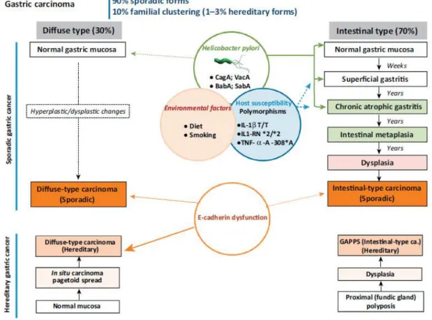
Figure 1 - Overall cancer incidence and mortality estimates worldwide in 2012, according to GLOBOCAN2. Figure 2 - Clinicopathological profiles of Diffuse and Intestinal Gastric Cancer11.
Figure 3 - The E-Cadherin-catenin complex (calcium ions are not represented). Interaction with the actin cytosketelon occurs via α-catenin. The possible N-glycosylation sites existent in E-Cadherin (Asn554, Asn566, Asn618 and Asn633) are here represented as the initial GlcNAc monosaccharide.
Figure 4 - Major classes of mammalian glycoconjugates.
Figure 5 - N-glycosylation process occurs in the endoplasmic reticulum and the Golgi compartment. The synthesis of the Dol-Pi-Pi-GlcNAc2Man9Glc3 structure is necessary for the action of oligosaccharyltransferase (OST)
that transfers the glycan from the activated dolichol carrier to the protein. The protein can then be folded correctly and go to the Golgi compartment, or targeted for degradation. In the Golgi, the action of several glycosyltransferases and glycosidases determines the final glycoprotein that is formed.
Figure 6 - O-mannosylation process initiates in the endoplasmic reticulum by the action of POMT1 or POMT2 that transfer a mannose residue to a nascent protein. The consequent action of several glycosyltransferases determines the type of structure that is formed. O-mannosyl glycans can be divided in cores M1, M2 and M3, according to the type of linkage between the mannose and the GlcNAc residues (adapted from 34).
Figure 7 - The importance of glycosylation in the formation of a primary tumor and in the metastatic process. Glycans are important in the cell-cell adhesion process, in cell-extracellular matrix interactions, as well as in several other processes.
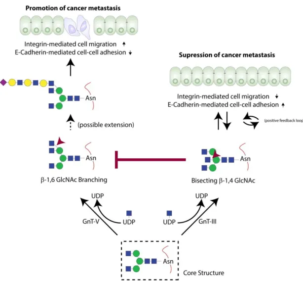
Figure 8 - Alterations in the structure and function of E-Cadherin occur due to the action of GnT-III and GnT-V. Enzymatic activity of these glycosyltransferases determines the type of glycans formed, which in turn contributes to the promotion or suppression of cancer metastasis (adapted from 67).
Figure 9 - Type 1 cadherins were identified by the Clausen group as major carriers of O-mannosyl glycans84. Figure 10 - Glycosites identified and predicted on E-Cadherin to date. There are a total of 4 N-glycosylation sites, 9 O-mannosylation sites (identified in 84) and 1 GalNAc site, distributed on the protein as depicted. A total of 6 O-mannosylation sites were also predicted by the Clausen’s group. The glycosylation sites represented were numbered considering the propeptide as part of E-Cadherin protein.
Figure 11 - Slow aggregation assays performed using the MDCK cell line using an E-Cadherin antibody (DECMA-1) and a T[α1-Man]-specific antibody allowed to understand the importance of O-mannosyl glycans in cell-cell adhesion. EGTA (blocks the Ca2+-dependent cadherin-mediated adhesion), IIH6 and VIA4-1 (antibodies directed against O-mannosyl epitopes linked to α-dystroglycan) were used as controls86
Figure 12 - MKN28 and KATO III cells morphological and phenotypical characteristics. (A) MKN28 cell line display an epithelial-like phenotype, forming a monolayer of adhesive cells while (B) KATO III cells are smaller and round-shaped.
Figure 13 - Immunofluorescence analysis on (A) MKN28 and (B) KATO III cell lines. MKN28 cells demonstrated a cell membranous pattern of E-Cadherin expression, whereas KATO III cells displayed a mislocalization of the protein. Figure 14 - Expression levels of POMT2 and the overall O-mannosylation profile. (a) Analysis by Western blot from total cell lysates of MKN28 and KATO III probed with antibodies against POMT2 and tubulin (loading control). POMT2 is expressed in MKN28 while in KATO III no visible band appears. (b) Lectin blot and Western blot analysis
from digested total cell lysates of MKN28 and KATO III cells with Con A and the antibody against actin (loading control). MKN28 presents higher content in O-mannosyl glycans when compared to KATO III, as the lectin blot presents higher reactivity in the MKN28 cell line lane.
Figure 15 - The overall O-mannosylation profile of MKN28 and KATO III. Lectin blot and Western blot analysis from digested total cell lysates of MKN28 and KATO III cells with Con A and the antibody against actin (loading control). MKN28 presents higher content in O-mannosyl glycans when compared to KATO III, as the lectin blot presents higher reactivity in the MKN28 cell line lane.
Figure 16 - O-mannosylation of E-Cadherin. (A) E-Cadherin was immunoprecipitated from MKN28 and KATO III cell lines and the existence of O-mannosyl glycans attached was assessed using simultaneously PNGase F digestion and Con A lectin blot. (B) O-mannosyl glycans were only detected on Cadherin in MKN28 cell line. Level of E-Cadherin was assessed in parallel using the antibody anti-E-E-Cadherin (clone 36) as a control of the immunoprecipitation. The levels of Con A density for MKN28 and KATO III were obtained after normalization to E-Cadherin.
Figure 17 - Levels of O-mannosyl glycans and branched N-glycans in total cell lysates of MKN28 and KATO III cell lines. Lectin blots were performed in fresh total cell lysates, and actin was used as a loading control for both of them. An initial step of digestion was necessary when using Con A lectin to assess the O-mannosylation status of the lysates.
Figure 18 - Cadherin O-mannosylation and branched N-glycans content in MKN28 and KATO III cell lines. (A) E-Cadherin was immunoprecipitated in order to study its O-mannosylation profile and β-1,6 glycans content. Whereas MKN28 E-Cadherin presents a huge level of mannosyl glycans, it seems that in KATO III E-Cadherin is not very O-mannosylated. The levels of L-PHA reactivity in both cell lines are very similar to each other.
Figure 19 - Relative expression of MGAT5 and POMT2 mRNA in MKN45 MOCK and MKN45+GnT-V cell lines. (left) MGAT5 is expressed approximately 30 times more in the MKN45 cell line transfected with GnT-V. (right) POMT2 mRNA levels are slightly superior in MKN45+GnT-V when compared to the cells transfected with the empty vector (MKN45 MOCK). Results were normalized for GAPDH mRNA expression levels. Relative expression of MKN45 MOCK was considered to be 1, and the expression of MKN45+GnT-V was compared in each condition (Student’s t-test: * P≤0.05; **** P≤0.001).
Figure 20 - E-Cadherin O-mannosylation and branched N-glycans content in MKN45 MOCK and MKN45+GnT-V cell lines. (A) E-Cadherin was immunoprecipitated in order to study its O-mannosylation profile and β-1,6 glycans content. The presence of GnT-V (MKN45+GnT-V) increases the number of β-1,6 branched N-glycans, when comparing to MKN45 MOCK. The level of reactivity of Con A is higher in MKN45 MOCK cells, which indicate a greater number of O-mannosyl glycans present on E-Cadherin, when compared to MKN45+GnT-V. (B and C) The levels of Con A and L-PHA densities for MKN45 MOCK and MKN45+GnT-V were obtained after normalization to Cadherin, being expressed as the fold change, compared with the association level of the same lectin with E-cadherin in MKN45 MOCK cells, which was taken as 1 (Student’s t-test: **P≤0.01; **** P≤0.001).
Figure 21 – Proposed model for the interplay between O-mannosylation and N-glycosylation. In a normal epithelial cell context, more O-mannosyl glycans are linked to E-Cad, as the protein does not have such bulky N-glycans due to the absence of GnT-V activity. On the other hand, in an adenocarcinoma cell context, GnT-V activity leads to the formation of huge complex N-glycan structures, which precludes the attachment of so many O-Man residues, contributing to a decrease in cell-cell adhesion and to a decreased stability of the E-Cad-catenins complex.
List of Abbreviations
α-DG – Alpha-dystroglycan aa. - Aminoacid Asn - Asparagine β-DG – Beta-dystroglycan Ca2+ - CalciumC1GalT1 - Core 1 Gal-transferase
C2GnT - Core 2 β1-6 N-acetylglucosaminyltransferase CMDs – Congenital Muscular Dystrophies
Con A – Concanavalin A
CPTM – Co- and post-translational modification Cys - Cysteine
Dol-Pi – Dolichol phosphate
E-Cad – E-Cadherin
ECM – Extracellular Matrix ECs – Cadherin domains
E-PHA - Biotinylated Phaseolus vulgaris Erythroagglutinin ER – Endoplasmic Reticulum
Fuc – Fucose Gal – Galactose
GalNAc – N-acetylgalactosamine
GalNAc-T - UDPGalNAc-polypeptide N-acetylgalactosaminyltransferase GC – Gastric Cancer
Glc – Glucose
GlcA – Glucuronic Acid
GlcNAc – N-acetylglucosamine Gln – Glutamine
GnTs – N-acetylglucosamyniltransferases IGF-I -Insulin-like Growth Factor I
IGF-IR – Insulin-like Growth Factor I Receptor IP – Immunoprecipitation
L-PHA - Biotinylated Phaseolus vulgaris Leucoagglutinin Man – Mannose
miR - MicroRNAs N-Cad – N-Cadherin Neu5Ac – Sialic Acid
ON – Overnight
OST – Oligosaccharyltransferase PBS – Phosphate-Buffered Saline
PBST – Phosphate-Buffered Saline Tween 20 0,05% PMT - Protein O-mannosyltransferase (in yeast) PNGase F - Peptide-N-glycosidase F
polyLacNAc - Poly-N-acetylactosamine
POMGnT1 - Protein O-linked-mannose β-1,2-N-acetylglucosaminyltransferase 1 POMGnT2 - Manα1-O- Ser/Thr β1,4GlcNAc-transferase
POMT - Protein O-mannosyltransferase (in mammalian organisms) POMT1 - Protein O-mannosyltransferase 1
POMT2 - Protein O-mannosyltransferase 2 Pro – Proline
PTM – Post-translational modification
R3A-5a - Rhodanine-3-acetic acid Derivative, Compound 5a RTK – Receptor Tyrosine-Kinase
Ser – Serine
TAA – Tumor Associated Antigen TCGA – The Cancer Genome Atlas Thr – Threonine
WB – Western Blot
WHO – World Health Organization WT – Wild type
1.1 Gastric Cancer
According to 2012 GLOBOCAN assessments, Gastric Cancer (GC) was the fifth most commonly diagnosed cancer-related malignancy worldwide and the third most common cause for cancer-related death in both sexes (Figure 1)1,2. Despite the fact that the incidence of GC has been declining over the past few decades, an increase in the total number of cases is expected due to the ageing population3. Focusing on Portugal statistics, GC takes the fifth place when accounting to overall incidence, and the third when considering the mortality rate2. However, GC presents a high incidence to mortality rate when compared to all other types of cancers, being supplanted only by lung cancer.
As GC is a complex and heterogeneous disease displaying different types of phenotypes, several methods were proposed to categorize its specific subtypes, namely the World Health Organization (WHO) and the Laurén classifications4-6. In September 2014 The Cancer Genome Atlas (TCGA) Research Network described a whole new way to classify GC into four distinct groups, taking into account its molecular characteristics, specifically: Epstein–Barr virus (EBV)-positive, microsatellite unstable (MSI), genomically stable (GS), and chromosomally unstable (CIN)7. However, the Laurén method is still the most described in the large majority of the publications and the most widely used by clinicians.
Figure 1 - Overall cancer incidence and mortality estimates worldwide in 2012, according to GLOBOCAN2.
According to Laurén classification, GC can be sub-divided in two major histological types (according to microscopic morphology of the tumor itself), the Intestinal and the Diffuse types4. The Intestinal type comprehends between 60 to 70% of the total number of GC cases3,6 and is characterized by the formation of autonomous tumors of glandular structure. Several risk factors are associated with this subtype of GC, for example Helicobacter pylori (H. pylori) infection. H. pylori has a central role in different stages of the precancerous processes that culminate in GC, the so-called Correa Cascade3,8. Sporadic Intestinal GCs are also usually associated with loss-of-function mutations in the adenomatous polyposis coli (APC) gene and gain-of-function mutations in the CTNNB1 gene (that codifies β-catenin), which results in an increased signaling via the Wnt pathway9. In contrast to the Intestinal type, Diffuse GC, which comprehends near 30% of GC cases, is characterized by the loss of cohesion between cells and the invasion of the gastric wall by individually infiltrating neoplastic cells (Figure 2). The development of Diffuse GC and the precise carcinogenic process is still
debatable, and occurs in the corpus and fundus portions of the stomach5. Diffuse GC can also occur due to hereditary conditions associated to genetic abnormalities, namely loss-of-function mutations in the tumor suppressor gene CDH1, which encodes for the cell-cell adhesion protein E-Cadherin (E-Cad)10,11. As a matter of fact, these mutations in the CDH1 gene also occur sporadically, which means that loss of E-Cad seems to be a key event in the development of Diffuse GC12.
Due to their specific phenotypical characteristics, GCs of the Diffuse sub-type are usually diagnosed at latter stages than Intestinal GCs. Usually for both types of GC, surgical resection provides the best approach for effectively remove the tumor. However, late diagnosis presents a problem for the patient, as tumors become very advanced and unresectable. At that time, chemotherapy is the main therapeutic option available, but the five year survival rate drops sharply, with patients surviving usually less than one year. Even though significant efforts are being made in order to improve the therapeutics and surgical approaches, the identification of precise molecular mechanisms and biomarkers is urgently needed to improve early diagnosis, prognosis and for the development of new therapeutic strategies3,13,14.
1.2 E-Cadherin at the adherens junctions: the importance in
cell-cell adhesion
The role of E-Cad in cell-cell adhesion molecule and the importance of calcium (Ca2+) in this process was first described in 1977 by Masatoshi Takeichi15. The protein itself was only described in 1980 by François Jacob and his group16, and it was only one year later, in a subsequent work by the same group, that they named it “uvomorulin”, due to its role in embryogenesis17. In 1984, Takeichi et al. proposed that the molecules responsible for cell-cell adhesion with the dependence of Ca2+, should be named “cadherins” in a more wide-ranging manner, as an alternative to “uvomorulin”, as they hypothesized that these molecules were present in a diversity of differentiated epithelial cells, instead of being specific of early embryonic cells18. Two years later, Takeichi’s group distinguished clearly two individual members of the cadherin family, E-Cad (“E” stands for epithelial) and N-Cad (“N” stands for neural)19.
E-Cad is a type I cadherin, and is considered to be the prototype of all cadherins due to the fact that it was the first protein from this family that was identified, and to all the efforts that were made for its characterization, which allowed a better understanding of the molecular behavior of E-Cad both in a normal and in a pathological condition. The gene which encodes for human E-Cad, CDH1, is located in chromosome 16q22.1, and it was first cloned by Berx et al. in 199520, which led to subsequent studies regarding the gene activation and silencing (reviewed in 21). It is interesting to know that Takeichi et al. were the first ones to be successful in the attempt to clone E-Cad cDNA (despite it being mouse’s cDNA22) observing profound alterations on the adhesive properties of the transfected cells. Moreover, the same authors predicted the importance of the E-Cad propeptide (formed by approximately 130 aminoacid (aa.) residues) as a short signal sequence for the import of the E-Cad precursor to the ER, as well as the length of E-Cad itself, which was estimated to be 728 aa. residues long, comprising a short cytoplasmic domain (151 aa.), a single transmembrane segment (26 aa.) and a large extracellular domain (551 aa.).
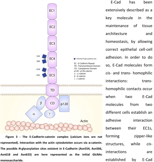
The extracellular domain is composed by five cadherin sub-domains (ECs) (EC1 to EC5). These ECs present on cadherins are sequences of 110 aa. residues, and are a key element for the classification of this family of proteins. In E-Cad, EC1 is the domain that is responsible for the Ca2+-dependent homophilic interactions between two cadherin molecules of adjacent cells, which is the reason why it is called the “strand swapping domain”. The normal conformation of E-Cad is only attained in the presence of Ca2+ in the surrounding microenvironment, which is endorsed with the fact that the Ca2+ -binding domains are extremely well conserved aa. sequences23. The other four domains (ECs 2-5) are called the “non-swapping cadherin domains”. EC5 is the domain closest to the transmembrane span, and its structure is distinct from the other E-Cad ECs, as it is characterized by the presence of additional four conserved cysteine (Cys) residues, which have an effect in the formation of strong cell-cell contacts24. Moreover, E-Cad has four possible N-glycosylation sites, two on EC4 (Asn554 and Asn566) and two on EC5 (Asn618 and Asn633)25, that are of crucial importance when considering E-Cad folding26 and cell-cell adhesion function27.
The cytoplasmic domain of E-Cad interacts with different types of catenins, namely α-, β- and p120-catenin. p120-catenin has central role in the maintenance of E-Cad at
the cell membrane, but it also influences the cytoskeleton organization, cell signaling processes and transcriptional regulation28. β-catenin is also a central molecule in the cytoskeleton organization, as it is the intermediate molecule between the cytoplasmic domain of E-Cad and α-catenin, which in turn is the responsible for the interaction with the actin cytoskeleton itself29(Figure 3).
E-Cad has been extensively described as a key molecule in the maintenance of tissue architecture and homeostasis, by allowing correct epithelial cell-cell adhesion. In order to do so, E-Cad molecules form
cis- and trans- homophilic
interactions: trans- homophilic contacts occur
when two E-Cad
molecules from two different cells establish an adhesive interaction between their EC1s, forming zipper-like structures, while cis-
interactions are established by E-Cad molecules in the same cell. The trans- and cis- complexes interact between themselves and with the catenins-actin complex in order to form mechanically stable E-Cad clusters at the adherens junctions, inhibiting cell motility and providing strong and normal associations between cells29,30.
Several mechanisms have been described to underlie E-Cad dysregulation. Genetic alterations (for example, the loss of exons 8 or 9) of the CDH1 gene were associated
Figure 3 - The E-Cadherin-catenin complex (calcium ions are not represented). Interaction with the actin cytosketelon occurs via α-catenin. The possible N-glycosylation sites existent in E-Cadherin (Asn554, Asn566, Asn618 and Asn633) are here represented as the initial GlcNAc monosaccharide.
with absence of a wild type (WT) E-Cad, with decreased cell-cell adhesion and increased cellular motility31-37. Epigenetic events, specifically hypermethylation in a CpG island of one 5’-CDH1 promoter38, are observable in several types of carcinomas and are have also been associated with loss of E-Cad gene expression. MicroRNAs (miR), such as members of the miR-200 family also regulate CDH1 expression by targeting transcriptional repressors (in this case, ZEB1 and ZEB2)39. All the alterations leading to decreased E-Cad mRNA expression and protein levels are usually associated with loss of cell-cell adhesion and a new capacity of epithelial cells to invade and metastasize, hence a poor clinical outcome 40,41. The functional loss of E-Cad is one of the best characterized molecular alterations during tumor progression of carcinoma cells (cancer cells arising from epithelial tissues), as emphasized by Hanahan and Weinberg in “Hallmarks of Cancer: The Next Generation”42. Moreover, there is a myriad of transcriptional repressors that act frequently on E-Cad promoter (for example, Snail, Slug and Twist21), that are specifically expressed at the invasive front of human cancers and are highly regulated by pathways that promote tumor progression, such as Wnt and TGF-β43. The protein levels of E-Cad can also be altered due to the action of several receptor tyrosine-kinases (RTKs), such as the endothelial growth factor recetor (EGFR) or the insulin-like growth factor I receptor (IGF-IR), that phosphorylate E-Cad, ultimately resulting in its ubiquitylation by Hakai ligase (and subsequent protein degradation)43-45.
All these types of alterations can be considered major causes of E-Cad dysfunction in GC41. Nevertheless, a significant percentage of GC patients harboring E-Cad dysfunction cannot be explained solely at the genetic or epigenetic levels46,47. Several studies have been pointing towards the impact of glycosylation alterations in the regulation of E-Cad in this disease context, as there are several cases that cannot be explained solely by genetic or epigenetic mechanisms3. The importance of these post-translational modifications (PTMs) in GC will be further addressed later.
1.3 Protein Glycosylation
Life as we know it cannot be explained solely by the flux of information from DNA to RNA and from RNA to proteins. Proteins can be modified in a wide variety of manners
by a wide variety of molecules, resulting in the huge biological complexity observed. Glycosylation is a universal process of the majority of eukaryotic cells and one of the most frequent occurring PTM, especially when considering transmembrane proteins, since their associated glycans contribute to numerous biological functions, such as cell adhesion, molecular trafficking, signal transduction pathways and endocytosis48-51. Glycans exist as the product of covalent attachment of carbohydrate structures, such as oligo- and polysaccharides, to proteins, lipids, carbohydrates or other organic compounds, and can be categorized in several different families of glycoconjugates (Figure 4). The attachment reaction is catalyzed by specific glycosyltransferases that use a specific set of sugars as donor substrates52. There are several types of glycans linked to proteins, and the vast majority of those are either N- or O-linked glycans50. N-glycosylation is characterized by the addition of oligosaccharides to asparagine(Asn) residues of the proteins, in a consensus sequence Asn-X-serine(Ser)/threonine(Thr), with X being any aa. other than proline(Pro)53,54, while in O-glycosylation the attachment reaction occurs in hydroxyl groups of either Ser or Thr residues55,56.
1.3.1 N-glycosylation
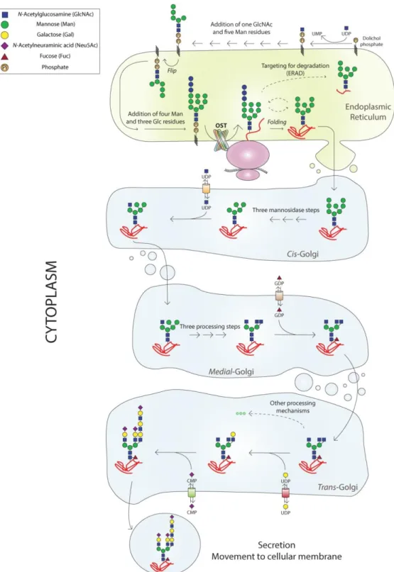
N-glycosylation (Figure 5) is initiated at the endoplasmic reticulum (ER) membrane
where dolichol phosphate (Dol-Pi) located on the cytoplasmic side of the ER functions
as a membrane anchor to the formation of the oligosaccharide structure composed by two N-acetylglucosamine (GlcNAc) and five mannose (Man) residues, Dol-Pi-Pi
-GlcNAc2Man5. Subsequently, this oligosaccharide structure is flipped to the luminal
side of the ER membrane, where four Man and three glucose (Glc) residues are added. This lipid-linked oligosaccharide precursor is then transferred to an Asn residue of a polypeptide chain, that is being translated and arising from the translocon, by the action of oligosaccharyltransferase (OST) enzyme complex50,57,58, which is why N-glycosylation is considered a co- and post-translational modification (CPTM). It is noteworthy that GlcNAc2Man9Glc3 acts as a ligand for chaperones from the lectin
family such as calnexin and calreticulin, and that the complex formed has a role in protein quality control and folding (the calnexin/calreticulin cycle)58. In that process, the Glc residues and one Man residue are removed in the ER by the action of glycosidase II and mannosidase, respectively. The glycoprotein can then continue to the Golgi or be degraded, with its fate depending essentially on its N-glycosylation and folding status59. In the cis compartment of the Golgi apparatus it is common that additional Man residues are removed from the glycoprotein, yielding a GlcNAc2Man5
glycan linked to the Asn residue. This is the main oligosaccharide structure that allows the sequential action of several N-acetylglucosamyniltransferases (GnTs) and glycosidases, resulting in the formation of a complex N-glycan. This assembly can then be further modified and extended by the addition of sugar residues such as GlcNAc, galactose (Gal), fucose (Fuc), Glc, and sialic acid(Neu5Ac). There is still a possibility of degradation of badly constructed structures at the trans-Golgi, especially the Man-6-Pi
-Receptor delivers a largely mannosylated N-glycan for the endosomes. Usually however, the final structure is moved from the trans-Golgi compartment to vesicles in order to be specifically delivered to the cellular membrane – in the case of transmembrane proteins such as E-Cad – or to be secreted49,50,54.
Figure 5 - N-glycosylation process occurs in the endoplasmic reticulum and the Golgi compartment. The synthesis of the Dol-Pi-Pi-GlcNAc2Man9Glc3 structure is necessary for the action of oligosaccharyltransferase (OST)
that transfers the glycan from the activated dolichol carrier to the protein. The protein can then be folded correctly and go to the Golgi compartment, or targeted for degradation. In the Golgi, the action of several glycosyltransferases and glycosidases determines the final glycoprotein that is formed.
1.3.2 O-glycosylation
The most common example of an O-glycosylated glycoprotein is the mucin, which has several N-acetylgalactosamine (GalNAc) monosaccharides covalently α-linked to several Ser and Thr residues in tandem repeats. This O-glycosidic bond formation is controlled by a member of the UDPGalNAc-polypeptide
N-acetylgalactosaminyltransferase (GalNAc-T) family60,61. This GalNAc residue can then be further extended with several monosaccharides such as Gal, GlcNAc, Fuc and Neu5Ac (whereas Man and Glc, for example, are not present in mucin-type O-glycans at all). This extension process occurs with the action of several distinct glycosyltransferases (for example, Core 1 Gal-transferase [C1GalT1] and Core 2 β1-6 N-acetylglucosaminyltransferase [C2GnT]) and originates eight O-GalNAc glycan core structures: cores 1 to 4 are the so-called common cores, and cores 5 to 8 are the additional cores61,62. There are several other types of glycosylation, namely, O-mannosylation, O-GlcNAcylation, and O-fucosylation. For the purpose of the present work, only O-mannosylation will be further explored.
1.3.3 O-mannosylation
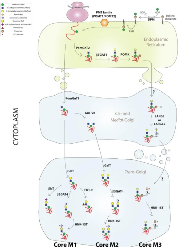
Protein O-mannosylation (Figure 6) consists in the covalent linkage of Man to Ser or Thr residues of nascent proteins, and that is the reason why it is considered, as N-glycosylation, a CPTM63. This process begins in the cytosolic face of the ER with the synthesis of Dol-Pi-Man from Dol-Pi and GDP-Man. The produced Dol-Pi-Man is then
flipped to the luminal side of the organelle, where the action of enzymes from the protein O-mannosyltransferase family - POMT in mammalian organisms, PMT in yeast (for example) - takes place. These enzymes are responsible for the transference of the Man monosaccharide from the activated lipid donor to the nascent protein, with the inversion of the anomeric configuration of the Man residue added (leading to the formation of an α-D-mannosidic linkage). In mammalian organisms, it seems that the two most important POMTs are POMT1 and POMT2, and that POMT1 and POMT2 genes coexpression is necessary in order to achieve the intended enzymatic activity64. This O-Man can then be elongated in the Golgi apparatus with a GlcNAc
monosaccharide, in a reaction catalyzed by glycosyltransferases such as protein O-linked-mannose β-1,2-N-acetylglucosaminyltransferase 1 (POMGnT1)65, Manα1-O- Ser/Thr β1,4GlcNAc-transferase (POMGnT2) or GnT-IX (also known as GnT-Vb). These
Figure 6 - O-mannosylation process initiates in the endoplasmic reticulum by the action of POMT1 or POMT2 that transfer a mannose residue to a nascent protein. The consequent action of several glycosyltransferases determines the type of structure that is formed. O-mannosyl glycans can be divided in cores M1, M2 and M3, according to the type of linkage between the mannose and the GlcNAc residues (adapted from 34).
enzymes do not have, however, a redundant purpose, as the linkage between the GlcNAc and Man sugar residues is not the same, which in turn allows the formation of several O-Man-glycans, divided from cores M1 to M3 (Figure 6). This nomenclature to the set of core O-Man structures was first proposed in 2013 in a publication from the Campbell laboratory66, and revised by Jeremy Praissman and Lance Wells in 201467, in order to approximate to the one that is accepted for O-GalNac glycans. It is also worth mentioning that not all these enzymes have been observed in every tissue studied, which can possibly imply a tissue-specific O-mannosylation pattern67.
This core structures can then be extended by the addition of a variety of monosaccharides, such as Gal, Neu5Ac, Fuc or glucuronic acid (GlcA). At least 23 different O-Man glycan structures have been described by now, the majority of which were detected in mammalian brain tissue67-70. The constant improvements of glycomic and glycoproteomic technologies will be of huge impact in the attempt to unravel novel O-Man structures that can, for instance, be used as important biomarkers.
1.4 Glycosylation in gastric cancer: the importance of
N-glycosylation of E-Cadherin in a gastric cancer context
Glycans have a particular significance in cell biology when considering the cell-extracellular matrix (ECM) interactions, cell-cell adhesion, cell signaling and communication, and in the immune system (Figure 7)51,71. It is well known that aberrant glycosylation occurs in all types of cancer, and that there are many specific glycosyl epitopes that constitute the tumor associated antigens (TAA), used often as biomarkers for cancer detection72-74. Alterations in the N-glycosylation pattern of cell-cell adhesion molecules such as cadherins3,25,27,75,76 have been correlated with cancer invasion and metastasis.
The frequent dysfunction of E-Cad expression observed in Diffuse-type GC patients was observed by Joo et al. in 200246. In effect, this decreased expression was found at earlier stages of Diffuse GC, while in the Intestinal subtype the alteration occurs typically at later stages. Moreover, when the alteration on E-Cad expression is observed in any sub-type of GC, it is usually associated with a poor prognosis for the
patient3. It has been described that approximately 70% of GC cases present any structural alteration in CDH1 gene, but rather a delocalization of E-Cad from the cell membrane to the cytoplasm47, which suggests that other mechanisms rather than genetic or epigenetic are regulating E-Cad functions in cancer77,78.
It is now widely accepted the fundamental role of glycosylation (particularly N-glycosylation) in the correct molecular organization of E-Cad in the adherens junctions76, as well as its impact in the process of E-Cad expression, and in the modulation of its biological functions27,79. As previously referred, E-Cad has four potential N-glycosylation sites, two in EC4 and two in EC5. The N-glycans that are attached to E-Cad depend on an intricate network of enzymes (glycosyltransferases) giving rise to different glycans depending on the tissue type and the physiopathological context. The effect of the different glycans on E-Cad activity has been demonstrated, for example, by the enzymatic competition between the
N-acetylglucosaminyltransferase-III and N-acetylglucosaminyltransferase-V (GnT-III/GnT-V) glycosyltransferases. GnT-III is encoded by the MGAT3 gene, and is responsible for the addition of a GlcNAc residue via β-1,4 linkage to the central Man of the [Asn]-GlcNAc2Man3(GlcNAc2) core structure, while GnT-V, encoded by the MGAT5 gene, adds
Figure 7 - The importance of glycosylation in the formation of a primary tumor and in the metastatic process. Glycans are important in the cell-cell adhesion process, in cell-extracellular matrix interactions, as well as in several other processes.
GlcNAc via β-1,6 linkage to an external α6-Man80. GnT-III is considered of vital importance in N-glycan biosynthesis, as its action precedes that of GnT-V. Therefore, the bisected oligosaccharide cannot be used as a substrate by GnT-V, precluding the synthesis of branching structures81,82.
In highly metastatic tumors, it is common to observe an increased activity of GnT-V and, consequently, an increased expression of β-1,6 GlcNAc branching N-glycan structures83. On the other hand, when GnT-V is not present (i.e. MGAT5-knockout mice) cancer metastasis is suppressed in a noticeable manner84. GnT-III has been described to exhibit an enzymatic priority, which contributes to the suppression of cancer metastasis by increasing the number of bisecting GlcNAc structures and, at the same time, by impeding the formation of branching GlcNAc structures85. In addition, an improvement in the cell-cell adhesion was documented when GnT-III was being expressed, as a result of an E-Cad arrest at the cell membrane and delayed turnover75. In 2008, the influence of the E-Cad-catenin complex on GnT-III expression was determined, suggesting that this complex may act as an inductor of GnT-III expression, which in turn allows the formation of bisected GlcNAc N-glycans77. The relationship between E-Cad overexpression and the inhibition of the RTKs IGF-IR and the insulin receptor (IR) was described by our group using MDA-MD-435 cancer cell line (that endogenously does not express E-Cad at the mRNA or protein levels)86, where stimulation of these cells with insulin or insulin-like growth factor I (IGF-I) induced a decrease in the quantity of bisecting GlcNAc N-glycans (in general and attached to E-Cad), that was accompanied by delocalization of E-Cad to the cytoplasm and with an alteration to a more fibroblastoid-like appearance86. Furthermore, Pierce et al. determined that the action of GnT-V on N-Cad’s EC2 and EC3 contributed to a decrease of homotopic cell-cell interactions and to the decrease of several intracellular (outside-in signal(outside-ing) pathways also (outside-involved (outside-in cell adhesion (levels of phosphorylated-ERK are enhanced when GnT-V was knockdown)87.
Taking into consideration that E-Cad dysfunction has a central role in GC development and progression, our group has been particularly interested in characterizing the role of glycosylation as a key molecular mechanism responsible for E-Cad impairment in cancer. In 2009, the group established a clear association between E-Cad expression and the upregulation of the GnT-III transcription levels.
Furthermore, the decrease of β-1,6 branched structures (catalyzed by GnT-V) was noticeable when a lectin specific for this type of structures was used (Biotinylated
Phaseolus vulgaris Leucoagglutinin [L-PHA]), together with a slight increase of bisecting
GlcNAc structures (catalyzed by GnT-III). The role of GnT-III in the regulation of E-Cad transcription was also studied: using siRNA to silence the gene that codifies for the enzyme, the group observed that the level of E-Cad’s mRNA remained unaltered, when compared to control cells (non-silenced). Nevertheless, silencing of GnT-III induced a significant alteration in the localization of the protein, with E-Cad exhibiting an increased cytoplasmic expression, and mislocalization from the membrane. Interestingly, the decrease in bisecting GlcNAc structures on E-Cad, detected by
biotinylated Phaseolus vulgaris Erythroagglutinin (E-PHA - lectin that specifically
recognize bisecting GlcNAc N-glycans), together with an increase of β-1,6 branched glycans, was observed when GnT-III was silenced. In vivo studies were performed in diffuse gastric carcinomas, and a significant mislocalization of E-Cad expression was observed in tumors, concomitantly with a decrease in bisecting GlcNAc structures and higher levels of the branching structures, which validated the in vitro results78. Moreover, our group also described the impact of GnT-V activity in E-Cad expression in a GC context, and how the absence of this glycosyltransferase (and its product) results in a normal distribution of E-Cad in the cell. Also, the importance of specific N-glycosylation sites was explored, with Asn554 as the selected site that is modified with β-1,6 GlcNAc N-glycans structures, thereby assuming a central role in the regulation of E-Cad biological functions, namely by interfering with the formation of the E-Cad-catenins complex27.
Considering all these results, it seems that there is a mutual regulatory mechanism between GnT-III expression and E-Cad-mediated cell-cell adhesion80, and that the modification of E-Cad by bisecting GlcNAc N-glycans induces the recruitment of catenins to the adherens junction, stabilizing the adhesion complex, and impairing cell invasion and metastasis (Figure 8). Accordingly, GnT-V activity in a GC background leads to the formation of tri- or tetra-antennary N-glycans that can be extended in a wide variety of ways, such as poly-N-acetylactosamine (polyLacNAc) - that is composed by a polymer of GlcNAc and Gal monosaccharides - or terminal Neu5Ac residues88. The modification of E-Cad with β-1,6 GlcNAc branched structures affects cell-cell adhesion
capacity27, by altering the cellular localization of E-Cad and by modifying the cell morphology.
1.5 O-mannosylation and E-Cadherin: evidences of a “new”
player in the adhesion game
Glycoproteins can be modified in a comprehensive variety of ways, such as the addition of O-Man monosaccharides to its Ser/Thr residues contributing to their stability and biological functions. This O-mannosylation process was thought to be for a long time, exclusive of organisms such as Saccharomyces cerevisiae (S. cerevisiae). The first time O-mannosylation was described was in 1968 by Sentandreu and Northcote, in a glycopeptide that composed the yeast cell wall89. It was only in 1993
Figure 8 - Alterations in the structure and function of E-Cadherin occur due to the action of GnT-III and GnT-V. Enzymatic activity of these glycosyltransferases determines the type of glycans formed, which in turn contributes to the promotion or suppression of cancer metastasis (adapted from 67).
that Strahl-Bolsinger et al. described the gene PMT1, which codifies for the protein PMT1 responsible for the initial step of the yeast O-mannosylation process90. The same group described a few years later the existence of five additional PMT genes in S.
cerevisiae, which led them to hypothesize the existence of different protein substrate
specificities to the different PMTs91.
In 1996, however, the idea that O-mannosylation was a PTM exclusive from yeast was abandoned, as Martín-Blanco and García-Bellido proved the existence of a protein in Drosophila melanogaster (D. melanogaster), rotated abdomen (rt), that presented a high homology to the S. cerevisiae PMTs92. It is interesting that in Drosophila and in
Saccharomyces, both rt and PMT, respectively, exhibit important roles in the
development of the organisms, and that mutations in those genes were potentially lethal to the organisms carrying them93.
In 1999, Jurado et al. identified a putative Human homolog of D. melanogaster’s rt, which they named Protein O-mannosyltransferase 1 (POMT1). At that time, they were capable of assigning the gene POMT1 to chromosome 9q34.1, and computer simulations allowed them to predict that the possible product POMT1 would be a transmembrane ER protein94. At that moment, and bearing in mind what was known in
Drosophila rt, the authors suggested that POMT1 gene mutations might be the
explanation for some uncharacterized forms of congenital muscular dystrophies. Later, the first review of the O-mannosylation process in mammalian organisms was published, and the molecular organization of the dystroglycan complex, which is composed by α-dystroglycan (α-DG), β-dystroglycan (β-DG), laminin, dystrophin and actin, was illustrated for the first time (this complex is of great importance, in a biological context, as it is responsible for connecting the ECM and the cellular cytoskeleton in skeletal muscle and in Schwann cells). Tamao Endo proposed that O-mannosylation should has an important role in the basic biological function of α-DG (the protein linker between laminin and β-DG) as this glycoprotein was depicted as being heavily O-mannosylated56. A lot of work has been done since, especially relating
O-mannosylation and α-DG in a pathological context, and it is now known that defects
in this process result in a heterogeneous group of congenital muscular dystrophies (CMDs)66,69,95,96. The most severe type of CMD is called the Walker-Warburg syndrome, and is essentially characterized by malformations of the brain of the subject, often
resulting in a fatal outcome97. The study of CMDs extensively contributed to the understanding of the O-mannosylation process. Nevertheless, in 2013 new insights about the O-mannosylation process were provided, but this time referring to the association between O-mannosylation and cadherins.
Vester-Christensen et al. modified a cell line (MDA-MB231, which is derived from a metastatic site of mammary gland, but has an epithelial morphology) to solely exhibit
O-Man in its O-glycoproteome98. The fact that the cells were unable to produce extended O-GalNAc glycans due to the knockout of COSMC (the chaperone necessary for C1GalT1 activity), or to further extend the O-Man monosaccharide due to the
knockout of POMGNT1, allowed them to look exclusively to the O-mannosylation sites
existent in all kinds of proteins present in the cell. In order to do that however, they first needed to get rid of the N-glycans and the short GalNAc O-glycans present in the cell samples, which they did by performing two enrichment steps, using peptide-N-glycosidase F (PNGase F - to remove the N-glycans) at first, and subsequently using a short Concanavalin A (Con A) column (Con A binds α-Man residues, and the O-GalNAc glycans are eluted). The analysis of the nanoflow liquid chromatography-mass spectrometry results confirmed the existence of a large number of O-Man glycosites on α-DG, as it was expected. But the most intriguing fact is, of a total of 52 glycoproteins identified, 37 of those belonged to the cadherin family of proteins (Figure 9), which accounted for approximately 56% of the total number of O-Man
glycosites covered on those 52 glycoproteins. On E-Cad, a type 1 cadherin, nine O-mannosylation sites were identified, distributed between EC2 (3), EC3 (2) and EC4 (4), and six sites were assigned as putative on ECs 2 to 5 (Figure 10).
Simultaneously, Lommel
et al. explored the importance of
O-mannosylation on E-Cad’s biological function99. In order to do that they used
Pomt2 deficient/null mice,
as the adhesion process (and E-Cad in particular) has a central role in the epithelial to mesenchymal transition during embryogenesis (in a previous study by the same group they demonstrated that the absence of Pomt1 is lethal to mouse embryos100). Pomt2+/+ WT and heterozygous Pomt2+/- mice developed normally, and in a very similar manner. However, intercrosses between Pomt2+/- mice did not generate offspring, as the Pomt2-/- inheritance is lethal even at the embryo phase. The authors isolated these embryos and cultivated them in vitro, which allowed them to perceive the inability of Pomt2-/- to generate the blastocyst. Additionally, the authors used an inhibitor of fungal POMTs rhodanine-3-acetic acid derivative, compound 5a (R3A-5a), to selectively inhibit the activity of POMT in Madin-Darby canine kidney (MDCK) cells. They observed using an antibody (VIA4-1) specific for an Man linked epitope on α-DG, that 50 µM of R3A-5a was enough to inhibit the
O-Figure 10 - Glycosites identified and predicted on E-Cadherin to date. There are a total of 4 N-glycosylation sites, 9 O-mannosylation sites (identified in 84) and 1 O-GalNAc site, distributed on the protein as depicted. A total of 6 O-mannosylation sites were also predicted by the Clausen’s group. The glycosylation sites represented were numbered considering the propeptide as part of E-Cadherin protein.
mannosylation of α-DG. Having this in mind, the group used R3A-5a when cultivating embryos, which arrest their development at the morula to blastocyst transition, with the phenotype observed being in everything similar to that of the Pomt2-/- embryos. To validate all these results, the authors generated an antibody, T[α1-Man], against a Thr
O-Man-conjugated epitope, to detect O-mannosylated proteins. The conjugated use of
T[α1-Man] with the inhibitor R3A-5a and Pomt2-/- mice allowed them to better understand the importance of O-mannosylation in cell-cell adhesion, and how that influences the development of the embryo. It seemed that, in the R3A-5a-treated embryos, the failure in the morula-to-blastocyst transition was due to an abnormal formation of cell-cell contacts, namely adherens junctions, which suggested that defects in the O-mannosylation of E-Cad could lead to an impairment of cell-cell adhesion. In fact, aggregation assays performed using MDCK cells (that express high endogenous levels of E-Cad) suggest that O-mannosyl glycans affect cadherin mediated cell adhesion, specifically due to the
high aggregation inhibition observed when using the T[α1-Man] antibody and an E-Cad blocking antibody (Figure 11).
The influence of glycosylation for E-Cad function has been extensively described, namely the influence of two specific glycosyltransferases – GnT-III and GnT-V – in the N-glycan landscape composing this transmembrane protein. Considering this relationship, and taking into account the evidences that indicate that E-Cad
O-mannosylation is essential for its correct
biological function, the main purpose of this project is to assess how O-mannosylation, and its interplay with N-glycosylation, regulate E-Cad adhesion function, and how this process ultimately influences cancer development and progression.
Figure 11 - Slow aggregation assays performed using the MDCK cell line using an E-Cadherin antibody (DECMA-1) and a T[α1-Man]-specific antibody allowed to understand the importance of O-mannosyl glycans in cell-cell adhesion. EGTA (blocks the Ca2+ -dependent cadherin-mediated adhesion), IIH6 and VIA4-1 (antibodies directed against O-mannosyl epitopes linked to α-dystroglycan) were used as controls86
Aims
The main purpose of this project is to understand the role of O-mannosylation and its interplay with N-glycosylation in the modulation of E-Cad functions in tumor development and progression.
In order to accomplish this goal, three specific aims were designed:
1) To evaluate the O-mannosylation profile in different GC cell line models
2) To determine the pattern of E-Cad O-mannosylation in a cancer context using in
vitro cellular models, in order to evaluate the importance of this PTM on E-Cad
biological function
3) To characterize the relationship between N-glycosylation and O-mannosylation of E-Cad in cancer
3.1 Cell lines and cell culture
MKN28 (JCRB0253) and KATO III (ATCC HTB-103) cell lines were cultured at 37ºC in an incubator with 5% CO2 in RPMI 1640 medium with Glutamax (Gibco, Invitrogen)
containing 10% of fetal bovine serum (FBS) (Gibco, Invitrogen), and 1% of the antibiotics penicillin/streptomycin (P/S; Invitrogen).
MKN45 MOCK (empty vector) and MKN45+GnT-V (vector containing N-acetylglucosaminyltransferase V) cell lines had already been previously stably transfected81,101, and were cultured at 37ºC in an incubator with 5% CO2 in RPMI 1640
medium with Glutamax (Gibco, Invitrogen) containing 10% of FBS (Gibco, Invitrogen), 1% of the antibiotics P/S (Invitrogen), and 500 µg/mL of the selection antibiotic G418 (Gibco, Invitrogen).
3.2 Immunofluorescence
Cells from MKN28 and KATO III cell lines were cultured in coverslips in 6 well plates. When confluent, the medium was removed and the cells were washed 3 times with PBS. After that, the 6 well plates were left on ice as the cells were fixed with methanol (previously stored at -20ºC). The cells in the coverslips were then washed 3 times with PBS, and subsequently blocked with a BSA 5% solution in PBS for 30 minutes at room temperature. After that, the primary mouse monoclonal anti-E-Cad IgG2a antibody (clone 36, BD Transduction Laboratories and Cell Signaling), diluted 1:200 in the BSA 5% solution in PBS, was used to incubate the coverslips for 1 hour at room temperature. The coverslips were then washed 3 times with PBS and, from this point forward, the procedure was made by avoiding light contact with the samples. Incubation with goat Alexa Fluor 488 anti-mouse (1:500 – Invitrogen) was followed by 6 consecutive washing steps with PBS and by the use of DAPI (1:100) as a nuclear marker. A VECTASHIELD mounting medium for immunofluorescence kit was used, and the slides were stored at -20ºC and in the dark, until visualization in a Carl Zeiss Apotome Axiovert 200 M Fluorescence Microscope.
3.3 PNGase F digestion
PNGase F digestion was performed in order to analyze O-linked α-Man glycans specifically, avoiding the interaction of the α-Man residues present in the N-linked glycans with the lectin used (Con A). PNGase F is an amidase that cleaves that cleaves between the innermost GlcNAc and Asn residues of high-Man, hybrid and complex oligosaccharides from N-linked glycans in proteins. Total cell lysates (10 to 30 µg) and immunoprecipitated samples were combined with denaturing buffer and incubated at 100ºC for 10 min. Samples were digested overnight (ON) with 1 unit of PNGase F kit (New England BioLabs) at 37ºC, 300 rpm. The deglycosylated proteins were loaded onto 7,5% SDS-PAGE and immunoblotting with anti E-Cad or Con A lectin. In order to control the digestion process, samples were incubated in the same exact conditions but without the enzyme.
3.4 Western-Blot and lectin blot analysis
After reaching confluence status, cell cultures were submitted to a washing step with autoclaved Phosphate-Buffered Saline (PBS). Cell protein lysates were obtained by lysing cell cultures using a cold PBS solution containing 1%(v/v) Triton X-100, 1%(v/v) NP40, cOmplete (a protease inhibitor cocktail – Roche, 1 tablet/50 mL buffer) and a phosphatase inhibitor cocktail (composed by Phenylmethanesulfonyl fluoride (PMSF) and Vanadate - Sigma, 1:100 dilution). This lysis buffer was applied on the cell monolayers (that were kept on ice) for the period of 15 minutes. After that, the cells were scrapped and centrifuged at 13200 rpm for 10 minutes at 4ºC. Prior to use in a Western-Blot (WB) or lectin Blot experiment, total protein content of each lysate was quantified using a BCA protein assay kit (Pierce). Equal amounts of total cell protein lysates (10-35 µg) were submitted to 7,5% SDS-PAGE electrophoresis after the addition of Elution buffer (Laemmli 4X, β-mercaptoethanol and Bromophenol Blue) to each sample and performing a denaturing step (98ºC, 5 minutes). After electrophoresis, the separated proteins were transferred during 2 hours at 50V to nitrocellulose membranes (Amersham Biosciences). Ponceau solution was then used for rapid


![Figure 11 - Slow aggregation assays performed using the MDCK cell line using an E-Cadherin antibody (DECMA-1) and a T[α1-Man]-specific antibody allowed to understand the importance of O-mannosyl glycans in cell-cell adhesion](https://thumb-eu.123doks.com/thumbv2/123dok_br/17611546.820358/36.892.507.764.534.932/aggregation-performed-cadherin-antibody-specific-antibody-understand-importance.webp)
