Joana Margarida Franco Dantas
Mestre em Bioquímica Estrutural e Funcional
Characterization of extracellular electron
transfer networks in
Geobacter
sulfurreducens
, a key bacterium for
bioremediation and bioenergy applications
Dissertação para obtenção do Grau de Doutora em Biotecnologia
Orientador: Prof. Doutor Carlos A. Salgueiro, Professor
Auxiliar com Agregação, Faculdade de Ciências e
Tecnologia, Universidade Nova de Lisboa
Júri:
Presidente:
Arguentes:
Vogais:
Prof. Doutor Fernando Jorge da Silva Pina
Prof. Doutor Douglas Vinson Laurents Prof. Doutor Brian James Goodfellow
Doutora Sofia Rocha Pauleta
Prof. Doutor Carlos Frederico de Gusmão Campos Geraldes
Universidade Nova de Lisboa
Joana Margarida Franco Dantas
Mestre em Bioquímica Estrutural e FuncionalCharacterization of extracellular electron transfer networks
in
Geobacter sulfurreducens
, a key bacterium for
bioremediation and bioenergy applications
Dissertação para obtenção do Grau de Doutora em Biotecnologia
Orientador: Prof. Doutor Carlos A. Salgueiro, Professor Auxiliar com Agregação,
Faculdade de Ciências e Tecnologia, Universidade Nova de Lisboa
iii Characterization of extracellular electron transfer networks in Geobacter sulfurreducens, a key
bacterium for bioremediation and bioenergy applications
“Copyright”
Joana Margarida Franco Dantas Faculdade de Ciências e Tecnologia Universidade Nova de Lisboa
Todos os capítulos foram parcialmente reproduzidos de artigos publicados sob permissão dos editores originais e sujeitos às restrições de cópia impostos pelos mesmos.
v
Agradecimentos
O trabalho apresentado nesta tese envolveu a participação direta ou indireta de várias pessoas a quem gostaria de expressar os meus agradecimentos. Em especial gostaria de agradecer:
Ao Prof. Doutor Carlos Salgueiro, excelente orientador que providenciou todas as condições para desenvolver todo o trabalho descrito nesta tese, bem como pelo magnifico exemplo de respeito e dedicação pela investigação científica e pelos seus alunos captando sempre o melhor de cada um. Agradeço também por todo o conhecimento transmitido, pelo companheirismo, paciência, pela disponibilidade que sempre manifestou e pelos momentos de descontração, assim como pela confiança e estímulo para prosseguir e nunca desistir, mesmo em situações complicadas.
À Doutora Marta Bruix, pelo acesso ao espectrómetro de Ressonância Magnética Nuclear (RMN) Bruker Avance 800 utilizado em algumas das experiências desta tese, bem como pela ajuda na interpretação dos resultados e todo o conhecimento transmitido. Agradeço ainda pelo excelente exemplo de dedicação, determinação e paixão pela investigação científica.
Ao Doutor P. Raj Pokkuluri e Doutor O. Kokhan pelo trabalho na determinação da estrutura de raios-X do citocromo PccH e em alguns cálculos de docking, bem como pela disponibilidade demonstrada na
interpretação dos resultados experimentais.
À Prof. Doutora Teresa Catarino pela ajuda incansável na realização, interpretação e discussão das experiências de cinética, bem como disponibilidade para utilização do equipamento de stopped-flow e da
câmara anaeróbica no ITQB.
Ao Prof. Doutor David L. Turner agradeço a sua ajuda nos cálculos da estrutura do OmcF no estado reduzido, bem como pelas discussões cruciais para a interpretação de alguns resultados experimentais.
À Prof. Doutora Isabel Couto pela ajuda e disponibilidade em questões relacionadas com biologia molecular.
À Doutora Leonor Morgado, por todas as discussões profícuas, pela partilha de conhecimento, pelo trabalho de equipa, pelo perfeccionismo demonstrado e dedicação pela investigação científica, bem como pelos momentos de lazer e boa disposição.
A todos os meus colegas de laboratório, bem como estudantes de licenciatura e mestrado que foram passando pelo Lab 6.11 durante os últimos anos, pelo dinamismo, diversidade, simpatia, boa disposição, admirável capacidade de trabalho e com os quais também aprendi imenso.
vi
À Fundação para a Ciência e Tecnologia pelo suporte e financiamento, através da bolsa de doutoramento, SFRH/BD/89701/2012, e pelos projetos PTDC/BBB-BQB/3554/2014 e UID/Multi/04378/2013 co-financiados por ERDF através de fundos nacionais, no âmbito do Acordo de Parceria PT2020 (POCI-01-0145-FEDER-007728).
Ao Laboratório de RMN da FCT-UNL, integrado na Rede Nacional de RMN e financiado pela Fundação para a Ciência e Tecnologia através do projeto, RECI/BBB-BQB/0230/2012, pelo acesso aos espectrómetros de RMN.
vii
Resumo
As bactérias do género Geobacter têm despertado interesse devido ao seu impacto no meio ambiente
e às suas aplicações biotecnológicas, incluindo a biorremediação de contaminantes orgânicos e inorgânicos, produção de bioenergia e bioeletrónica. Além da capacidade de transferência de eletrões para aceitadores terminais, estas bactérias conseguem também aceitar eletrões de elétrodos, um processo que é atualmente explorado na área da eletrossíntese microbiana. Estas aplicações baseiam-se num processo de troca de eletrões entre as células e o seu exterior, designado de transferência extracelular de eletrões (TEE). No entanto, os mecanismos subjacentes a estes processos encontram-se ainda em discussão. Com base em estudos genéticos e proteómicos identificaram-se citocromos do tipo c essenciais no processo de
TEE, localizados na membrana interna, periplasma e membrana externa da bactéria G. sulfurreducens.
Nesta Tese vão ser estudados alguns destes citocromos, tais como, o MacA, associado à membrana interna, os citocromos periplasmáticos PpcA-E e PccH, e o citocromo OmcF localizado na membrana externa.
As interações moleculares entre as proteínas PpcA-E e os seus possíveis parceiros redox, nomeadamente, MacA, PccH e um análogo dos ácidos húmicos, foram avaliadas por espectroscopia de ressonância magnética nuclear (RMN), cinética e docking. A afinidade entre cada complexo redox foi
determinada por experiências de perturbação de desvio químico. Estes resultados demonstraram que as moléculas formam complexos reversíveis de baixa afinidade, compatíveis com uma transferência de eletrões rápida e seletiva, tipicamente observada entre parceiros redox.
A técnica de RMN foi igualmente utilizada para determinar a estrutura em solução do citocromo OmcF no estado reduzido, as alterações conformacionais dependentes da variação de pH e a sua dinâmica em solução.
O citocromo PccH foi estudado a nível bioquímico e estrutural, recorrendo às técnicas de dicroísmo circular, espectroscopia de UV-visível e RMN. Com base na sua estrutura, determinada por cristalografia de raios-X, demonstrou-se que esta é única entre os citocromos monohémicos do tipo c. De igual modo,
os potenciais de redução, determinados por titulações redox a diferentes valores de pH, são invulgarmente baixos em comparação com outros valores descritos para este tipo de proteínas. Considerando as suas
características estruturais e funcionais o citocromo PccH representa uma nova subclasse de citocromos monohémicos.
Os resultados obtidos e descritos nesta Tese constituem um importante contributo para a compreensão dos mecanismos de transferência eletrónica na bactéria G. sulfurreducens.
Palavras-chave: Transferência eletrónica, Geobacter sulfurreducens, Proteínas hémicas, RMN,
ix
Abstract
Geobacter bacteria have awakened significantly attention because of their impact on natural
environments and biotechnological applications that include the bioremediation of organic and inorganic contaminants, bioenergy production and bioelectronics. In addition to electron transfer towards extracellular terminal acceptors, Geobacter cells can also accept electrons from electrodes, in
current-consuming biofilms, a process that is currently explored in microbial electrosynthesis. These practical applications rely on an efficient transfer of electrons between the cell and its exterior, a process designated extracellular electron transfer (EET). However, the precise mechanisms underlying EET processes are still under debate. Genetic and proteomics studies have identified several c-type
cytochromes as key components for EET in G. sulfurreducens. These proteins are located at the
inner-membrane (IM), periplasm and outer-inner-membrane (OM). Examples of such cytochromes include the IM-associated cytochrome MacA, periplasmic cytochromes PpcA-E and PccH, as well as, the OM cytochrome OmcF, which were studied in this Thesis.
Molecular interactions between PpcA-E and their putative redox partners, including a humic substance analogue molecule, MacA or PccH, were probed by NMR spectroscopy, stopped-flow kinetics and molecular docking. For the interacting pairs, their binding affinity was also determined by NMR chemical shift perturbation experiments. The results obtained showed that the interacting molecules establish reversible low-binding affinity complexes in specific regions of the proteins to warrant a rapid and selective electron transfer, a typical feature observed for electron transfer reactions between redox partners.
In addition, NMR spectroscopy was also used to determine the solution structure of OmcF in the reduced state, its pH-dependent conformational changes and backbone dynamics.
A biochemical and structural characterization of the cytochrome PccH was also carried out using circular dichroism, UV-visible and NMR spectroscopic techniques. The structure of PccH determined by X-ray crystallography showed that it is unique among the monoheme c-type cytochromes. The
reduction potentials determined for PccH at different pH values by visible redox titrations are unusually low compared to those reported for other monoheme c-type cytochromes. Considering the structural and
functional features of PccH it was proposed that this protein represents a first characterized example of a new subclass of monoheme c-type cytochromes.
Overall, the results obtained constitute an important contribute to the current understanding of the G. sulfurreducens extracellular electron transfer mechanisms.
Keywords: Electron transfer; Geobacter sulfurreducens; Heme proteins, NMR; Protein interactions;
xi
Table of contents
1. Introduction ... 1.1 The Geobacter sulfurreducens bacteria ...
1.2 Cytochromes ... 1.3 Multiheme cytochromes ... 1.4 Extracellular electron transfer pathways in G. sulfurreducens ...
1.4.1 Characterization of cytochromes c ...
1.4.1.1 Structural characterization ... 1.4.1.2 Thermodynamic characterization ... 1.5 Current-consuming biofilms ... 1.6 Objectives and Thesis outline ... 1.7 References ...
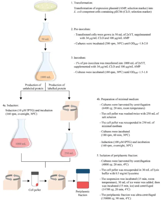
2. Materials and methods ... 2.1 Expression vectors and site directed mutagenesis ... 2.2 Heterologous expression ... 2.2.1 Unlabeled protein expression ... 2.2.2 13C, 15N uniformly labeled protein expression ...
2.3 Isolation of the periplasmic fraction ... 2.4 Protein purification ... 2.5 Protein purity evaluation ... 2.6 UV-visible analysis, quantification and molar extinction coefficient ... 2.7 References ...
3. Molecular interaction studies between the triheme cytochromes from G. sulfurreducens and
the redox active humic substances analog ... 3.1 Results and discussion ...
3.1.1 Equilibrium and kinetic studies probed by UV–visible spectroscopy ... 3.1.2 PpcAox-AQDS interaction studies ...
3.1.2.1 NMR chemical shift perturbation experiments ... 3.1.2.2 Molecular docking calculations ... 3.1.3 PpcAred- AH2QDS interaction studies ...
3.1.3.1 NMR chemical shift perturbation experiments ... 3.1.3.2 Molecular docking calculations ... 3.1.3.3 Effect of binding of AQDS on the thermodynamic properties of the redox centers in the
cytochrome PpcA ... 3.1.4 Interaction studies between AQDS and PpcB, PpcD and PpcE cytochromes ... 3.1.4.1 Backbone and side chain assignment of PpcB, PpcD and PpcE cytochromes ... 3.1.4.2 NMR chemical shift perturbation experiments ...
xii
3.1.4.3 Molecular docking calculations ... 3.2 Conclusions ... 3.3 Materials and methods ... 3.3.1 NMR studies ... 3.3.1.1 NMR samples preparation and experiments ... 3.3.1.2 Protein backbone assignment ... 3.3.1.3 Molecular interactions ... 3.3.1.4 Thermodynamic characterization ... 3.3.2 Kinetic studies ... 3.3.3 Molecular docking calculations ... 3.4 References ...
4. NMR interaction studies between inner membrane-associated and periplasmic cytochromes .... 4.1 Results and discussion ... 4.2 Conclusions ... 4.3 Materials and methods ... 4.3.1 NMR samples preparation and experiments ... 4.3.2 Complex interface and binding affinity ... 4.3.3 Molecular docking calculations ... 4.4 References ...
5. Solution structure and dynamics of the outer membrane cytochrome OmcF ... 5.1 Results and discussion ... 5.1.1 Heme spin state and axial ligands of OmcF ... 5.1.2 Sequential assignment and structure calculations ... 5.1.3 Quality and analysis of the structures ... 5.1.4 Comparison of cytochrome OmcF reduced and oxidized structures ... 5.1.5 Backbone dynamics ... 5.1.6 pH-linked conformational changes ... 5.2 Conclusions ... 5.3 Materials and methods ... 5.3.1 NMR samples preparation and experiments ... 5.3.2 Assignment of the heme proton signals ... 5.3.2.1 Reduced state ... 5.3.2.2 Oxidized state ... 5.3.3 Solution structure determination ... 5.3.4 Data Bank accession number ... 5.3.5 Backbone dynamics ... 5.3.6 pH-linked conformational changes probed by NMR ... 5.4 References ...
xiii
6. Biochemical and structural characterization of PccH... 6.1 Results and discussion ... 6.1.1 Purification of cytochrome PccH and PccH mutants ... 6.1.2 Spectroscopic characterization ... 6.1.3 Specific assignment of PccH heme axial ligands ... 6.1.4 Temperature dependence of PccH heme methyl signals ... 6.1.5 Crystal structure ... 6.1.6 Redox Bohr center ... 6.1.7 Analysis of PccH protein sequence and comparison with other cytochromes ... 6.1.8 Functional clues for PccH ... 6.2 Conclusions ... 6.3 Materials and methods ... 6.3.1 Molecular mass determination and heme quantification ... 6.3.2 Circular dichroism analysis ... 6.3.3 Redox titrations followed by UV-visible spectroscopy ... 6.3.4 NMR samples preparation and experiments ... 6.4 References ...
7. Future perspectives ... 7.1 Periplasmic triheme cytochromes ... 7.1.1 Backbone NMR resonance assignment in the reduced state ... 7.1.2 Solution structure determination of PpcD in the reduced state ... 7.2 Outer membrane cytochromes ... 7.2.1 Transmembrane helices and signal peptide prediction ... 7.2.2 Construction of the expression vectors ... 7.3 Materials and methods ... 7.3.1 NMR samples preparation and experiments ... 7.4 References ...
8. Conclusions...
A. Appendix ... A.1 Supplementary Figures ... A.2 Supplementary Tables ... A.2.1 NMR assignment of PpcB, PpcD and PpcE proteins in the oxidized state ... A.2.2 NMR assignment of the OmcF, PpcB, PpcD and PpcE proteins in the reduced state ……...…
xv
List of figures
1. IntroductionFig 1.1 Schematic representation of bacterial direct and indirect electron transfer mechanisms to the extracellular electron acceptors. ...3
Fig 1.2 Time line of the important discoveries associated with Geobacter sulfurreducens. ...5 Fig 1.3 Schematic representation of a c-type heme and the correspondent polypeptide binding motif. ...7 Fig 1.4 Time line of important discoveries related with the G. sulfurreducens cytochromes addressed in this
Thesis. ...13
Fig 1.5Structures of G. sulfurreducens cytochromes obtained in the oxidized state. ...15 Fig 1.6 Electronic distribution scheme and potentiometric redox titrations followed by visible spectroscopy for
monoheme (OmcF) and triheme (PpcA) cytochromes. ...17
2. Materials and methods
Fig 2.1 Overview of the Site-Directed mutagenesis method. ...32
Fig 2.2 Overview of the production and isolation scheme for the unlabeled and 13C, 15N labelled proteins ...33
3. Molecular interaction studies between the triheme cytochromes from G. sulfurreducens and the redox
active humic substances analog
Fig 3.1 Alignment of the amino acid sequences of periplasmic triheme cytochromes from G. sulfurreducens…44 Fig 3.2 UV–visible absorption spectra of PpcA and AQDS/AH2QDS. ...45
Fig 3.3 Assays of AH2QDS oxidation coupled to PpcA reduction and AQDS reduction coupled to PpcA
oxidation at 25 °C and pH 7. ... 47
Fig 3.4 Region of the backbone NH signals in the 2D 1H, 15N HSQC NMR spectra of PpcA in the oxidized and
reduced state at 298 K and pH 7.1. ... 51
Fig 3.5 Overlay of the 2D 1H, 15N HSQC NMR spectra of 15N-enriched PpcAox in the presence of increasing
amounts of AQDS. ... 52
Fig 3.6 Binding isotherms for PpcAox-AQDS interaction and selected regions from overlaid 2D 1H, 15N HSQC
NMR spectra. ... 53
Fig 3.7 Surface map of significantly perturbed residues in PpcA upon AQDS binding. ... 54
Fig 3.81H chemical shift changes of the heme methyls of PpcAox. ... 55
Fig 3.91H chemical shift changes |δPpcA- δPpcA+AQDS| in the βCH2 groups of the heme axial histidines ………....56
Fig 3.101H chemical shift changes of the AQDS signals in presence of PpcAox. ... 57
Fig 3.11 Reversibility of AQDS binding to PpcAox monitored by 1D 1H NMR. ... 58
Fig 3.12 PpcA–AH2QDS docked complexes calculated in DOCK 6.4 ... 58
Fig 3.13 Overlay of the 2D 1H, 15N HSQC NMR spectra of 15N-enriched PpcA in the presence of increasing
amounts of AH2QDS. ... 59
Fig 3.14 Variation of the 1H chemical shift of the PpcAred heme substituents... 60
Fig 3.15 Selected region of 2D 1H, 1H NOESY NMR spectra of PpcA acquired in the absence and presence of
AH2QDS. ... 61
Fig 3.16 Selected region of 2D 1H, 1H NOESY NMR spectra of PpcA acquired in the presence and absence of
AH2QDS. ... 62
xvi
Fig 3.18 Binding isotherms for PpcA:AH2QDS interaction and selected regions from overlaid 2D 1H, 15N HSQC
NMR spectra. ... 64
Fig 3.19 PpcA–AH2QDS docked complexes calculated in DOCK 6.7 ... 65
Fig 3.20 Expansions of 2D 1H, 1H EXSY NMR spectra obtained for PpcA in the absence and the presence of
AH2QDS. ... 66
Fig 3.21 Fitting of the thermodynamic model to the experimental data for PpcA in the presence of AH2QDS... 67
Fig 3.22 Oxidized fractions of the individual hemes and molar fractions of the 16 individual microstates for PpcA in the presence and absence of quinol at pH 7.5. ... 69
Fig 3.23 Overlay of the 2D 1H,15N HSQC NMR spectra of 13C,15N-enriched PpcB, PpcD and PpcE in the
presence of increasing amounts of AQDS. …... 72
Fig 3.24 Chemical-shift perturbation map of PpcB, PpcD and PpcE in the presence of AQDS. ... 74
Fig 3.25 Binding isotherms for PpcB, PpcD and PpcE redox complex with AQDS and selected regions from overlaid 2D 1H,15N HSQC NMR spectra. ... 75
Fig 3.26 Selected regions of 2D 1H,13C HMQC of PpcB, PpcD and PpcE in the absence and presence of
AQDS... 77
Fig 3.27 Variation of the 1H chemical shift of the heme methyls of PpcB, PpcD and PpcE. ... 78
Fig 3.281H chemical shift changes of the AQDS signals in presence of PpcB, PpcD and PpcE. ... 79
Fig 3.29 Docked complexes between PpcB, PpcD and PpcE and AQDS calculated with HADDOCK 2.2
webserver. ... 80
Fig 3.30 Strategy to specifically assign the backbone NH signals in 2D 1H,15N HSQC. ... 84
4. NMR interaction studies between inner membrane-associated and periplasmic cytochromes
Fig 4.1 1D 1H NMR spectra of cytochromes PpcA and MacA in the oxidized state. ... 97
Fig 4.2 1D 1H chemical shift changes of the heme methyls of PpcA. ... 98
Fig 4.3 Overlay of the 2D 1H,13C HMQC NMR spectra of PpcA obtained in the absence and in the presence of
MacA at 298 K, pH 7 and 20 mM final ionic strength. ... 99
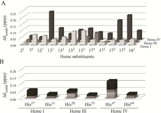
Fig 4.4 Variation of the combined chemical shift changes determined from the 1H and 13C signals of the PpcA
heme substituents and heme axial ligands. ... 100
Fig 4.5 Chemical shift perturbation on PpcA backbone NH signals. ... 101
Fig 4.6 PpcA-MacA docked complex calculated using the HADDOCK2.2 Web server. ... 102
Fig 4.7 Elution profile for the molecular exclusion chromatography of PpcA and MacA after the NMR chemical shift perturbation experiments. ... 103
Fig 4.8 Electrostatic properties of MacA monomer and PpcA. ... 104
Fig 4.9 Variation of the PpcA 1H heme methyl chemical shifts. ... 105
5. Solution structure and dynamics of the outer membrane cytochrome OmcF
Fig 5.1 Amino acid sequence of OmcF. ... 113
Fig 5.2 1D 1H NMR spectra of the reduced and oxidized OmcF obtained at 298 K, pH 7 and final ionic strength
100 mM. ... 114
Fig 5.3 Temperature dependence of the OmcF heme methyl signals in the oxidized form at pH 7 and 100 mM ionic strength. ... 116
Fig 5.4 2D 1H,15N HSQC spectra of OmcF in the fully reduced state. ... 117
Fig 5.5 Sequential NOE connectivities involving NH, Hα and Hβ protons observed in the 2D 1H,1H NOESY
spectrum for reduced cytochrome OmcF. ... 118
xvii
Fig 5.7 Average pairwise backbone and heavy atom rmsd values per residue of the family of 20 conformers
obtained for the cytochrome OmcF solution structure. ... 121
Fig 5.8 Solution structure of OmcF in the reduced state. ... 122
Fig 5.9 Comparison of OmcF lowest-energy solution structure with OmcF crystal structure. ... 123
Fig 5.1015N relaxation parameters for the cytochrome OmcF backbone in the reduced state. ... 124
Fig 5.11 pH-linked conformational changes in cytochrome OmcF. ... 126
Fig 5.12 Diagram of heme c numbered according to the IUPAC-IUB nomenclature ... 130
6. Biochemical and structural characterization of PccH Fig 6.1 Amino acid sequence of mature cytochrome PccH. ... 137
Fig 6.2 Purification of cytochrome PccH. ... 139
Fig 6.3 MALDI-TOF mass spectrum of PccH. ... 140
Fig 6.4 Spectroscopic characterization of PccH by circular dichroism. ... 141
Fig 6.5 UV-visible spectral features of cytochrome PccH as purified and in the reduced state. ... 142
Fig 6.6 1D 1H NMR spectra of the reduced and oxidized PccH obtained at 298K and pH 7. ... 144
Fig 6.7 Portion of a 2D 1H, 1H EXSY NMR spectrum of cytochrome PccH obtained for a partially oxidized sample. ... 145
Fig 6.8 2D 1H, 13C HMQC NMR spectrum of cytochrome PccH in the oxidized state. ... 146
Fig 6.9 Temperature dependence of the four PccH heme methyl 1H signals. ... 147
Fig 6.10 Crystal structure of cytochrome PccH from G. sulfurreducens. ... 149
Fig 6.11 Redox titrations followed by visible spectroscopy for PccH at pH 8 and pH 6. ... 150
Fig 6.12 Effect of the pH on the heme reduction potential values of cytochrome PccH. ... 151
Fig 6.13 pH dependence of the heme propionate and methyl 1H chemical shifts. ... 152
Fig 6.14 Polar interactions involving the heme propionates including a zinc ion. ... 153
Fig 6.15 Electrostatic potential displayed on surface representation of PccH. ... 154
Fig 6.16 Sequence alignment of the top six hits returned for the amino acid sequence of the mature PccH using the basic local alignment search tool. ... 155
Fig 6.17 Amino acid sequence comparison of G. sulfurreducens PccH cytochrome c with prokaryotic c-type monoheme cytochromes. ... 156
Fig 6.18 Comparison between PccH and Chlorobaculum tepidum cytochrome c554 structures. ... 157
Fig 6.19 A gallery of representative bacterial class I monoheme cytochromes including PccH shown as Cα cartoons ... 159
Fig 6.20 Expansions of the low-field region of 1D 1H NMR spectra obtained for cytochrome PpcD in the oxidized form in the presence of increasing amounts of PccH at 298 K, pH 7, 100 mM final ionic strength…... 160
Fig 6.21 Overlay of the 2D 1H,15N HSQC spectra of 15N-enriched PpcD in the presence of PccH in a 1:1 molar ratio. ... 161
Fig 6.22 Schematic representation of possible electron transfer pathways that involve PccH in G. sulfurreducens cells grown using graphite cathode as a sole electron donor and fumarate as terminal electron acceptor....162
7. Future perspectives Fig 7.1 2D 1H,15N HSQC spectra of reduced PpcB, PpcD and PpcE, acquired in 45 mM sodium phosphate buffer with NaCl to 100 mM ionic strength, 298 K. ... 174
xviii
Fig 7.3 Gel electrophoresis of PCR products in 0.8% agarose gel in TAE buffer. ... 181
8. Conclusions
Fig 8.1 Time line of the important outcomes associated with the biochemical characterization of electron transfer components of G. sulfurreducens. ... 187
A. Appendix
xix
List of tables
1. IntroductionTable 1.1 Analysis of proteome and phenotypes of different deletion mutant strains of G. sulfurreducens for cells
grown in the presence of the acetate and Fe(III) oxide or Fe(III) citrate... 9
Table 1.2 Data set of G. sulfurreducens c-type cytochromes participating in extracellular electron transfer
pathways... 19
Table 1.3 Heme reduction potentials and pairwise interactions of the fully reduced and protonated forms of PpcA, PpcB, PpcD and PpcE. ... 19
Table 1.4 Macroscopic pKa values of the redox-Bohr center for PpcA, PpcB, PpcD, and PpcE at each stage of
oxidation... 20
2. Materials and methods
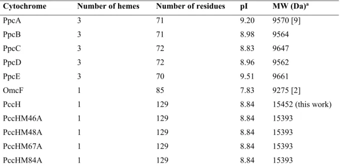
Table 2.1 Isolectric point and molecular weight of mature proteins targeted in this Thesis. ... 35
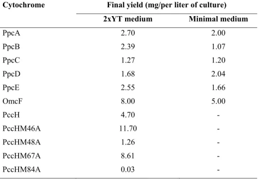
Table 2.2 Final yields of each cytochrome produced and purified during the time course of this Thesis. ... 37
3. Molecular interaction studies between the triheme cytochromes from G. sulfurreducens and the redox
active humic substances analog
Table 3.1 UV-visible maximum absorption bands in the electronic absorption spectra of cytochromes PpcA, PpcB, PpcD, PpcE, AQDS and AH2QDS in oxidized and reduced forms. ... 46
Table 3.2 Rate constants obtained from the fit of normalized stopped-flow kinetic data with exponentials. ... 48
Table 3.3 Global and heme midpoint reduction potentials of cytochromes PpcA, PpcB, PpcD and PpcE from G. sulfurreducens. ... 49 Table 3.4 Thermodynamic parameters determined for PpcA in the presence of quinol. ... 68
Table 3.5 Most affected NH signals in PpcB, PpcD and PpcE in the presence of AQDS. ... 73
Table 3.6 Equilibrium dissociation constants values calculated from the NH signals of PpcB, PpcD and PpcE showing the highest chemical shift perturbation in presence of AQDS. ... 76
Table 3.7 NMR experiments and acquisition details for the PpcB, PpcD, PpcE backbone resonance assignment in the oxidized state. ... 83
4. NMR interaction studies between inner membrane-associated and periplasmic cytochromes Table 4.1 Equilibrium dissociation constants for the complex formed between MacA and PpcA at 298 K, pH 7
and 20 mM final ionic strength ... 103
5. Solution structure and dynamics of the outer membrane cytochrome OmcF
Table 5.1 Summary of restraint violations and quality analysis for the final family of solution structures for cytochrome OmcF from G. sulfurreducens. ... 119 Table 5.2 NMR experiments and acquisition details for OmcF backbone resonance assignment in the reduced
state. ……….... 129
6. Biochemical and structural characterization of PccH
Table 6.1 UV-visible absorption bands and molar extinction coefficient of cytochrome PccH. ... 142
Table 6.2 Heme accessibility and reduction potential for representative bacterial class I c-type cytochromes.. 154
xx
7. Future perspectives
Table 7.1 Percentage of NH backbone signal assignment of PpcB, PpcD and PpcE in reduced state. ... 175
Table 7.2 Amino acid sequences of OmcE, OmcS and OmaB proteins. ... 178
Table 7.3 DNA templates and primers used to clone the genes encoding for OmcE, OmcS and OmaB. ... 180
Table 7.4 Quantity of each expression vector containing the gene sequence encoding for OmcE, OmcS and OmaB, measured in the NanoDrop. ... 181
Table 7.5 NMR experiments and acquisition details for the PpcB, PpcD and PpcE backbone resonance assignment in the reduced state, at 298 K. ... 183
A. Appendix
Table A.1 Backbone assignment of PpcB from G. sulfurreducens in the oxidized state. ... 194 Table A.2 Backbone assignment of PpcD from G. sulfurreducens in the oxidized state ... 195 Table A.3 Backbone assignment of PpcE from G. sulfurreducens in the oxidized state ... 196 Table A.4 Percentage of the backbone assignment of PpcB, PpcD and PpcE in oxidized state. ... 197
Table A.5 Assignment of 1H, 13C and 15N signals of OmcF from G. sulfurreducens in the reduced state. ... 198
Table A.6 Backbone assignment of PpcB from G. sulfurreducens in the reduced state. ... 207 Table A.7 Assignment of 1H, 13C and 15N signals of PpcD from G. sulfurreducens in the reduced state. ... 208
xxi
Abbreviations, symbols and constants
2xYT 2x Yeast extract – tryptone
AH2QDS Anthrahydroquinone-2,6,-disulphonate Amp Ampicillin
AQDS Anthraquinone-2,6-disulphonate
BMRB Biological Magnetic Resonance Data Bank Ccm Cytochrome c maturation
CD Circular dichroism Clo Chloramphenicol COSY Correlation spectroscopy
DMRB Dissimilatory metal reducing bacteria
Eapp Macroscopic apparent midpoint reduction potential eapp Microscopic apparent midpoint reduction potential
EDTA Ethylenediamine tetraacetic acid EET Extracellular electron transfer EXSY Exchange spectroscopy
HMQC Heteronuclear multiple quantum coherence HP MacA high potential heme
HS Humic substances
HSQC Heteronuclear single quantum coherence
IM Inner membrane
Imc Inner membrane cytochrome
IPTG Isopropyl-β-D-thiogalactopyranoside
IUPAC-IUB International Union of Pure and Applied Chemistry and International Union of Biochemistry
Kd Equilibrium dissociation constant
LB Luria-bertani medium Lols lower distance limits Lov lower limit volume LP MacA low potential heme
MALDI-TOF-MS Matrix-assisted laser desorption−ionization time-of-flight mass spectrometry Mac Membrane associated cytochrome
MFC Microbial fuel cells MHC Multiheme cytochromes c
MW Molecular weight
NHE Normal hydrogen electrode NMR Nuclear magnetic resonance NOE Nuclear Overhauser effect
NOESY Nuclear Overhauser effect spectroscopy OD600 Optical density at 600 nm
OM Outer membrane
Omc Outer membrane cytochrome PCR Polymerase chain reaction PDB Protein data bank
pI Isolectric point
Ppc Periplasmic cytochrome
xxii
Ppm Parts per million
rmsd Root mean square deviation Rpm Rotations per minute
S2 Order parameter
SDS-PAGE Sodium dodecyl sulfate – polyacrylamide gel electrophoresis
T1 Longitudinal relaxation T2 Transverse relaxation
TOCSY Total correlation spectroscopy TMBZ Tetramethylbenzidine
Tris Tris – tris(hydroxymethyl)aminomethane Upls Upper distance limits
Upv Upper limit volume UV-visible Ultraviolet-visible
Chemical shift
τc Correlation time
τm Global rotational diffusion correlation time Δδcomb Combined chemical shift
Amino acid abbreviations
Alanine Ala A
Arginine Arg R
Asparagine Asn N
Aspartate Asp D
Cysteine Cys C
Glutamate Glu E
Glutamine Gln Q
Glycine Gly G
Histidine His H
Isoleucine Ile I
Leucine Leu L
Lysine Lys K
Methionine Met M
Phenylalanine Phe F
Proline Pro P
Serine Ser S
Threonine Thr T
Tryptophan Trp W
Tyrosine Tyr Y
1
1. INTRODUCTION
2
1. Introduction ... 3 1.1 The Geobacter sulfurreducens bacteria ... 4 1.2 Cytochromes ... 5 1.3 Multiheme cytochromes ... 8 1.4 Extracellular electron transfer pathways in G. sulfurreducens ... 8 1.4.1 Characterization of cytochromes c ... 12 1.4.1.1 Structural characterization ... 13 1.4.1.2 Thermodynamic characterization ... 16 1.5 Current-consuming biofilms ... 20 1.6 Objectives and Thesis outline ... 21 1.7 References ... 22
1. INTRODUCTION
3
1.
Introduction
Dissimilatory metal reduction is a widespread process that occurs in the major groups of Bacteria and is fundamental for the geochemistry of aquatic sediments, submerged soils, and terrestrial subsurfaces [1, 2]. Unlike other respiratory pathways, where the final electron acceptor is a freely diffusible gas or a readily soluble molecule that can be reduced inside the cell, dissimilatory metal reducing bacteria (DMRB) can sustain their energy requirements by coupling the oxidative metabolism of organic compounds to the reduction of extracellular metals [2].
The respiratory reduction of extracellular electron acceptors can be achieved by two broad mechanisms that involve direct or indirect reduction [3, 4]. In the first case, direct electron transfer is achieved by the contact between redox proteins located at the surface of the bacteria or via electrically conductive appendages that have been called biological nanowires (Fig 1.1). In the indirect mechanism, reduction of acceptors can be mediated by small redox-active molecules that are either secreted by the cell into the environment (endogenous electron shuttles) or are already available in the environment (exogenous electron shuttles) or mediated by bacterial-produced organic ligands (also designated as chelators), which solubilize metals prior to their reduction (Fig 1.1).
Fig 1.1 Schematic representation of bacterial direct and indirect electron transfer mechanisms to the extracellular electron acceptors. SOx and SRed correspond to oxidized and reduced electron shuttles,
respectively. The action of chelators is illustrated by circles: blue (chelator); gray (insoluble acceptor); white (solubilized acceptor) [5]. This figure was adapted from [5].
1. INTRODUCTION
4
Enterococcus gallinarum, Shewanella putrefaciens and Shewanella oneidensis [6]) belong to the group of exoelectrogens, as they can also couple their oxidative metabolism to the electron transfer toward electrode surfaces from which electric current can be harvested in microbial fuel cells (MFC) [7, 8]. Particularly in the case of Geobacter species, highly cohesive protein filaments with metallic-like conductivity can be produced, offering the possibility to design microbial-based electronic sensors and other devices [9].
Currently much research is focused on alternative and renewable energy sources to offset world-wide concerns regarding global warming and fossil energy depletion [10]. Understanding the electron transfer processes by which exoelectrogens can couple the oxidation of organic compounds to the electron transfer to electrodes is a crucial step to allow the optimization of current production by MFC. Although, the present MFC power densities are too low to be considered as a viable alternative energy source, substantial improvements have been made in the last few years [11]. Given the broad range of biotechnological applications covered by DMRB it is important to understand the functional mechanism of their respiratory pathways. This would allow the scientific community to improve and optimize DMRB-based practical applications. Of these, the most studied DMRB belong to the Shewanellaceae and Geobacteraceae families, particularly S. oneidensis and G. sulfurreducens. The work developed and presented in this Thesis is focused on the structural and functional characterization of electron transfer components from G. sulfurreducens and for this reason the current knowledge of this bacterium is summarized in the next sections.
1.1
The
Geobacter sulfurreducens
bacteria
The G. sulfurreducens bacteria are comma-shaped rods are Gram-negative and can be found in contaminated soils and sediments where Fe(III) is an important respiratory terminal electron acceptor. However, their respiratory capability is not confined to iron reduction since they can also utilize a large diversity of electron donors and acceptors making them important agents in several biogeochemical cycles [12]. The important discoveries associated with G. sulfurreducens bacteria are summarized in Fig 1.2.
1. INTRODUCTION
5
bioremediation of these pollutants [12, 14]. Moreover, these bacteria can be used to develop bioenergy applications as they grow on several electrode surfaces [12].
G. sulfurreducens was the first Geobacter species to have its genome fully sequenced, it is amenable to genetic manipulation and has been used as a model organism [15, 16]. This bacterium was previously classified as a strict anaerobe, but it has been verified that it can also grow in presence of low atmospheric oxygen levels [17, 18]. G. sulfurreducens tolerated exposure to oxygen for at least one day and grew with oxygen as the sole electron acceptor until concentrations of 10% in headspace. Additionally, the analysis of its genome revealed the presence of genes encoding typical enzymes for oxygen detoxification, such as catalase, cytochrome c oxidase or superoxide dismutase [18]. The analysis of the genome sequence of G. sulfurreducens revealed that it encodes 111 cytochromes c, 73 of which are multiheme cytochromes [15].
Fig 1.2 Time line of the important discoveries associated with Geobacter sulfurreducens. The time line
presented in this figure was adapted from [12].
1.2
Cytochromes
Cytochromes are important proteins involved in a diversity of biological functions including electron transfer, oxygen transport and storage, catalysis, gas sensing and gene regulation [19]. The functional diversity of cytochromes is further extended by combining heme groups with other cofactors, including flavins and/or metal ions (e.g. molybdenum or copper). Different variables related with the type of heme groups and neighbor amino acids, modulate this functional versatility, namely (i) the ability of the protein environment to tune heme reactivity, (ii) the number and nature of protein-donated axial ligands to iron, (iii) the heme solvent exposure, (iv) the heme accessibility to exogenous ligands, (v) the distribution of polar and charged groups in the heme neighborhood, and (vi) specific properties of the heme-binding site in the protein [19, 20].
1. INTRODUCTION
6
their heme type letter, in italic, and a number in subscript depending on some intrinsic characteristics related to the protein axial ligands coordination, number of heme groups, optical or functional properties [21]. For example, the cytochrome c551, involved in dissimilative denitrification of Pseudomonas aeruginosa, is a monoheme cytochrome axially coordinated by one histidine and one methionine that shows a band in the optical spectra with absorption maximum at 551 nm (band) in the reduced state [22].
Cytochromes c are heme proteins containing a c-type heme and often function as electron carriers. In most cases, the polypeptide chain of these proteins is covalently bound to one or several c-type heme groups through thioether linkages established with the sulfhydryl groups of two cysteine residues in a conserved binding motif sequence CXXCH, where the X represents any amino acid (Fig 1.3) [19, 23]. This sequence is highly variable and all amino acids have been found in the “XX” segment in nature, except for the amino acid cysteine. In some cases, this segment can be larger and contain up to 15 amino acids (e.g. cytochrome MccA from Wolinella succinogenes) [19, 24].
The heme group displays a central role in the functional modulation of these proteins. It is constituted by four pyrrole subunits connected by methane bridges (protoporphyrin IX) and in the center, the iron ion is equatorially coordinated by four nitrogen atoms (Fig 1.3). The Iron (0) is a transition metal with 26 electrons arranged in the electronic configuration 1s22s22p63s23p63d64s2, where the normal number correspond to the orbitals (s, p and d) and the superscripts represent the electron occupancies. This configuration is also represented as [Ar]3d64s2, to emphasize that it is the occupancy of the higher energy 4s and 3d orbitals, superimposed on an argon core, that determines the electronic properties and the chemical behavior of the heme iron. Generally, in electron transfer cytochromes, the iron exists in the ferrous (Fe(II)) and ferric (Fe(III)) states, with the electronic configurations [Ar]3d64s0 and[Ar]3d54s0, respectively. Iron(IV) ([Ar]3d44s0) is a less common state but often found in catalytic cycles of some enzymes, such as catalases or peroxidases [23].
1. INTRODUCTION
7
Fig 1.3Schematic representation of a c-type heme and the correspondent polypeptide binding motif. The
axial coordination position labeled with A can be free or occupied by ligands such the side chain of methionine, histidine, asparagine or tyrosine residues, which are the most common. The IUPAC nomenclature for tetrapyrroles is illustrated in gray [27]. This figure was adapted from [5].
As depicted in Fig 1.3, four out the six heme iron coordination positions are equatorially occupied by nitrogen atoms. In most cases, one of the two axial coordination positions of cytochromes c is occupied by the side chain of a histidine in the binding motif sequence, which is also designated as proximal ligand. On the contrary, the distal ligand is more variable and can be the side chain of a (i) methionine, which predominates in monoheme cytochromes c, (ii) histidine, particularly in multiheme cytochromes c or (iii) asparagine or tyrosine, although less frequently [21, 28]. The distal position of the heme can be also transiently vacant, as observed in cytochromes with enzymatic activity [5, 21, 28].
1. INTRODUCTION
8
1.3
Multiheme cytochromes
Multiheme cytochromes c (MHC) are crucial components of several biological processes where they drive electron transfer pathways. However, MHC can also display enzymatic activity [31, 32] or participate in signal transduction events [33]. In these complex cytochromes, the heme groups are arranged along the polypeptide chain and extend the protein global working redox potential ranges, because of the contribution of each individual heme redox potential. Additionally, these proteins might be able to receive or donate multiple electrons in a cooperative way, a process that can be modulated by the intrinsic properties of neighboring hemes (redox interactions) or protonatable centers (redox-Bohr effect) [34]. As well, these proteins can also putatively work as electron biocapacitors and contribute to the enhancement of the bacterial electron-storage capacity [35-37]. Typically, the heme iron-iron distances between adjacent hemes in MHC do not exceed 16 Å. This allows a fast electronic exchange between the redox centers, a crucial feature to assure efficient redox reactions [3, 28, 38]. The high number of hemes in these proteins not only provides the cell with a remarkable electron storage capacity but also allows them to transfer electrons through large distances without the need for successive binding events. For the reasons presented above, the precise tuning of the heme redox potentials in MHC is of crucial importance. These are affected by several factors that include: (i) the differences in the free energy between the oxidized and reduced states resulting from molecular interaction changes; (ii) the modulation of the electrostatic interactions within the protein or with the solvent; (iii) the heme solvent accessibility; (iv) the extent to which the heme group is distorted from planarity; (v) the protonation state of the heme propionate groups; (vi) the type of axial ligands and heme iron coordination [23, 39-45]. In most of the cases, MHC have a lower amino acid residue to heme ratio compared to monoheme cytochromes and, therefore, are more solvent exposed [23, 41]. The heme solvent exposure and its axial coordination (typically His-His) strongly contribute to the lower redox potential values generally observed in MHC.
1.4
Extracellular electron transfer pathways in
G. sulfurreducens
1. INTRODUCTION
9
Table 1.1 Analysis of proteome and phenotypes of different deletion mutant strains of G. sulfurreducens
for cells grown in the presence of the acetate (electron donor) and Fe(III) oxide or Fe(III) citrate (electron acceptor). “IM” stands for inner membrane, “P” for periplasm and “OM” for outer membrane.
Locus ID Gene name Predicted cellular
location Analysis of the proteome
Phenotype during growth on
the electron acceptors Ref Fe(III) oxide Fe(III) citrate
GSU0274 cbcL IM - Impaired
growth Increased growth rate [47]
GSU0364 ppcB P Detected in Fe(III) citrate
and Fe(III) oxide cultures No phenotype Impaired growth [46, 48]
GSU0365 ppcC P Detected in Fe(III) citrate
and Fe(III) oxide cultures No phenotype Impaired growth [46, 48]
GSU0466 macA
IM-associated More abundant during growth with Fe(III) oxides
vs Fe(III) citrate. Involved
in the regulation of OmcB
Impaired
growth Impaired growth [46, 48]
GSU0612 ppcA P Detected in Fe(III) citrate
and Fe(III) oxide cultures No phenotype Impaired growth [46, 48, 49]
GSU0618 omcE OM More abundant during
growth with Fe(III) oxide
vs Fe(III) citrate
Impaired
growth No phenotype [48, 50]
GSU1024 ppcD P More abundant during
growth with Fe(III) oxides
vs Fe(III) citrate
No phenotype Increased Fe(III) reduction
[46, 48]
GSU1760 ppcE P Detected in Fe(III) citrate
cultures No phenotype Increased growth rate [46, 48]
GSU1996 - P More abundant during
growth with Fe(III) citrate
vs Fe(III) oxide
- - [46]
GSU2076 omcZ OM More abundant during
growth with Fe(III) citrate
vs Fe(III) oxide
No phenotype No phenotype [46, 48, 51]
GSU2432 omcF OM Involved in the regulation
of OmcB, OmcC and OmcS
Impaired
growth Impaired growth [48, 52, 53]
GSU2504 omcS OM More abundant during
growth with Fe(III) oxides
vs Fe(III) citrate
No growth No phenotype [48, 50,
54]
GSU2731 omcC OM More abundant during
growth with Fe(III) oxides
vs Fe(III) citrate
No phenotype No phenotype [48, 55]
GSU2737 omcB OM Detected in Fe(III) citrate
and Fe(III) oxide cultures Impaired growth Impaired growth [48, 55]
GSU3259 imcH IM - Increased
1. INTRODUCTION
10
The electron transfer mechanisms in G. sulfurreducens and the proteins implicated in each pathway are still under investigation. It is consensual that NADH electrons are transferred to the quinone pool via an IM NADH dehydrogenase. However, from this point the electron transfer mechanism(s) toward the final electron acceptors are still under debate.
The first studies envisioned that the IM-associated diheme cytochrome MacA (GSU0466, the naming scheme of the genome is in accordance with http://www.genome.jp/kegg/genome.html) with 35 kDa molecular weight, might be part of an electron transport chain moving electrons from the inner membrane, through the periplasmic cytochromes, to the outer surface of the cell [57, 58]. Proteomic studies revealed that MacA is more abundant during growth with Fe(III) oxides vs Fe(III) citrate (Table 1.1) [46]. Then, gene knock-out studies implicated the deletion of macA in the inhibition of the expression of the omcB gene. Expression of the outer membrane cytochrome OmcB in the macA-deficient mutant restored the capacity for Fe(III) reduction [53]. Additionally, the similarity in expression patterns and mutant phenotypes between MacA and OmcB suggests that they may function in the same or similar routes of electron transfer [48]. Cytochrome MacA was also identified as a peroxidase and capable of exchange electrons with the periplasmic c-type cytochrome PpcA [59].
More recently, it was suggested that depending upon the redox potential of the final electron acceptor different proteins are involved in the quinone pool regeneration [47, 56]. At least, two different routes were identified in response to the amount of energy available in a metal or electrode distant from the cell [56]. This is supported by several studies that revealed a complex transcriptional response by G. sulfurreducens to different electron acceptors [46, 54, 60, 61]. Indeed, the large number of c-type cytochromes [15] and the examination that no single deletion eliminates electron transfer to all electron acceptors further confirm the complexity of the EET in G. sulfurreducens [48, 62].
Studies examining electron transfer from G. sulfurreducens to poised graphite electrodes demonstrated that at least two IM proteins are implicated in two different EET pathways, i) CbcL-dependent pathway, for potentials below -100 mV vs NHE (Fe(III)- oxides, such as ferrihydrite), and ii) ImcH-dependent pathway, for electrodes poised at redox potentials above -100 mV vs NHE (Fe(III) citrate, Fe(III)-EDTA) [47, 56]. These observations confirmed that at least another quinone oxidoreductase is active in the bacteria to support growth at low redox potentials even when the imcH gene is deleted [56].
1. INTRODUCTION
11
transfer pathways that support lower cell yield and slower growth rates, but still allow some respiration.
With this new perspective, G. sulfurreducens bacterium can also be used as a redox sensor to report the redox potential of an extracellular electron acceptor and represents an array of thermodynamic opportunities, where these bacteria rapidly alter their respiratory strategy to take advantage of the available extracellular electron acceptor [63]. However, the electron transfer pathways from the reduced quinone pool to the final electron acceptor are still far from being understood.
In the currently proposed model for G. sulfurreducens, the electrons from IM cytochromes are then transferred to periplasmic c-type cytochromes. The most studied periplasmic cytochrome is PpcA (GSU0612) that belongs to a family composed by five low molecular weight (~10 kDa) periplasmic triheme cytochromes with approximately 70 residues each. The other four cytochromes of this family are designated PpcB (GSU0364), PpcC (GSU0365), PpcD (GSU1024), PpcE (GSU1760) and share 77 % (PpcB), 62 % (PpcC), 57 % (PpcD) and 65 % (PpcE) amino acid sequence identity with PpcA. Genetic studies revealed that these cytochromes are involved in different electron transfer pathways. PpcA was detected in G. sulfurreducens cultures that grow in presence of Fe(III) citrate and Fe(III) oxide [46]. Deletion of the gene encoding for PpcA affected the reduction of Fe(III) and U(VI) [49, 64]. Genes encoding for PpcB and PpcC belong to the same locus and the double deletion of these genes affected U(VI) reduction. Both cytochromes were detected in Fe(III) citrate and Fe(III) oxide cultures [46, 64]. PpcD is more abundant during growth with Fe(III) oxides vs Fe(III) citrate [46]. The deletion of its gene affected the reduction of U(VI) [64]. PpcE was only detected in cultures with Fe(III) citrate and its deletion also affected U(VI) reduction [46, 64]. Due to the cellular location of these five cytochromes it was proposed that they are the likely reservoir of electrons destined for the cell outer surface and bridge the electron transfer between the cytoplasm and the cell exterior [49, 60].
Also located in the periplasm of G. sulfurreducens, the dodecaheme cytochrome GSU1996 (42.3 kDa) is composed by four similar triheme domains, designated A, B, C and D (see section 1.4.1.1). and the 12-heme groups might allow the cytochrome to act as a periplasmic electron capacitor [36, 37].
1. INTRODUCTION
12
OmcF (GSU2432) is the smallest monoheme cytochrome c (11 kDa) and it was predicted to be localized at the OM of G. sulfurreducens [15]. The deletion of the omcF gene impaired Fe(III) citrate reduction and affected the expression of the OM cytochromes OmcB, OmcC and OmcS [52]. The deletion of omcF resulted in a loss of expression of omcB and omcC, and overexpression of omcS, during growth on Fe(III) citrate [66]. More recent studies demonstrated that a omcF-deficient strain was unable to grow in presence of Fe(III) oxide and showed a significant decrease in current production [48, 66].
OmcS (47 kDa) and OmcZ (30 kDa) are extracellular c-type cytochromes from G. sulfurreducens containing six and eight heme groups, respectively [51, 67]. It was demonstrated that OmcS is associated with conductive pili [67] and OmcZ is well suited for promoting electron transfer in current-producing G. sulfurreducens biofilms [68]. The deletion of the gene encoding OmcS cytochrome affected the bacterial growth in the presence of Fe(III) oxides (Table 1.1). It was also observed that OmcS is more abundant during growth with Fe(III) oxides vs Fe(III) citrate (Table 1.1). The opposite effect was observed for OmcZ (Table 1.1) and also, in vitro studies demonstrated that OmcZ transfers electrons to a diversity of potential extracellular electron acceptors, such as Fe(III) citrate, U(VI), Cr(VI), Au(III), Mn(IV) oxide, and AQDS, but not Fe(III) oxide [46, 51].
OmcE (GSU0618) is a tetraheme c-type cytochrome (26 kDa) located in the exterior of the OM of the bacterium. This cytochrome is more abundant during growth with Fe(III) oxide than in cells grown on Fe(III) citrate (Table 1.1). Previous studies revealed that the omcE-deficient mutant did slowly adapt to reduce Fe(III) oxide suggesting that one or more, as undetermined yet, components can partially compensate for the loss of OmcE [50].
1.4.1
Characterization of cytochromes
c
1. INTRODUCTION
13
Fig 1.4 Time line of important discoveries related with the G. sulfurreducens cytochromes addressed in
this Thesis.
1.4.1.1
Structural characterization
Three-dimensional structures of biological macromolecules can be determined by X-ray crystallography and NMR at near atomic resolution. The structures obtained by NMR represent the average over oriented molecules in solution, whereas diffraction data represent an average over molecules arranged in a periodic crystal lattice. Despite their similarities and differences, X-ray crystallography and NMR are well established complementary high-resolution methods to analyze protein structure-function relationships.
Structures of heme proteins are extremely under-represented in structural databases (~4%, data from Protein Data Bank, http://www.rcsb.org/pdb/, 2017), which constitute a severe bottleneck in the elucidation of their structural–functional relationships. Further, structures of heme proteins determined by NMR are very few (~1%). Mostly because of the presence of heme groups, which not only contribute with their proton-containing groups (four methyl groups, four meso protons, two thioether protons, two thioether methyls and two propionates; see Fig 1.3), but also affect the polypeptide chain NMR signals, particularly in the paramagnetic state, due to the magnetic properties of the heme iron that further spread and broaden the signals [70, 71].
1. INTRODUCTION
14
Out of cytochromes found in Geobacter, PpcA was the first structurally characterized by X-ray crystallography in the oxidized state [77]. In the following years the crystal structures of the other four periplasmic triheme cytochromes PpcB-E belonging to the PpcA-family were also determined in the oxidized state [78, 79]. More recently, the solution structures of PpcA in the reduced and oxidized forms were also determined by NMR [80, 81]. The structures of these cytochromes are similar (Fig 1.5) but important local variations are observed. In these proteins, an antiparallel β-sheet is conserved at the N-terminal of all structures, and is followed by distinct helical regions in the different proteins. The heme core structures are similar, with hemes I and IV roughly parallel to each other and with both nearly perpendicular to heme III [77-80]. Within the five structures, in general heme I shows the highest solvent exposure, while heme III is the lowest exposed. On the other hand, heme IV shows the largest positive electrostatic surface due to the considerable number of neighboring lysine residues, except for PpcE. This positively charged surface around heme IV is the most conserved region, while the lowest similarity region is located near heme I [79]. The spatial arrangement of the hemes is superimposable with those of the structurally homologous tetraheme cytochromes c3, with the sole difference being the absence of heme II and the corresponding polypeptide segment. For this reason, the three heme groups in triheme cytochromes are numbered I, III and IV [82]. All members of the PpcA-family contain three low-spin heme groups with His–His axial coordination [79].
Structures for other G. sulfurreducens cytochromes were also determined by X-ray crystallography, namely those of cytochromes OmcF [83, 84] and GSU1996, both in the oxidized state [36], and MacA in the reduced, oxidized and intermediate states [59] (Fig 1.5).
1. INTRODUCTION
15
Fig 1.5Structures of G. sulfurreducens cytochromes obtained in the oxidized state. The solution structure of
PpcA (lowest energy; PDB ID: 2MZ9 [81] and the crystal structures of PpcB (chain A, PDB ID: 3BXU [78]), PpcC (PDB ID: 3H33 [79]), PpcD (chain A, PDB ID: 3H4N [79]), and PpcE (PDB ID: 3H34 [79]) are represented in blue. Crystal structures of OmcF (PDB ID: 3CU4 [84]), GSU1996 (PDB ID: 3OV0 [36]) and MacA (PDB ID: 4AAL [59]) are represented in cyan, gray and green, respectively. Roman numerals indicate the hemes (in red) in their order of attachment to the CXXCH motif in the polypeptide chain. The structures were drawn using the PyMOL molecular graphics system [85]. This figure was adapted from [5].
1. INTRODUCTION
16
MacA forms two globular domains, each holding a c-type heme group. The hemes are named as high potential (HP) and low potential (LP), because of the electric potential difference between them. The LP heme is located at the N-terminal domain whereas the C-terminal domain harbors the HP one.
1.4.1.2
Thermodynamic characterization
In the same way, the presence of several heme groups constitutes the major bottleneck in the detailed thermodynamic characterization of MHC. For monoheme cytochromes only the reduced and oxidized states may coexist in solution. However, for MHC several one-electron reversible transfer steps convert the fully reduced state into the fully oxidized one, yielding additional intermediate oxidation stages (Fig 1.6). Therefore, when the values of redox potentials of each heme group are not sufficiently different, the global redox potentials measured by voltammetry or potentiometric redox titrations are macroscopic in nature and only describe the overall redox behavior of the MHC [68, 84, 86, 87]. Although these parameters contain information on the working functional ranges of these cytochromes, in most cases they are insufficient to provide mechanistic information on the electron transfer pathways. This can only be achieved when the fractional contribution of each possible microstate during the oxidation of the protein is obtained.
1. INTRODUCTION
17
Fig 1.6 Electronic distribution scheme and potentiometric redox titrations followed by visible spectroscopy for monoheme (OmcF) and triheme (PpcA) cytochromes. The inner circles represent heme groups, which can either be reduced (red circles) or oxidized (white circles). The microstates are grouped per the number of oxidized hemes in each oxidation stage, connected by consecutive one-electron redox steps. P0H and
P0 represent the reduced protonated and deprotonated microstates, respectively. PijkH and Pijk, indicate, respectively, the protonated and deprotonated microstates, where i, j, and k represent the heme(s) that are
oxidized in that particular microstate. In the lower panels, potentiometric redox titrations followed by visible spectroscopy of OmcF and PpcA (pH 7) are indicated. Solid lines indicate the result of the fits for the Nernst equation (OmcF) and for a model of three consecutive reversible redox steps between the different oxidation stages (PpcA) [78, 84]. This figure was adapted from [88].
1. INTRODUCTION
18
Thus, to completely characterize the redox centers in a MHC it is necessary to determine the heme reduction potentials, the redox interactions and the properties of the redox-Bohr center(s). To calculate these thermodynamic parameters, it is necessary to monitor the stepwise oxidation of each heme oxidation at several pH values. This information can be achieved by NMR spectroscopy, which allows probing the individual heme oxidation profiles due to the different spectral signatures of the NMR spectra of low spin MHC in the reduced and oxidized states.
In conditions of fast intramolecular electron exchange (between the different microstates within the same oxidation stage) and slow intermolecular electron exchange (between different oxidation stages) on the NMR time scale [78], the heme oxidation fraction can be determined from the chemical shifts of the heme substituents in the different oxidation stages. Combining the information from redox titrations monitored by UV-visible spectroscopy and this NMR data it is possible to determine the absolute thermodynamic parameters and evaluate the contribution of each microstate and their relevance to the electron transfer mechanism.
This strategy was firstly described in the study of tetraheme cytochrome c3 from Desulfovibrio vulgaris (13 kDa) by Turner and co-workers [89]. The increase in the protein molecular weight and/or the number of hemes leads to a decrease of the NMR spectral quality, as a result of signal overlapping, and usually impairs the monitoring of the individual heme oxidation profiles. To date, the detailed thermodynamic characterization of multiheme cytochromes has been limited to proteins containing up to four heme groups and 64 kDa molecular weight [90]. Therefore, the redox characterization of larger multiheme proteins can only be accomplished at the macroscopic level. This characterization was already performed for some cytochromes from G. sulfurreducens (Table 1.2).
1. INTRODUCTION
19
Table 1.2 Data set of G. sulfurreducens c-type cytochromes participating in extracellular electron transfer
pathways.Eapp stands for the midpoint reduction potential correspondent to the point at which the oxidized and reduced fractions are equal, and ‘NHE’ for normal hydrogen electrode. The potential window was determined from potentiometric redox curves considering 1-99% range for protein reduction/oxidation. The redox potential
values were determined at pH 7.
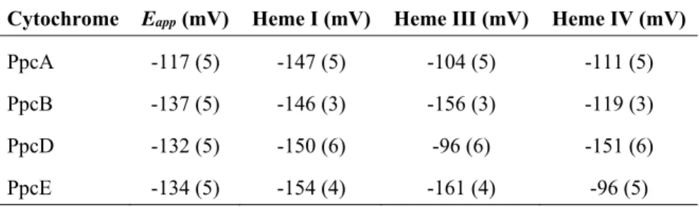
Protein Heme axial ligands Eapp (mV) vs NHE Potential window (mV)
MacA His-Met -188 [59] 250
GSU1996 His-His; His-Met -124 [91] 320
PpcA His-His -117 [78] 285
PpcB His-His -137 [78] 270
PpcC His-His -143 [91] 265
PpcD His-His -132 [92] 275
PpcE His-His -134 [92] 280
OmcZa Not determined -220 [68] 405
OmcF His-Met +180 [84] 240
OmcS His-His -212 [86] 320
a OmcZ is present in large (OmcZL = 50 kDa) and small (OmcZs = 30 kDa) forms. The data
presented refer to OmcZs, which is the predominant extracellular form of OmcZ and retains the
eight heme groups.
Except for PpcC, which presented multiple conformations in solution that impaired the monitorization of its heme oxidation profiles, the complete thermodynamic parameters were determined for the triheme cytochromes from PpcA-family (Tables 1.3 and 1.4) [92, 93]. The heme reduction potentials are negative, differ from each other, and cover different functional ranges. These reduction potentials are strongly modulated by heme-heme interactions and by interactions with protonated groups (the redox-Bohr effect), yielding different cooperative networks for each protein.
Table 1.3Heme reduction potentials and pairwise interactions (mV) of the fully reduced and protonated forms of PpcA, PpcB, PpcD and PpcE [92].
Heme redox potentials Redox interactions Redox-Bohr interactions
I III IV I-III I-IV III-IV I-H III-H IV-H
PpcA -154 -138 -125 27 16 41 -32 -31 -58
PpcB -150 -166 -125 17 8 32 -16 -9 -38
PpcD -156 -139 -149 46 3 14 -28 -23 -53
1. INTRODUCTION
20
The macroscopic pKa values associated with the redox-Bohr center in each of the four oxidation stages are indicated in Table 1.4. The larger redox-Bohr effect is observed for PpcA and PpcD, 2.1 and 1.8 pH units, respectively. The ∆pKa value is lower for PpcB and much smaller for PpcE (1.1 and 0.3 pH units, respectively) [92].
Table 1.4Macroscopic pKa values of the redox-Bohr center for PpcA, PpcB, PpcD, and PpcE at each stage
of oxidation [92].
pKa
Oxidation stage PpcA PpcB PpcD PpcE
0 8.6 7.4 8.7 7.7
1 8.0 7.1 8.1 7.6
2 7.2 6.8 7.4 7.5
3 6.5 6.3 6.9 7.4
∆pKa 2.1 1.1 1.8 0.3