Vítor Manuel Monteiro Borges
Mestre em Ciências Farmacêuticas
Evolutionary dynamics of
Chlamydia trachomatis
genome and identification of molecular patterns of
hypothetical protein coding genes
Dissertação para obtenção do Grau de Doutor em Biologia
Orientador: Doutor João Paulo dos Santos Gomes, Investigador
Auxiliar, Instituto Nacional de Saúde Doutor Ricardo Jorge
Co-orientadora: Doutora Maria José Gonçalves Gaspar Borrego,
Investigadora Auxiliar, Instituto Nacional de Saúde Doutor Ricardo
Jorge
Co-orientadora: Doutora Ana Madalena Peres de Drummond
Ludovice Mendes Gomes, Professora Auxiliar, Faculdade de
Ciências e Tecnologia, Universidade Nova de Lisboa
Júri :
Presidente: Prof. Doutora Ana Isabel Nobre Martins Aguiar Oliveira Ricardo Arguentes: Doutor João André Nogueira Custódio Carriço
Doutora Isabel Maria dos Santos Leitão Couto
Vogais: Doutora Maria Aida da Costa e Silva da Conceição Duarte Doutora Isabel Maria Godinho de Sá Nogueira
Doutor João Paulo dos Santos Gomes
iii
Vítor Manuel Monteiro Borges
Mestre em Ciências Farmacêuticas
Evolutionary dynamics of
Chlamydia trachomatis
genome and identification of molecular patterns of
hypothetical protein coding genes
Copyright © Vítor Borges
A Faculdade de Ciências e Tecnologia e a Universidade Nova de Lisboa têm o direito, perpétuo e sem limites geográficos, de arquivar e publicar esta dissertação através de exemplares impressos reproduzidos em papel ou de forma digital, ou por qualquer outro meio conhecido ou que venha a ser inventado, e de a divulgar através de repositórios científicos e de admitir a sua cópia e distribuição com objetivos educacionais ou de investigação, não comerciais, desde que seja dado crédito ao autor e editor.
v
Agradecimentos
De um modo sincero, agradeço:
Ao meu orientador Dr. João Paulo Gomes, Responsável da Unidade de Investigação do Departamento de Doenças Infecciosas do Instituto Nacional de Saúde Doutor Ricardo Jorge (INSA), por me ter integrado no seu grupo de investigação, por todo o saber científico que me transmitiu, pelo apoio incondicional, pela liderança, pela permanente disponibilidade e ajuda, pelo encorajamento e amizade.
À minha co-orientadora Dra. Maria José Borrego, Responsável do Laboratório Nacional de Referência das Infecções Sexualmente Transmissíveis do INSA, pelos ensinamentos científicos, pela total disponibilidade, pela constante simpatia, por todo o apreço, apoio e amizade.
A todos os restantes membros da Comissão de Acompanhamento de Tese (CAT), pela discussão científica e por todas as sugestões concedidas. Em especial, à minha co-orientadora Dra. Ana Madalena Ludovice, Professora Auxiliar da Faculdade de Ciências e Tecnologia da Universidade Nova de Lisboa (FCT/UNL) por ter aceitado ser minha orientadora, pela simpatia e disponibilidade; e ao Dr. Jaime Mota (FCT/UNL) pela profícua discussão e colaboração científica, apoio e disponibilidade. Agradeço, ainda, ao Professor Miguel Viveiros do Instituto de Higiene e Medicina Tropical (UNL) e à Professora Cristina Lobo Vilela da Faculdade de Medicina Veterinária da Universidade Técnica de Lisboa.
A todos os colegas de investigação do INSA. Em particular, à Alexandra Nunes, ao Carlos Florindo e à Rita Ferreira, pela entreajuda e solidariedade, pela partilha de ideias e pela discussão científica. Foi e tem sido um prazer trabalhar convosco. À Minia Antelo, à Mafalda Sousa-Uva, à Vera Damião e ao Miguel Pinto, pelo companheirismo e colaboração.
A todos os membros do grupo do Dr. Jaime Mota, em especial ao Filipe Almeida pela entreajuda e profissionalismo.
Ao Dr. Miguel Pinheiro da Universidade de St. Andrews (Escócia, Reino Unido), pelos ensinamentos na área da bioinformática, pelo constante encorajamento e suporte, e, consequentemente, pelo seu importante contributo para o desenvolvimento dos trabalhos do último ano de Doutoramento.
vi Ao Instituto Nacional de Saúde Dr. Ricardo Jorge, em particular ao Departamento de Doenças Infecciosas, na pessoa do seu coordenador, Dr. Jorge Machado, por ter disponibilizado todas as condições para a melhor prossecução deste trabalho de Doutoramento.
À Faculdade de Ciências e Tecnologia da Universidade Nova de Lisboa (FCT/UNL), em particular à Professora Isabel Sá Nogueira, Coordenadora do Programa Doutoral de Biologia.
À Fundação para a Ciência e Tecnologia, Ministério da Educação e Ciência (FCT/MEC), pelo financiamento concedido através da Bolsa Individual de Doutoramento (SFRH/BD/68527/2010).
vii
Resumo
A bactéria intracelular obrigatória Chlamydia trachomatis é um agente patogénico para o Homem com importante impacto em termos de saúde pública. As estirpes podem ser classificadas em 15 principais serotipos (A a L3) que causam preferencialmente infecções oculares (A-C), infecções genitais (D-K) ou linfogranuloma venéreo (LGV) (L1-L3), contudo a base molecular que justifica o seu distinto tropismo, sucesso ecológico e patogenicidade não está bem definida. A investigação neste organismo exige normalmente a sua cultura em linhas celulares eucariotas, mas é desconhecida a existência de um processo de adaptação laboratorial.
Pretendemos, essencialmente através de estudos de genómica e transcriptómica, investigar os padrões evolutivos subjacentes à adaptação de C. trachomatis aos diferentes tecidos humanos, dando ênfase aos perfis moleculares de genes que codificam proteínas com função desconhecida, e procurámos compreender o processo adaptativo inerente à transição de C. trachomatis de in vivo para in vitro.
Os nossos resultados indicam que a bactéria C. trachomatis tem uma evolução mediada por eventos de seleção positiva orientada para a adaptação aos diferentes tecidos humanos. Observámos que proteínas que interagem com o hospedeiro têm sido importantes alvos de selecção, nomeadamente proteínas efectoras e da membrana da inclusão, sendo que algumas destas apresentam padrões de expressão relacionados com o nicho biológico que as estirpes infectam. Identificaram-se, ainda, potenciais pseudogenes específicos das estirpes oculares e genes que estarão mais sujeitos a mutações adaptativas associadas ao LGV. Verificámos que a constituição genética de estirpes provenientes do ambiente in vivo não é substancialmente afectada pela sua propagação laboratorial a longo termo. Pelo contrário, a introdução de C. trachomatis em laboratório pode potencialmente levar à atenuação da sua virulência. De facto, observou-se uma rápida inactivação do gene de virulência CT135, fenómeno que parece ser restrito às estirpes que causam infecções genitais, o que pode ter impacto na interpretação de dados provenientes de estudos que requerem cultura.
Globalmente, os resultados apresentados nesta tese contribuem para a melhor compreensão da evolução adaptativa de C. trachomatis e fornecem novos dados sobre o papel biológico de proteínas com função desconhecida. Estes dados poderão servir de base para estudos futuros de funcionalidade focados em clarificar os determinantes das diferenças de tropismo, virulência ou patogenicidade entre estirpes de C. trachomatis.
ix
Abstract
The obligate intracellular bacterium Chlamydia trachomatis is a human pathogen of major public health significance. Strains can be classified into 15 main serovars (A to L3) that preferentially cause ocular infections (A-C), genital infections (D-K) or lymphogranuloma venereum (LGV) (L1-L3), but the molecular basis behind their distinct tropism, ecological success and pathogenicity is not well-defined. Most chlamydial research demands culture in eukaryotic cell lines, but it is not known if stains become laboratory adapted.
By essentially using genomics and transcriptomics, we aimed to investigate the evolutionary patterns underlying the adaptation of C. trachomatis to the different human tissues, given emphasis to the identification of molecular patterns of genes encoding hypothetical proteins, and to understand the adaptive process behind the C. trachomatis in vivo to in vitro transition.
Our results highlight a positive selection-driven evolution of C. trachomatis towards niche-specific adaptation, essentially targeting host-interacting proteins, namely effectors and inclusion membrane proteins, where some of them also displayed niche-specific expression patterns. We also identified potential "ocular-specific" pseudogenes, and pointed out the major gene targets of adaptive mutations associated with LGV infections. We further observed that the in vivo-derived genetic make-up of C. trachomatis is not significantly compromised by its long-term laboratory propagation. In opposition, its introduction in vitro has the potential to affect the phenotype, likely yielding virulence attenuation. In fact, we observed a "genital-specific" rampant inactivation of the virulence gene CT135, which may impact the interpretation of data derived from studies requiring culture.
Globally, the findings presented in this Ph.D. thesis contribute for the understanding of C.
trachomatis adaptive evolution and provides new insights into the biological role of C. trachomatis hypothetical proteins. They also launch research questions for future functional studies aiming to clarify the determinants of tissue tropism, virulence or pathogenic dissimilarities among C. trachomatis strains.
xi
Table of contents
Agradecimentos v
Resumo vii
Abstract ix
Table of contents xi
Figure index xiii
Table index xv
List of abbreviations xvii
Notes of the author: thesis organization, format and outline xxi
1. Chapter I: General Introduction 1
1.1.The genus Chlamydia: host preference and pathogenicity 3
1.2. Chlamydia trachomatis 4
1.2.1. Molecular epidemiology, tissue tropism and impact on human health 4 1.2.2. Biology: a specialized life-cycle of host-cell dependence and manipulation 5 1.2.3. Evolution, genetic diversity and genotype-phenotype associations 7 1.2.4. Genomic research: historical hurdles and ongoing advances 10
1.3. Aims and General Research Plan 11
2. Chapter II: Directional evolution of Chlamydia trachomatis towards niche-specific adaptation
13
2.1.Abstract 15
2.2. Introduction 15
2.3. Materials and Methods 16
2.4. Results 19
2.5. Discussion 29
3. Chapter III: Polymorphisms in Inc proteins and differential expression of inc genes among Chlamydia trachomatis strains correlate with invasiveness and tropism of lymphogranuloma venereum isolates
33
3.1.Abstract 35
3.2. Introduction 35
3.3. Materials and Methods 36
3.4. Results 40
3.5. Discussion 52
4. Chapter IV: Identification of type III secretion substrates of Chlamydia trachomatis using Yersinia enterocolitica as a heterologous system
55
4.1.Abstract 57
4.2. Introduction 57
4.3. Materials and Methods 59
4.4. Results 61
4.5. Discussion 72
xii 6. Chapter VI: Deep comparative genomics among Chlamydia trachomatis
lymphogranuloma venereum isolates highlights genes potentially involved in pathoadaptation
83
6.1.Abstract 85
6.2. Introduction 85
6.3. Materials and Methods 86
6.4. Results and Discussion 89
6.5. Concluding remarks 106
7. Chapter VII: Effect of long-term laboratory propagation on Chlamydia trachomatis genome dynamics
107
7.1.Abstract 109
7.2. Introduction 109
7.3. Materials and Methods 110
7.4. Results 115
7.5. Discussion 121
8. Chapter VIII: Chlamydia trachomatis in vivo to in vitro transition reveals mechanisms of phase variation and down-regulation of virulence factors
127
8.1.Abstract 129
8.2. Introduction 129
8.3. Materials and Methods 131
8.4. Results 136
8.5. Discussion 150
9. Chapter IX. Final overview, concluding remarks and future directions 155
References 163
xiii
Figure index
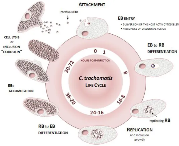
Figure 1.1. Schematic representation of the C. trachomatis developmental cycle. 6
Figure 2.1. Chromosomal mapping of loci involved in the directional evolution of Chlamydia trachomatis.
20
Figure 2.2.Evidence for Muller’s ratchet phenomenon. 21
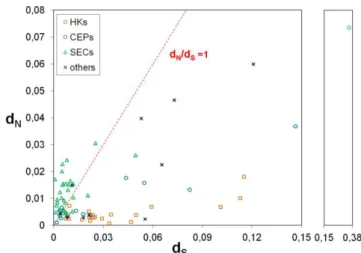
Figure 2.3. dNversus dS by gene functional category. 22
Figure 2.4. Genetic variability versus type of mutation. 23
Figure 2.5. Non-random distribution of both nonsynonymous and synonymous mutations according to tropism and ecological success.
24
Figure 2.6. Positive selection driving the directional C. trachomatis evolution towards niche-specific adaptation.
25
Figure 3.1. Type III secretion (T3S) signals in C. trachomatis Inc proteins. 42
Figure 3.2. Polymorphisms in C. trachomatis Inc proteins. 44
Figure 3.3. Evolutionary dynamics of inc genes. 46
Figure 3.4. mRNA levels of inc genes during the developmental cycle of different C. trachomatis strains.
49
Figure 3.5. Identification of LGV-specific nucleotides in the promoter regions of ct192 and ct214 and within the ct059-ct058 transcript.
51
Figure 4.1. The first 20 amino acids of known C. trachomatis T3S substrates (IncA or IncC) are sufficient to efficiently drive T3S of TEM-1 hybrid proteins by Y. enterocolitica.
63
Figure 4.2. Identification of T3S signals in C. trachomatis proteins using Y. enterocolitica as a heterologous system.
64
Figure 4.3. Analysis of the T3S of C. trachomatis full-length proteins by Y. enterocolitica. 66
Figure 4.4. Translocation of C. trachomatis proteins into the cytoplasm of HeLa cells by Y. enterocolitica.
68
Figure 4.5. mRNA levels of newly identified putative effectors during the developmental cycle of C. trachomatis prototype strain L2/434.
69
Figure 4.6. Comparison of the expression profiles of newly identified genes encoding putative effectors during the developmental cycle of C. trachomatis ocular, epithelial-genital and LGV strains.
71
Figure 4.7. Identification of LGV-specific genetic features in the putative promoter region of ct105.
72
Figure 5.1. Integration of the genetic backbone of the sequenced and annotated genome of the C. trachomatis serovar C strain TW-3 in the species phylogeny and diversity.
81
Figure 6.1. Genetic diversity among C. trachomatis LGV isolates. 90
Figure 6.2. Distribution of the intra-LGV variant sites occurring in coding regions. 92
xiv Figure 7.1. Impact oflong-term laboratory passaging on the C. trachomatis plasmid load. 118
Figure 7.2. Evaluation of the attachment/entry rate of strains before and after their long-term in vitro propagation.
119
Figure 7.3. Prediction of the rate of appearance of variant clones. 120
Figure 7.4. Mathematical modeling of fluctuations in clone frequency 121
Figure 8.1. Phase variation mediated by variable homopolymeric tracts. 137
Figure 8.2. Mutational scenario throughout experimental evolution. 140
Figure 8.3. Schematic representation of the CT135 deletion in the serovar D strain. 142
Figure 8.4. Proposed schematic representation of the adaptive process underlying the inactivation of the CT713/porB.
144
Figure 8.5. Impact of in vitro passaging on the C. trachomatis growth kinetics. 145
Figure 8.6. CT135 mRNA decay analysis. 146
Figure 8.7. Comparative analysis of global gene expression (RNA-seq) between D/CT135-positive and D/CT135-negative populations.
147
Supplemental Figure S3.1. Examples of variation of mRNA levels (profiles of expression) of inc
genes throughout the developmental cycle of the indicated C. trachomatis strains.
200
Supplemental Figure S3.2. Differences in the mRNA levels of inc genes throughout the developmental cycle of C. trachomatis C/TW3, E/Bour, and L2/434.
201
Supplemental Figure S3.3. LGV-specific nucleotide differences in the promoter region of ct059,
coding sequence of ct059, ct059-ct058 intragenic region, and first codons of ct058.
202
Supplemental Figure S3.4. LGV-specific nucleotide differences in the promoter region of ct192 and of
ct214.
203
Supplemental Figure S8.1. Genome make-up of the studied C. trachomatis strains and evaluation of
putative mosaic structures.
207
xv
Table index
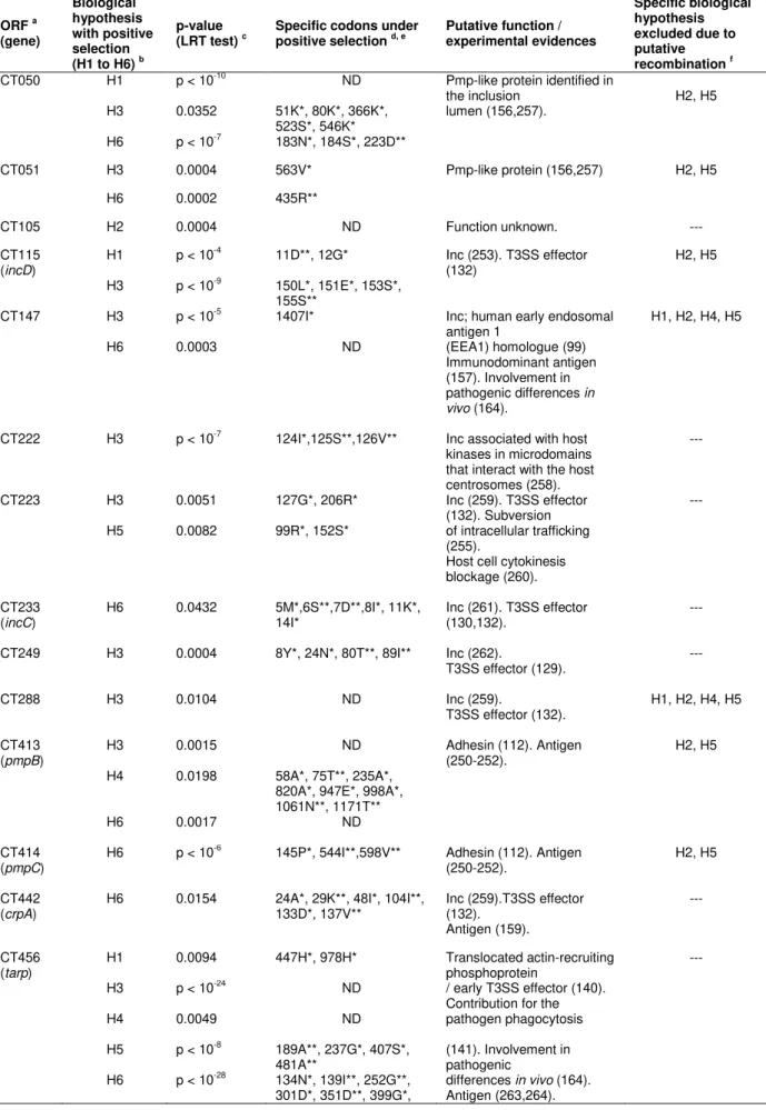
Table 2.1. Positively selected genes and the inferred codons putatively involved in specific adaptive evolution based on the branch-site test of positive selection by PAML.
26
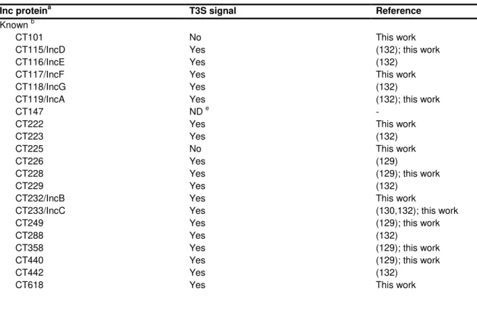
Table 3.1. Summary of T3S signals found in known and putative Inc proteins of C. trachomatis analyzed in this work.
40
Table 3.2. Amino acids residues within Inc proteins encoded by genes likely under positive selection that are specific of C. trachomatis disease groups.
47
Table 6.1. Global trends of genetic variability among LGV strains. 91
Table 6.2. Incs (or putative Incs) with variant amino acids among LGV strains. 96
Table 7.1. Genomic alterations after long-term in vitro passaging. 117
Table 8.1. Genomic alterations throughout in vitro passaging. 138
Table 8.2. Genes and a non-coding RNA found to be down-regulated in the serovar D CT135-negative strain.
148
Supplemental Table S2.1. List of strains studied. 193
Supplemental Table S2.2. Identification of genes and corresponding functional category. 194
Supplemental Table S2.3. Oligonucleotide primers used for PCR and sequencing. 195
Supplemental Table S3.1. Accession numbers of C. trachomatis genomic sequences used in this study.
198
Supplemental Table S3.2. Primers used in the analysis of transcriptional linkage (TL) and in the
determination and analysis of the transcriptional start sites (TSSs). 198
Supplemental Table S3.3. Deletion and insertion events, and pseudogenes in inc genes of C.
trachomatis.
198
Supplemental Table S3.4. Distribution of the segregation into disease groups displayed by amino acid-based phylograms of Inc proteins, Pmps, and housekeeping proteins (HKs).
199
Supplemental Table S3.5. Non-synonymous and synonymous substitutions between inc, pmp, and housekeeping genes among 51 C. trachomatis strains.
200
Supplemental Table S3.6. Real-time quantitative PCR (RT-qPCR) of mRNA levels of inc genes during the developmental cycle of Chlamydia trachomatis.
200
Supplemental Table S4.1. Plasmids used and constructed in this work. 203
Supplemental Table S4.2. Primers used in this work for construction of plasmids. 203
Supplemental Table S4.3. Summary of results obtained in analyses of T3S signals in proteins of
Chlamydia trachomatis and comparison to in silico prediction methods.
203
Supplemental Table S6.1. List of Chlamydia trachomatis LGV strains used for comparative
genomic analyses.
204
Supplemental Table S6.2. Genetic variability among LGV strains. 204
Supplemental Table S7.1. Summary of sequencing and assembly data. 204
xvii
List of Abbreviations
ABD Actin binding domain
ATCC American Type Culture Collection BEB Bayes Empirical Bayes
BLAST Basic Local Alignment Search Tool cDNA Complementary Deoxyribonucleic acid
D Aspartic acid
dN Number of nonsynonymous substitutions per nonsynonymous site
DNA Deoxyribonucleic acid
dS Number of synonymous substitutions per synonymous site
DUB Deubiquitinating
CDS Coding sequence
CEP Cell Envelope Protein
CPAF Chlamydial Protease/proteasome-like Activity Factor
EB Elementary body
FCT/MEC Fundação para a Ciência e Tecnologia, Ministério da Educação e Ciência FCT/UNL Faculdade de Ciências e Tecnologia, Universidade Nova de Lisboa FHA Forkhead associated
G Glycine
GCIP Grap2 cyclin D-interacting protein
h hour
HIV Human Immunodeficiency Virus
HK Housekeeping gene
HPV human papillomavirus
IM Inner membrane
Inc Inclusion membrane Indel Insertion/deletion IFN-γ Interferon-gamma IFU Iinclusion-forming unit IGR Intergenic region
LGV Lymphogranuloma venereum LPS Lipopolysaccharide
LRT Likelihood-ratio test
xviii mRNA Messenger ribonucleic acid
MSM Men who have sex with men MOMP Major outer membrane protein MoPn Mouse Pneumonitis
NCBI National Center for Biotechnology Information
OM Outer membrane
ORF Open reading frame ORI Origin of replication
PAML Phylogenetic Analysis by Maximum Likelihood PBS phosphate-buffered saline
PCR Polymerase chain reaction pi post-infection
Pmp Polymorphic membrane protein qPCR Real-time quantitative PCR
RB Reticulate body
RNA Ribonucleic acid
rRNA Ribosomal ribonucleic acid RT Reverse Transcription SD Standard deviation
SDS-PAGE Sodium dodecyl sulfate polyacrylamide gel electrophoresis SEC Secreted protein
SEM Standard error of the mean serovar serological variant
SNARE Soluble N-ethylmaleimide-sensitive attachment protein receptors SNP Single nucleotide polymorphism
sRNA small RNA
STI sexually transmitted infection T3S Type III secretion
T3SS Type III secretion system
Tarp Translocated actin-recruiting phosphoprotein TCA Tricarboxylic acid
TepP Translocated early phosphoprotein TER Termination region
TM Transmembrane
tRNA Transfer ribonucleic acid
xix
trpRBA Tryptophan synthase operon
Tsp Tail-specific protease TSS Transcriptional start site WHO World Health Organization
µbp mutation rate per base pair per replication
xxi
Notes of the author: thesis organization, format and outline
The main body of this Ph.D. dissertation is based on seven manuscripts (listed below) that are presented as individual chapters (II to VIII). Six of them have already been published (the remaining is submitted for publication at the time this thesis was completed) in peer reviewed international journals, being presented in this thesis essentially as a reproduction of the content that was published. In this context, the chapter presentation order does not perfectly reflect the chronological order of the manuscripts' publication, as several studies were developed simultaneously and the time elapsing between the manuscript submission and publication largely depends on the journal and on the inherent revision requirements. The chapters were organized so that they follow a rational order taking into account the objectives delineated for this Ph.D. work, and in agreement with the association between the scientific subjects addressed in each one, as the results obtained during one study influenced the progress of others, and vice-versa. Each manuscript-based chapter is preceded by a title page describing the reference of the publication, the specific contributions of the author of the present Ph.D. thesis, and, where applicable, the add-ons relative to the published content (alterations regarding style adjustments are referred as "minor changes"). Besides these manuscript-based chapters, each one including extensive and specific introduction and discussion sections, the present doctoral dissertation includes a general introduction (chapter I) and a conclusive overview (chapter IX). In brief, each chapter includes the following contents:
Chapter I. This chapter consists of an general introduction that intents to provide the reader with the state of the art in the subjects addressed in the this doctoral dissertation around the major human pathogen Chlamydia trachomatis. On behalf of this, it is firstly given a global overview of the major aspects of C. trachomatis taxonomy, biology, molecular epidemiology and impact on human health, followed by insights into the evolution, genetic diversity and some already established genotype-phenotype associations. Historical hurdles and ongoing advances on the genomics research in C. trachomatis are also reviewed in this chapter. It ends with the description of the main objectives of this Ph.D. project, and includes the specific research questions that drove the investigations carried out on behalf of each chapter.
Chapter II. Borges V., Nunes A., Ferreira R., Borrego M.J., Gomes J.P. 2012. Directional evolution of Chlamydia trachomatis towards niche-specific adaptation. Journal of Bacteriology, 194:6143-6153.
xxii using Yersinia enterocolitica as a heterologous system. BMC Microbiology, 14:40.
Chapter V. Borges V., Pinheiro M., Vieira L., Sampaio D.A., Nunes A. Borrego M.J. Gomes J.P. 2014. Complete genome sequence of Chlamydia trachomatis ocular serovar C strain TW-3. Genome
Announcements, 2:e01204-13.
Chapter VI. Borges V., Gomes J.P. 2015. Deep comparative genomics among Chlamydia trachomatis lymphogranuloma venereum isolates highlights genes potentially involved in pathoadaptation. Infection, Genetics and Evolution, 32:74-88.
Chapter VII. Borges V., Ferreira R., Nunes A., Sousa-Uva M., Abreu M., Borrego M.J., Gomes J.P. 2013. Effect of long-term laboratory propagation on Chlamydia trachomatis genome dynamics. Infection, Genetics and Evolution, 17:23-32.
Chapter VIII. Borges V., Pinheiro M., Antelo M., Sampaio D.A., Vieira L., Ferreira R., Nunes A., Almeida F., Mota L.J., Borrego M.J., Gomes J.P. Chlamydia trachomatis in vivo to in vitro transition reveals mechanisms of phase variation and down-regulation of virulence factors (submitted
manuscript).
Chapter IX. This chapter provides a global overview of the subjects addressed throughout the chapters, highlighting the main results and conclusions achieved in this Ph.D. dissertation. New research questions raised with this work that can be addressed in the future follow-up of these investigations are also presented.
1
Chapter I
Chapter I
3
1. General Introduction
1.1. The genus Chlamydia: host preference and pathogenicity
The family Chlamydiaceae (order Chlamydiales, phylum Chlamydiae) comprises a single genus, Chlamydia, which enrolls 11 currently recognized obligate intracellular bacterial species known to be important pathogens of humans and/or animals: C. trachomatis, C. pneumoniae, C. psittaci, C.
abortus, C. felis, C. pecorum, C. suis, C. avium, C. gallinacea, C. caviae and C. muridarum (1-3). These species present relevant differences in terms of host range, tissue tropism, and disease pathology and outcomes. The most relevant human pathogens are C. trachomatis, which infects exclusively the human being causing mostly ocular and genital infections (4-6), and C. pneumoniae, which, besides infecting animals such as horses, marsupials or frogs, has been implicated in acute human respiratory infections, and also in chronic diseases such as obstructive pulmonary disease, atherosclerosis or even type 2 diabetes (7-10). Other species which may have impact on human health due to their potential for zoonotic transmission are C. psittaci and C. abortus, and to a lesser extent C. felis (11). C. psittaci is the pathogenic agent of an avian respiratory disease (avian chlamydiosis or psittacosis), but severe cases of human respiratory infections have been described (12-15). On the other hand, C. abortus infects a wide range of animals (such as sheep, cattle or goats) being mostly known as a major cause of ovine enzootic abortion. Nevertheless, pregnant women are also susceptible to C. abortus infections, as this pathogen targets the placenta, potentially leading to abortion (16-18). C. felis is a causative agent of infections in the upper respiratory tract and eyes of cats and also displays zoonotic potential as there have been some reports of human conjunctivitis after contact with infected cats (19-21). Although the remaining species of the genus Chlamydia are not of recognized direct concern to human health, they may cause a wide diversity of animal diseases with significant economic impact. This particularly stands for C. pecorum, which is a pathogen of a wide range of animals (e.g., cattle and other ruminants, sheep, swine and koala) (22-24), and for C.
suis, which is a natural pathogen of pigs (25,26). Of note, the recent description of two new species, C. avium and C. gallinacea, displaying ability to infect birds (1) and potentially cause respiratory disease (27) implies that their zoonotic potential and economic impact cannot be discarded, and that the etiology of cases of avian chlamydiosis needs to be revisited (27). Finally, C. caviae may cause conjunctivitis and genital tract infections in guinea pig (28), whereas C. muridarum [the most genetically related species to C. trachomatis (29)] is the natural mouse chlamydial pathogen, being able to cause pneumonitis, but also infections in the urogenital tract in mice (30). These two species have been used to model chlamydial urogenital infections. In particular, the model "mouse - C.
4
1.2. Chlamydia trachomatis
1.2.1. Molecular epidemiology, tissue tropism and impact on human health
C. trachomatis strains have been traditionally classified into 15 main serological variants (serovars) based on the differential immunoreactivity of its major outer membrane protein (MOMP) or based on the polymorphism of the gene ompA (which codes for MOMP) (34). The serological profiles match the corresponding ompA genotypes, so that the "serovar" designations still persisted in the era of the molecular typing. Although both the serological relatedness of MOMP serovars and ompA phylogeny fail to reflect either the tropism of strains, or its pathogenicity and clinical prevalence (34-40), the applied nomenclature strongly links to those properties. In fact, strains from serovars A to C typically infect the epithelial cells from the conjunctival mucosa (and are normally named as "ocular strains") potentially leading to trachoma, a chronic eye diseasethat may lead to irreversible blindness (6,41-43). In turn, strains from serovars D to K are usually associated with localized infections of the epithelial surface of the urogenital (and also ano-rectal) mucosa [being commonly designated as "(uro)genital" or "epithelial-genital" strains], and are the major cause of bacterial sexually transmitted infections (STIs) worldwide (4,44). Finally, strains from serovars L1-L3 are the causative agents of lymphogranuloma venereum (LGV), which is an inguinal syndrome normally characterized by genital ulceration and painful inguinal lymphadenopathy (inguinal buboes) (45,46). The capacity of these so-called "LGV strains" to cause that particular clinical presentation (the typical bubonic LGV) relies on their ability to infect mononuclear phagocytes (upon genital or rectal entry) and disseminate to regional lymph nodes (46,47). Since 2003, an atypical LGV clinical presentation characterized by severe ulcerative proctitis (anorectal syndrome) and primarily caused by L2b strains has emerged in Europe and North America (5,48,49). Of note, as both A-C and D-K strains are normally epitheliotropic and mucosae-restricted, they have been historically grouped in a specific biovar designated as "trachoma biovar", in opposite to the "LGV biovar" that is composed by the LGV strains (50). Additionally, despite being an atypical outcome, strains from genital serovars can occasionally be detected in the ocular tract and vice-versa (51,52).
Chapter I
5 inflammatory disease and other severe complications, including chronic pain, ectopic pregnancy and infertility (women) (57-59). Over 100 million cases of STIs due to C. trachomatis are believed to arise annually (44), and, despite being effectively treatable with antibiotics (azithromycin and doxycycline are first choices), cases of antibiotic resistance could be increasing (58). Most worldwide cases of epithelial-genital tract infections in heterosexual populations have been found to be related to serovars E and F, whereas serovar D, G, and J seem to be more frequently detected in non-LGV rectal infections in men who have sex with men (MSM) (60-65). Noteworthy, the ongoing epidemics of LGV is also raising special concern to health authorities in the Western World. In fact, the underlying serovar L2b ano-rectal infections, which mostly affects MSM [usually co-infected with the human immunodeficiency virus (HIV) and other sexually transmitted pathogens] (66,67), may progress to the typical bubonic LGV (5,67-69), and cases of infections in woman (66,70-72) or treatment failure (doxycycline is the antibiotic of choice) have been described (5,73,74). Finally, although the hallmark of C. trachomatis is its ability to cause disease affecting three distinct human tissues (ocular mucosa, genital mucosa and lymph nodes), other diseases have been associated with C. trachomatis infections, particularly the Chlamydia-related reactive arthritis (75-78). Increasing evidence has been also implicated C. trachomatis infection as a risk factor for HIV acquisition (58,79-81) or human papillomavirus (HPV) infection (82,83), and a pro-carcinogenic role of C. trachomatis cannot be discarded (84-86).
1.2.2. Biology: a specialized life-cycle of host-cell dependence and manipulation
6
interferon-gamma (IFN-γ) lead to a state of persistence, where the bacteria remains viable but not cultivable, being characterized by morphologically enlarged, aberrant and non-replicative RBs (96,97).
Figure 1.1. Schematic representation of the C. trachomatis developmental cycle.
C. trachomatis undergoes a temporal program of gene expression essentially marked by early, mid-cycle and late genes, which perfectly links to the progression of the developmental cycle (98-101). Early genes, including some immediately early (< 1 h post-infection), are transcribed within about three hours of EB uptake, and are believed to be important for establishing the intracellular infection. Mid-cycle genes, which constitute the large majority of the chlamydial genes, code for proteins essentially involved in bacterial replication and in subversion of host-cell functions for intracellular growth maintenance. Finally, late genes are first expressed or up-regulated at the end of the developmental cycle, with some of them (called very-late or "tardy" genes) encoding proteins that have been shown to be pre-packaged into EBs for playing relevant roles in the subsequent round of host-cell invasion and infectivity (98-103). Genes from the three major temporal classes can be transcribed by the major chlamydial RNA polymerase sigma factor 66 [the homolog of
Escherichia coli main factor (70)], although
Chapter I
7 protein OmcB (107,108) and polymorphic membrane proteins (Pmps) (109-112); ii) the histone-like proteins HctA and HctB that mediate the chromosomal condensation in the differentiation of RBs and EBs (113,114); iii) porins that ensure nutrient transport (e.g., MOMP and PorB) (115,116); or iv) global transcriptional regulators, such as the small RNA (sRNA) IhtA, which was suggested to control the timing of RB to EB transition (117,118), and the repressor of late genes EUO (119,120).
Throughout all life-cycle, C. trachomatis intensively interacts and exploits the host cell by subverting critical cellular functions (such as, host cytoskeleton assembly, cytokinesis, apoptosis, nutrient transport, membrane trafficking pathways or immune responses), thus ensuring its specialized intracellular growth (121-126). This remarkable capacity to manipulate the host is essentially carried out by host-interacting proteins that are translocated into the host cytosol (so called “effectors”) or are localized into the inclusion membrane (103,127,128). The latter, designated as inclusion membrane (Inc) proteins, are inserted into the vacuolar membrane likely due to the possession of an amino acid bilobed hydrophobic motif (129-132). With the exception of some relevant translocated proteases, such as the chlamydial protease-like activity factor (CPAF) and the tail-specific protease (Tsp) (likely secreted via a sec-dependent pathway) (133-136), most of the so far described C. trachomatis effectors and Incs were found to be transported by using a type III secretion (T3S) system (103,127,129-132,137,138). This mechanism is widely used by many bacterial pathogens to manipulate eukaryotic host cells by injecting virulence proteins into their cytosol and membranes (139). Two C. trachomatis T3S substrates that have been raising special attention in the chlamydial research field include: the translocated actin-recruiting phosphoprotein (Tarp), an early-secreted effector known to play a critical role during the host-cell invasion (140,141); andIncA, an Inc protein involved in the subversion of the intracellular trafficking by inducing the homotypic fusion of intracellular inclusions and protecting them from the endolysosomal pathway (142-145). Although the biology of C. trachomatis clearly supports its capacity to promote virulence, the factors underlying the differential tissue tropism and disease severity displayed by distinct C. trachomatis strains are not completely understood. Nevertheless, during the complex host-pathogen "arms race" that takes place during in vivo infections, it is expected that variable host-related factors (e.g., genetics and/or immunological) (146,147) and dissimilar genetic backbones of the C. trachomatis infecting strains determine the type and fate of each infection.
1.2.3. Evolution, genetic diversity and genotype-phenotype associations
full-8
sequencing of C. trachomatis genome in 1998 (115) revealed that this bacterium likely reached the final stages of its reductive evolution (148). In fact, apart from its ~7,5 kb plasmid (identified in 1980) (154), C. trachomatis has a small circular chromosome (~1 Mb) (%G/C of ~41,3) marked by a high coding density (~90%) and expectedly few pseudogenes and non-essential genes (115). It also reveals no evidence of the occurrence of recent genetic exchange with other non-chlamydial bacterial species, neither transposons or prophages, nor any evidence for the recent acquisition of novel genes (155). Globally, its genome structure enrolling about 900 genes is likely reaching stability, contrarily to other organisms sharing a similar reductive evolution, namely Rickettsia prowazekii, whose chromosome contains a low coding content (about 75%) and multiple remnants of ancient genes that are potentially under the elimination process (149). The minimal genome of C. trachomatis, together with its extraordinary capacity to promote complex interactions with the host cellular machinery, further implies that it encodes a high density of virulence-related proteins (115,156). On this behalf, it is remarkable that ~32% of the C. trachomatis genome enrolls genes encoding proteins with unknown function (generally called "hypothetical proteins"), where only ~4% of them are believed to occur in other bacteria ("conserved hypothetical proteins") (115). Although the proportion of hypothetical proteins is overestimated because experimental proofs are not being systematically updated in genome annotations, cumulative data have been pointing relevant biological roles for these uncharacterized proteins in the complex cascade of host-pathogen interactions that take place during
C. trachomatis infections. In fact, they seem to be overrepresented by host-interacting proteins, namely effectors and Incs (103,132), and some of them were found, for instance, to encode strong antigens (157-162) or to be highly polymorphic among C. trachomatis strains (46,163-165). Therefore, studies aiming to assign and characterize their molecular profiles may certainly contribute to better understand the biology and pathogenesis of C. trachomatis, particularly the underlying features that justify the dissimilar tropism, virulence and ecological success displayed by different strains.
Studies focusing on the genetic diversity among C. trachomatis strains from the three “disease
Chapter I
9 (173,174,176), point mutations are likely the primary evolutionary driving force for strains’
diversification, whereas recombination events have also certainly played a relevant role in the never-ending adaptive process of bacterial fitness improvement(11,167).
Collectively, this scenario of genetic diversity within the C. trachomatis species sustains that the detection of genes with molecular signatures concordant with the different tissue tropism, ecological success or pathogenic differences among same-niche infecting strains is of crucial relevance. In fact, the search for the biological basis behind such phenotypes has been a permanent goal of the research on C. trachomatis. As a result of this, there are already some well-established genetic features that distinguish C. trachomatis strains with distinct tissue tropism (146,167,177,178), particularly the differences enrolling the tryptophan synthase operon (trpRBA) and the cytotoxin locus (51,179,180). In this regard, whereas the typical ocular strains display a non-functional trpRBA operon, as a result of the disruption of one of the genes encoding the alpha and beta subunits of the tryptophan synthase (CT171/trpA and CT170/trpB, respectively), the genital strains harbor a predicted functional enzyme, which enable them to biosynthesize tryptophan from exogenous sources, like the indole (51,180). Given that indole is abundantly produced by competing flora on genital tract, such ability is thought to function as an important mechanism for the survival of genital strains under tryptophan-limiting conditions caused by host IFN-γ immune response (a primary anti-chlamydial human immune response), which acts by induction of the tryptophan-catabolizing enzyme indoleamine-2,3-dioxygenase (51,180,181). Regarding the gene encoding the C. trachomatis cytotoxin (CT166), which is involved in promoting the disassembly of cytoskeleton actin filaments during
bacterial internalization (‘‘cytophatic effect’’) (51,182-184), it may be active, truncated or deleted among C. trachomatis strains. It is known that the epithelial-ocular and epithelial-genital strains retain remnants of a larger ancestral cytotoxin gene, which is still intact in other Chlamydia species (11,177,185,186). However, whereas oculotropic isolates only retain an UDP-glucose binding domain, and thus are non-cytotoxic, the typical epithelial-genital isolates harbor a functionally active CT166, since they encode an additional functional glycosiltransferase domain (179,187). In fact, as seen for the well-known microbial toxin homologs, like the clostridial glucosyltransferases, both domains are expectedly required for the enzymatic activity of CT166 in C. trachomatis (179,185,187,188). In the case of the LGV strains, CT166 is completely deleted (179). Finally, it is worth noting that C. trachomatis genomes contain a variable in size ~20-25 kb region (locus ~CT152-CT176) called "plasticity zone", which is known to display not only a high level of genetic variation between C.
10
1.2.4. Genomicresearch: historical hurdles and ongoing advances
Animal models have been central for the study of C. trachomatis infections, and thus, for the advances towards the vaccine development (33). Still, none of the available animal models, including the C. trachomatis mouse model (used to study genital infection) or the C. trachomatis non human primates model (used to study ocular infection), are believed to perfectly reproduce the immune responses and pathogenesis that occurred during human C. trachomatis infections (33,190,191). Besides the studies focusing the C. trachomatis immunopathology, crucial advances on C.
trachomatis research have taken advantage of the ability of C. trachomatis to be cultured using eukaryotic cell lines. Nonetheless, more than a half century since T'ang and et al (192) successfully isolated C. trachomatis through the inoculation of the yolk sack of an embryonated hen egg, the obligate intracellular life-style and the complex biphasic developmental cycle of this bacterium still make its laboratory maintenance as a laborious procedure. In fact, despite the recent advances that have been recently done towards the development of host-cell free (axenic) growth systems (94), C.
trachomatis only proliferates inside eukaryotic cells (the HeLa229 and McCoy are the most common cell lines used). Moreover, C. trachomatis purification procedures, which will ultimately concentrate it in a viable form, generally involve several time-consuming centrifugation steps (193). Also, contrarily to studies conducted with free-living bacteria, there is always the risk of contaminating experiments with host cell proteins, DNA or RNA, although important progresses were recently done towards the whole-genome sequencing of C. trachomatis directly from clinical samples (194-197) or the enrichment of the ratio of pathogen RNA to host RNA (101,102). Nonetheless, as C. trachomatis culture is mandatory for multiple research purposes, hundreds of strains have been isolated from clinical specimens. Still, most studies (either in vitro or in vitro) have been carried out using "prototype" (or "reference") strains, which were isolated up to 60 years ago and that have been laboratory maintained since then (63). In this regard, the use of these laboratory-propagated strains has been frequently questioned in Chlamydia research, as a result of the assumption that culture propagation may affect the genomic make-up of strains, or even, its transcriptomic or virulence traits (32,195,198-201). Therefore, research is required to characterize the adaptive process underlying the in vivo to in vitro transition of C. trachomatis, and also the impact of the long-term in vitro propagation of this bacterium. Despite these culture-associated issues, the advent and continuous improvement of techniques for tissue culture growth or bacterial isolation have made tremendous advancements in C.
trachomatis research. For instance, isolation of null mutants of the gene CT135 through plaque formation assays followed by in vivo experiments allowed to further implicate this uncharacterized gene as a critical virulence factor (202,203).
Chapter I
11 of plasmidless strains with plasmid shuttle vectors with deletions in each of the eight C. trachomatis plasmid coding regions (207). This study revealed the plasmid-encoded Pgp4 as a transcriptional regulator of multiple virulence genes, and consolidated the role of the plasmid in the C. trachomatis pathogenesis. In fact, plasmid deficient strains, which occurrence both in vivo or in vitro is considered to be a rare phenomenon (208-211), are highly attenuated in vivo (203). Meanwhile, multiple forward and genetic approaches were developed and the list of plasmid-based gene delivery vehicles is progressively growing (212-220), demonstrating the genetic tractability of Chlamydia. The most remarkable approach combines chemical mutagenesis, whole genome sequencing and a system of DNA exchange within infected cells (217,220,221).These advances are considered major steps towards the elucidation of the biology/pathobiology of C. trachomatis. Nevertheless, so far, these techniques were mostly used to confirm previously established genotype-phenotype associations (as proof of principle), and their routinely application is only available in very few laboratories.
Bioinformatic analyses have also been a powerful tool to predict virulence factors of intracellular pathogenic bacteria, taking advantage of homology features with proteins in other bacteria, namely model organisms such as Escherichia coli and Bacillus subtilis (222). Even so, as
Chlamydiaceae are phylogenetically deeply separated from other eubacteria, the inferences of gene function from the sequence are not straightforward because the level of homology with known proteins may be considerably low (223). On the other hand, the use of comparative genomics to identify inter and intra-species genetic differences has the potential to generate important research questions for further studies aiming to link genotype with phenotype (e.g., functional or epidemiological studies) (177). In this regard, the release of dozens of C. trachomatis genomes in 2012 (37) opened avenues for further analyses aiming to comprehend the mutational dynamics behind the strains' divergence, to establish novel and/or strongest genotype-phenotype associations and, ultimately, to identify key genes involved in pathogenesis.
1.3. Aims and General Research Plan
According to the described background, the major aims of this thesis can be pointed out as follows:
- To elucidate the evolutionary patterns underlying the separation of C. trachomatis as a species, and its adaptation to the different human tissues (eye, genitalia and lymph nodes). We gave special emphasis to the identification of proteins whose genomic and transcriptomic profiles sustain their contribution to the differential tissue tropism and/or the dissimilar disease outcomes and clinical prevalence of C. trachomatis strains.
12
In order to pursue these general objectives, we carried out several studies (each one constituting one distinct chapter of this thesis) attempting to answer to the following main research questions:
i) Which are the mutational trends behind C. trachomatis speciation and strains´s radiation? Which was the role of selection on these evolutionary processes? Which type of genes were particularly targeted by selection? (Chapter II);
ii) Does the polymorphism and/or the differential expression of putative virulence genes (mostly substrates of the virulence-associated T3S system) correlate with the tissue tropism of strains? (Chapter III and IV);
iii) To what extent the complete genome sequencing of a C. trachomatis serovar C strain may increase our knowledge of the genetic variability among ocular strains and, thus, our global understand of the pathogen factors responsible for the diverse trachoma-related clinical outcomes? (Chapter V);
iv) Which are the global trends of genetic variability among C. trachomatis strains capable to infect macrophages? Are there L2b-specific genetic features that may enable the epidemic L2b strains to exhibit wider tropism and transmission skills than strains from the remainder LGV serovars? (Chapter VI);
v) During the C. trachomatis in vivo to in vitro transition, which are the genomic and transcriptomic events underlying the adaptation? Is the adaptive dynamics dependent on the tissue-tropism of the infecting strains? (Chapter VII and VIII).
In this context, this doctoral dissertation includes cumulative reports from several investigations (which can be read separately) continuously marked by insights into the evolutionary dynamics of C.
13
Chapter II
Directional evolution of Chlamydia trachomatis
towards niche-specific adaptation
Manuscript with minor changes published in
2012, Journal of Bacteriology, 194:6143-6153.
Borges V., Nunes A., Ferreira R., Borrego M.J., Gomes J.P.
Directional evolution of Chlamydia trachomatis towards niche-specific adaptation. (http://www.ncbi.nlm.nih.gov/pubmed/22961851)
Reproduced with the authorization of the editor and subjected to the copyrights imposed.
Personal contribution
Chapter II
15
2. Directional evolution of Chlamydia trachomatis towards niche-specific adaptation
2.1. Abstract
On behalf of host–pathogen arms race, a cutting-edge approach for elucidating genotype-phenotype relationships relies on the identification of positively selected loci involved in the pathoadaptation. We studied the obligate intracellular bacterium Chlamydia trachomatis, for which same-species strains display a nearly identical core and pan genome, while presenting a wide range of tissue tropism and ecological success. We aimed to evaluate the evolutionary patterns underlying species separation (divergence) and C. trachomatis serovar radiation (polymorphism), and to establish genotype/phenotype associations. By analyzing 60 Chlamydia strains, we detected traces of Muller’s ratchet as a result of speciation, and identified positively selected genes and codons hypothetically involved in infection of different human cell types: columnar epithelial cells of ocular or genital mucosae, and mononuclear phagocytes; and also, events likely driving pathogenic and ecological success dissimilarities. In general, these genes code for proteins involved in immune response elicitation, proteolysis, subversion of host-cell functions, and also proteins with unknown function. Several genes are potentially involved in more than one adaptive process, suggesting multiple functions or a distinct modus operandi for a specific function, and thus should be considered as crucial research targets. Additionally, six out of the nine genes encoding the putative antigens/adhesins polymorphic membrane proteins seem to be under positive selection along specific serovars, which sustains an essential biological role of this extra-large paralogues family in chlamydial pathobiology. This study provides insight into how evolutionary inferences illuminate ecological processes such as adaptation to different niches, pathogenicity, or ecological success driven by arms races.
2.2. Introduction
Genomic changes of microbial pathogens are directly linked to the evolutionary arms race that takes place between microbe and host during the infectious process, as a result of the antagonistic interaction, and they are a consequence of polymorphisms accumulated after selective pressure from
16
core genes are equally subjected to positive selection as pathogen specific accessory genes (230), suggesting that blind genomic-scale analysis should be performed.
For a species such as Chlamydia trachomatis with a wide range of tissue tropism and ecological success, but presenting a nearly identical core and pan genome, and a DNA sequence similarity of > 98% (37), the few existing polymorphisms are expected to be extremely informative of the adaptive evolution process. However, an excess of nonsynonymous substitutions alone is not sufficient to invoke positive selection, as it requires an increase in fitness caused by the corresponding amino-acid replacement. Otherwise, it may represent the accumulation of slightly deleterious mutations (not severe deleterious as these will not become fixed because they render their bearers non-viable) to the pathogen on behalf of the Muller’s ratchet theory (150,151). This is predicted to operate in intracellular replicating bacteria (as C. trachomatis, which replicates within a host vacuole named inclusion) that are subject to recurrent bottlenecks and replicate in small populations, with little opportunity for recombination and few back or compensatory mutations (153). Although it was recently shown (37) that recombination events affect much more chromosome regions than previous suspected in C. trachomatis, the frequency and the relative weight of recombination and mutation calculated for this pathogen (ρ/θ < 0.07 and r/m < 0.71, respectively) (174,176) indicates the point mutation events as the major evolutionary driving force.
In the present study, we used comparative genomics over 59 C. trachomatis strains (comprising all serovars) to clarify the mutational dynamics underlying both the separation of C.
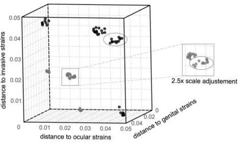
trachomatis as species, and the pathoadaptation driven by arms race. We identified positively selected genes and codons that are hypothetically involved in the evolutionary adaptation of C. trachomatis serovars to different cell types: mucosal cells from the eye conjunctiva (responsible for trachoma) (serovars A-C), from the genitalia (primarily yielding cervicitis) (serovars D-K), and mononuclear phagocytes (yielding invasive diseases such as hemorrhagic proctitis and suppurative lymphadenitis) (serovars L1-L3). Finally, we also detected positive selection events likely driving pathogenic and ecological success dissimilarities.
2.3. Materials and Methods
2.3.1. C. trachomatis strains, cell culture and DNA extraction
The present study encompasses data from 59 C. trachomatis strains and the Chlamydia
Chapter II
17
2.3.2. Selection of loci and sequencing
Based on available in silico full-genome sequence data, we searched for polymorphic genes among C. trachomatis strains through the progressiveMauve algorithm (232) of the Mauve software v2.3.1. A detailed evaluation of polymorphism of each locus was further performed by using Lasergene® 9.0. (DNASTAR, Madison, Wisconsin, USA) and MEGA5 (233). Chromosome loci revealing an extremely low polymorphism were discarded from the present analysis as their use would hamper the accurate application of likelihood tests. We ended up with 75 top-ranked polymorphic genes (listed in the Supplemental Table S2.2). These were categorized according to their functional role, involving 20 housekeeping genes (HKs), 14 genes encoding well-known cell envelope proteins (CEPs), 31 genes coding for secreted proteins (SECs), and 10 genes coding for proteins with unknown function or for which the biological role is not consensual. The SEC category involves proteins secreted [either by the Type III Secretion System (T3SS) - a machinery used by many bacterial pathogens to manipulate eukaryotic host cells by injecting virulence proteins - or by an undefined mechanism], into the cytosol of the host cells or to the inclusion membrane. For analyses enrolling divergence versus polymorphism, the corresponding orthologous genes of the Chlamydia
muridarum Nigg strain were identified (by NCBI-BLAST search) and sequences were collected from the full-genome annotated in the GenBank database (accession number NC_002620) (29). For the strains that we needed to propagate as no in silico data was available, the 75 genes were amplified and sequenced by using standard procedures (111). The sequences and location of primers, as well as the amplicon sizes are listed in the Supplemental Table S2.3. Automated sequencing was achieved using BigDye Terminator v1.1 Cycle Sequencing chemistry, according to the manufacturer’s
instructions (Applied Biosystems) in an Applied Biosystems 3130xl Genetic Analyzer. Sequence reads were assembled using SeqBuilder software (DNASTAR) and alignments were generated using the ClustalW algorithm implemented in both the MegAlign software (DNASTAR) and MEGA5. A concatenated alignment of the 75 genes was also constructed for all C. trachomatis and C. muridarum strains. As the ClustalW program generates alignment artifacts in the presence of insertion/deletion
(indel) events by disrupting codons, we edited “by hand” the amino acid alignments rather than only automate the process before editing the corresponding nucleotide sequences. When strain-exclusive single nucleotide polymorphisms (SNPs), indel events and pseudogenes were identified, resequencing was performed from a newly extracted DNA, and new sequences reads were generated for comparative purposes.
2.3.3. Phylogenomic analysis
18
evolutionary distances were computed using the Kimura 2-parameter method (235). For all these analyses, the pairwise-deletion option was selected as it excludes sites containing alignment gaps or missing data from the analysis only when necessary in the pairwise distance estimation. Truncated genes, which are expected to encode non-functional proteins, were excluded from the phylogenetic and evolutionary analyses, except for the strains with non-disrupted sequences, as their biological role may be phenotype specific.
2.3.4. Global analysis of molecular evolution
The nonsynonymous/synonymous substitution rate ratio (dN/dS) among related protein-coding
DNA sequences, where dN refers to the number of nonsynonymous substitutions per nonsynonymous
site and dS is defined as the number of synonymous substitutions per synonymous site, may be
suggestive of the selective pressures driving the mutational trends (236). Initially, for a global analysis of these trends, we estimated dN and dS values with MEGA5 by using the Kumar model (237). For
each gene, dN/dS was calculated over all C. trachomatis sequence pairs and between the sequences
of the two species (C. trachomatis and C. muridarum). More, in order to reinforce the comparison between the amount of evolutionary variation within the C. trachomatis species (polymorphism) and the variation between C. trachomatis and C. muridarum (divergence), we also applied the McDonald-Kreitman (MK) test (238,239). However, as it has been assumed that the results from the MK test cannot directly discriminate the type of selection acting on genes (240), the subjacent MK test algorithm was only used to clarify the neutral and amino acid-altering mutational trends underlying the C. trachomatis speciation process. This kind of analysis is suitable for tracing the Muller’s ratchet phenomenon, which is commonly observed in niche-restricted pathogens.
2.3.5. Evaluation of the directionality in C. trachomatis evolution
In order to search for genes on which positive selection putatively operates, two distinct approaches were applied. First, as a statistical support of the dN and dS estimations within C.
trachomatis strains, the codon-based Z-test of selection was computed by MEGA5 using the Kumar method (237), where bootstrapping (1000 replicates) was used for estimation of the variation in the statistic test. This test calculates the probability of rejecting the null hypothesis of strict-neutrality (dN =
dS) in favor of one of two alternative hypothesis: positive selection (dN > dS) or purifying selection (dN <
dS). Results with P values less than 0.05 were considered significant at the 5% level. On a second
Chapter II
19 when several lineages in the phylogeny may have been subjected to distinct selective pressures (241,242). The statistical significance of the presence of positive selection along the branch of interest was addressed by the likelihood-ratio test (LRT) (245). In the branch-site test 2, the LRT compares the twice of the log likelihood difference (2Δl) between two models (alternative and null models) with the chi-square distribution with one degree of freedom for p-value calculation (242). The alternative model allows positive selection (dN/dS ≥ 1) for the foreground branch, whereas the null model assumes the
dN/dS ratios < 1 or = 1 for all site classes in all branches in the phylogeny. When positive selection
acting on a specific gene was suggested by a significant LRT (P < 0.05), a Bayes empirical Bayes analysis (246) was used to identify the specific positively selected sites within that gene along the foreground branches. Therefore, the branch-site model requires an a priori definition and labeling of the foreground branches to be tested for positive selection, which should rely on well-defined biological hypotheses (241). Thus, based on the assumption that some genes might be involved in C.
trachomatis phenotypic dissimilarities as a result of targeted positive selective pressures, we created six comprehensible biological hypotheses (H1-H6). The hypotheses evaluate the existence of genes under positive selection that may be involved in the following biological processes: specific cell-appetence to columnar epithelial cells of ocular (H1) or genital mucosae (H2), and to mononuclear phagocytes (H3); pathogenic diversity among strains causing ocular disease (H4), genital disease (H5), or hemorrhagic proctitis and suppurative lymphadenitis (H6). Only the genes for which the phylogeny supported any of these scientific hypotheses were tested.
Finally, as recombination may bias the results of positive selection, we used published data on recombination analysis enrolling all C. trachomatis genes (37,172,174) to inspect whether the genes selected for the present study showed evidences of recombination. Consequently, for the genes showing incongruent trees where unequivocal recombination was detected within a specific branch, the analysis of positive selection was excluded a priori for the corresponding biological hypothesis. On the other hand, genes yielding congruent trees but for which recombination had been previously detected (37,172,174) were still subjected to positive selection analysis and are properly identified in the present study.
2.3.6. Nucleotide sequences accession number
The nucleotide sequences determined in the present study were submitted to the GenBank database (http://www.ncbi.nlm.nih.gov/Genbank/index.html) and are currently available for consulting through the accession numbers: JQ066324 - JQ066722.
2.4. Results
2.4.1. Polymorphism significance of the selected genes