Enterococcus faecalis
Jacqueline Abranches1,2., Pamella Tijerina1,2., Alejandro Avile´s-Reyes1,2., Anthony O. Gaca1,2 , Jessica K. Kajfasz1, Jose´ A. Lemos1,2*
1Center for Oral Biology, University of Rochester Medical Center, Rochester, New York, United States of America,2Department of Microbiology and Immunology, University of Rochester Medical Center, Rochester, New York, United States of America
Abstract
Enterococcus faecalisis an opportunistic nosocomial pathogen that is highly resistant to a variety of environmental insults, including an intrinsic tolerance to antimicrobials that target the cell wall (CW). With the goal of determining the CW-stress stimulon ofE. faecalis, the global transcriptional profile ofE. faecalisOG1RF exposed to ampicillin, bacitracin, cephalotin or vancomycin was obtained via microarrays. Exposure to the b-lactams ampicillin and cephalotin resulted in the fewest transcriptional changes with 50 and 192 genes differentially expressed 60 min after treatment, respectively. On the other hand, treatment with bacitracin or vancomycin for 60 min affected the expression of, respectively, 377 and 297 genes. Despite the differences in the total number of genes affected, all antibiotics induced a very similar gene expression pattern with an overrepresentation of genes encoding hypothetical proteins, followed by genes encoding proteins associated with cell envelope metabolism as well as transport and binding proteins. In particular, all drug treatments, most notably bacitracin and vancomycin, resulted in an apparent metabolic downshift based on the repression of genes involved in translation, energy metabolism, transport and binding. Only 19 genes were up-regulated by all conditions at both the 30 and 60 min time points. Among those 19 genes, 4 genes encoding hypothetical proteins (EF0026, EF0797, EF1533 and EF3245) were inactivated and the respective mutant strains characterized in relation to antibiotic tolerance and virulence in theGalleria mellonellamodel. The phenotypes obtained for two of these mutants,DEF1533 andDEF3245, support further characterization of these genes as potential candidates for the development of novel preventive or therapeutic approaches.
Citation:Abranches J, Tijerina P, Avile´s-Reyes A, Gaca AO, Kajfasz JK, et al. (2013) The Cell Wall-Targeting Antibiotic Stimulon ofEnterococcus faecalis. PLoS ONE 8(6): e64875. doi:10.1371/journal.pone.0064875
Editor:Willem van Schaik, University Medical Center Utrecht, The Netherlands
ReceivedJanuary 25, 2013;AcceptedApril 19, 2013;PublishedJune 3, 2013
Copyright:ß2013 Abranches et al. This is an open-access article distributed under the terms of the Creative Commons Attribution License, which permits unrestricted use, distribution, and reproduction in any medium, provided the original author and source are credited.
Funding:PT was supported by National Institutes of Health-National Institute of General Medical Sciences grant R25 GM064133. AOG and AAR were supported by National Institutes of Health-National Institute of Dental and Craniofacial Research grant T90 DE021985. The funders had no role in study design, data collection and analysis, decision to publish, or preparation of the manuscript
Competing Interests:The authors have declared that no competing interests exist.
* E-mail: [email protected]
.These authors contributed equally to this work.
Introduction
Enterococci are normal inhabitants of the gastrointestinal (GI) tract of humans and animals. While typically harmless to healthy individuals, two enterococcal species, Enterococcus faecalis and E. faecium, are the leading organisms involved in hospital-acquired infections such as catheter-associated urinary tract infections, endocarditis, and surgical and burn wound infections [1,2,3]. Notably, the risk of death for patients infected with multidrug resistant strains, such as vancomycin-resistant enterococci (VRE), is considerably higher than for those infected with antibiotic susceptible strains [4]. In addition, multidrug resistant enterococci pose an additional health care threat as these strains may function as reservoirs for the dissemination of antibiotic resistance determinants to other opportunistic pathogens [5].
Different from Gram-positive pathogens such as Streptococcus pyogenes and Staphylococcus aureus, enterococci do not appear to possess virulence factors such as pro-inflammatory toxins or immune modulators. The virulence of both E. faecalis and E. faeciumis directly associated with the organism’s ability to survive hostile conditions, including an intrinsic resistance against host immune responses, antagonistic products produced by the
competing microbial flora, and stresses imposed by eventual treatments with antimicrobials. When compared to closely related streptococcal and lactococcal species, enterococci are generally more resistant to a variety of stresses that damage the cell envelope, including nearly complete resistance to lysozyme and cephalosporins, and a relatively high tolerance to desiccation, detergents, and other inhibitors of peptidoglycan biosynthesis [1,2,3].
The intrinsic resistance to clinically usefulb-lactam antibiotics (in particular cephalosporins and penicillinase-resistant penicillins) is likely to provide an additional selective advantage for bothE. faecalis and E. faecium. In fact, treatment with broad-spectrum second and third generation cephalosporins has been long considered a risk factor for nosocomialE. faecalisbacteremia [6]. Although low-level resistance tob-lactams in enterococci has been associated with the increased synthesis of a low-affinity penicillin-binding protein (PBP) [7], many of the molecular events that result in the intrinsic resistance ofE. faecalisand E. faeciumto cell wall-stressing agents are poorly understood.
bacteria respond to antimicrobial challenges and to identify potential new targets for antimicrobial drug discovery [8,9,10,11,12]. In E. faecalis, microarrays have been used to investigate the transcriptional responses of this bacterium to growth in blood or urine [13,14], to antibiotics that inhibit protein synthesis [12,15], and to environmental stress such as amino acid starvation [16], iron excess [17] and to bile salts and SDS [18]. More recently, a functional genomic approach, termed Micro-array-based Transposon Mapping (M-TraM), was used to identify
E. faecium genes that contribute to ampicillin tolerance [19]. Despite the clinical relevance of cell wall-targeting agents in enterococcal infections, the genome-wide transcriptional profile of
E. faecalis in response to this class of antibiotics has not been determined. In this study, microarray analyses of cells treated with four different cell wall-targeting antibiotics were conducted to unravel the scope of the cell wall (CW)-stress stimulon ofE. faecalis. Based on the list of genes that belong to the CW-stress regulon, we selected six genes encoding conserved hypothetical proteins that appear to be unique toEnterococcusor restricted to Gram-positive bacteria for subsequent mutational analysis. Four of these six genes were successfully inactivated and their respective mutant strains characterized in relation to their tolerance to antibiotics and virulence in theGalleria mellonellamodel.
Results
Overview of the Microarray Analysis
To identify genes involved in the CW stress responses ofE. faecalis, a whole genome transcriptional profile was performed using antibiotics that target different CW biosynthesis steps: theb -lactams ampicillin and cephalotin, which competitively inhibit the final transpeptidation step of the peptidoglycan synthesis; bacitra-cin, a polypeptide antibiotic that interferes with dephosphorylation of the lipid carrier responsible for moving the peptidoglycan precursors through the cytoplasmic membrane to the CW; and vancomycin, a glycopeptide that disrupts cross-linkage of pepti-doglycan layers by preventing the incorporation ofN -acetylmura-mic acid- and N-acetylglucosamine-peptide subunits into the peptidoglycan matrix. The initial step in evaluating the responses of E. faecalis OG1RF to CW-inhibiting antibiotics was to determine the MIC for ampicillin, bacitracin, cephalotin (a first generation cephalosporin) and vancomycin. Then, for each antibiotic, a concentration corresponding to 1.256the MIC was
added to exponentially-grown cultures (OD600 0.3), and aliquots were incubated aerobically at 37uC in the presence of the antibiotics for 30 and 60 min. The rationale for using 1.256the MIC was to impose a stress without significantly affecting cell viability during the first few hours of antibiotic exposure. Under all tested conditions, OG1RF showed similar, and minimal, growth over the first 60 min incubation period indicating that all samples were at nearly identical growth phases (Fig. S1). These samples were then further analyzed by microarrays.
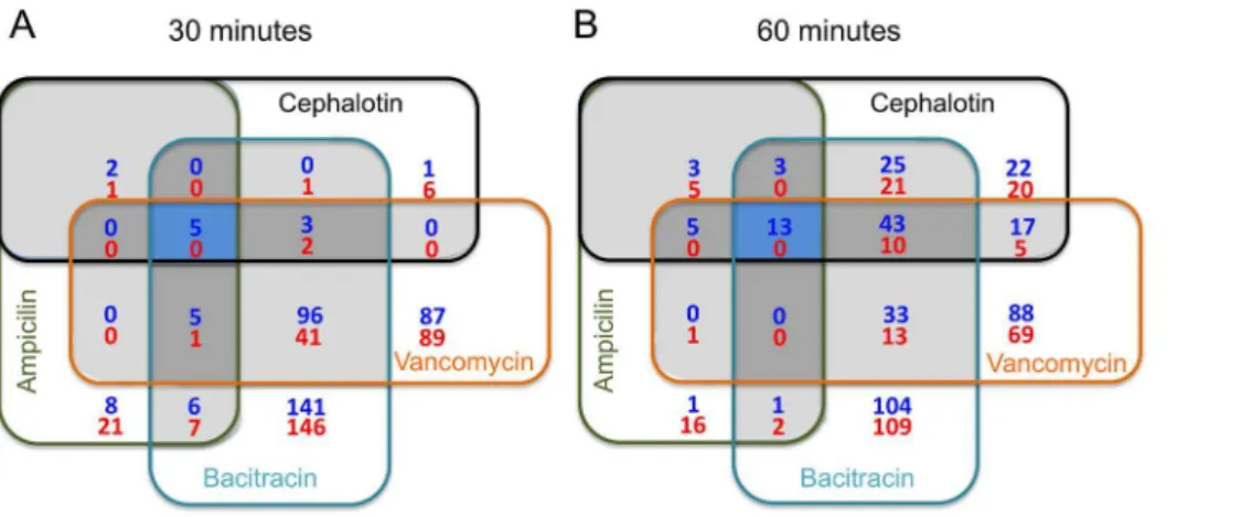
Combining all test conditions in both 30 and 60 min time-points, 861 genes showed significant changes in total transcript amounts as compared to untreated control (P#0.01) but only a small subset of common genes appeared with altered expression under all conditions tested (Fig. 1). Additional Venn diagrams depicting the overlaps in gene expression for each antibiotic regimen between the two tested points are provided as part of the supplemental material (Fig. S2). The complete list of genes with altered expression under all four antibiotic treatments is provided in Table S1. Given that the E. faecalisV583 genome annotation [20] has been widely used in the literature, we adopted the V583 gene designation throughout the text and tables. However,
differentially expressed genes present in the OG1RF genome but absent in V583 were presented with the original OG1RF designation [21]. The majority of genes found differentially expressed encoded for hypothetical proteins of unknown function (157 and 101 genes up- and down-regulated, respectively). Not surprisingly, cell envelope-related genes (65 up- and 39 down-regulated), and genes involved in transport and binding processes (45 up- and 86 down-regulated) were also well represented (Table S1).
To validate our microarray data, the expression of 9 selected genes identified with altered expression in the microarrays was verified by qRT-PCR. All genes showed the same expression trends as observed in the microarrays (Table S2).
Exposure to Bacitracin and Vancomycin, but not tob -lactams, Induced Major Alterations in the Transcriptome ofE. faecalisOG1RF
Among the four antibiotics tested, treatment with bacitracin resulted in the highest number of genes with altered expression (454 and 377 genes differently expressed 30 and 60 min post-treatment, respectively) (Table S1). In addition to the expected large number of cell envelope-related genes, expression of several genes encoding products that participate in central intermediary metabolism and proteins involved in transport and binding processes was also affected by bacitracin treatment. This large number of genes with altered expression could be related to the fact that bacitracin interference with CW biosynthesis occurs at early stages but also because bacitracin can inhibit isopentenyl pyrophosphate (IPP) biosynthesis. IPP is the central precursor of isoprenoids, via the mevalonate pathway [22]. The mevalonate pathway is the sole pathway for IPP biosynthesis inE. faecalis[23], and it is well known that isoprenoids are involved in a wide variety of vital biological processes such as electron transport and peptidoglycan biosynthesis [24]. Of note, four out of the six genes encoding enzymes of the mevalonate pathway were induced by bacitracin treatment (Table S1).
Exposure to vancomycin also resulted in major changes in the transcriptome of OG1RF, with 329 and 297 genes with altered expression at 30 and 60 min, respectively. In particular, a large number of genes (n= 40) encoding enzymes associated with cell envelope metabolism were up-regulated in vancomycin-treated cells (Table S1). Interestingly, genes from the mevalonate pathway were also induced by vancomycin, suggesting an association between IPP biosynthesis and CW homeostasis.
Unlike bacitracin and vancomycin, expression of a much smaller number of genes was affected by treatment with the b -lactams ampicillin and cephalotin. Treatment with cephalotin affected the expression of 21 and 192 genes at 30 and 60 min, respectively. Likewise, exposure to ampicillin for 30 and 60 min affected the expression of 56 and 50 genes, respectively. Even though the number of altered genes varied significantly among conditions, when the data were considered as groups of genes falling into particular functional categories, we observed a remarkably similar gene expression pattern for all four antimicro-bials (Fig. 2), with an overrepresentation of genes encoding hypothetical proteins followed by genes encoding proteins associated with transport and binding and cell envelope metab-olism.
E. faecalis. From this list of 19 genes, we selected 6 genes coding for hypothetical proteins that are unique to enterococci or closely Gram-positive bacteria (EF0026, EF0708, EF0797, EF1258, EF1533 and EF3245) for subsequent mutational analysis.
Antimicrobial Susceptibility of Targeted Mutants
Despite multiple attempts, we were not able to isolate mutants lacking the EF0708 and EF1258 genes. Both genes are predicted to encode small conserved hypothetical proteins (63 and 68 amino acids, respectively). Interestingly, EF0708 is only found in selected
Figure 1. Venn diagrams depicting overlaps in gene expression among ampicilin, cephalotin, bacitracin and vancomycin after 30 and 60 min exposure.Blue shaded areas indicated overlaps among all four antibiotics. Dark grey areas depicts overlaps among three antibiotics and light grey between two antibiotics. Blue numbers represent upregulated genes whereas red numbers indicate downregulated genes. doi:10.1371/journal.pone.0064875.g001
Figure 2. Pie chart of overrepresented functional categories with altered expression after 60 min of exposure to (A) ampicillin, (B) bacitracin, (C) cephalotin, and (D) vancomycin.
Gram-positive bacteria (e.g., Enterococci, Bacilli, Lactobacilli and Geobacilli) whereas EF1258 appears to be unique to E. faecalis
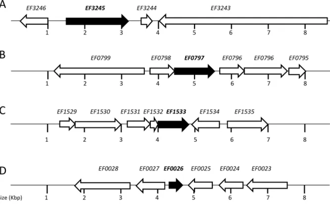
strains. Markerless deletions of the remaining genes were readily obtained, and the deletions were confirmed by PCR sequencing. BLAST search analysis indicated that the putative hypothetical proteins encoded by the EF0026, EF0797 and EF1533 genes are relatively conserved among Gram-positive bacteria. Interestingly, EF3245 is unique toE. faecalismostly because it appears to encode a dual function protein possessing two conserved domains, PAP2 and LytR-CpsA-Psr. PAP2 is a superfamily of phosphatases and haloperoxidases that may act as a membrane-associated lipid phosphatase whereas LytR-CpsA-Psr is a superfamily of cell envelope-related transcriptional attenuators. The genetic organi-zation of the four mutated genes is shown in Figure 3.
Under standard growth conditions, e.g. brain heart infusion (BHI) broth at 37uC under aerobic conditions, all mutants grew as well as the parent strain OG1RF (Table 2). With the exception of
DEF1533, which formed longer cell chains when grown in broth, all other mutants had no obvious morphological cell differences (data not shown). To evaluate the susceptibility of theDEF0026,
DEF0797, DEF1533 and DEF3245 strains to antibiotics that inhibit CW biosynthesis, the minimum inhibitory concentration (MIC) and growth under sub-MIC of each strain to ampicillin, bacitracin, cephalotin and vancomycin was assessed. When compared to the parent strain, all mutants displayed the same MIC to ampicillin and cephalotin (16mg ml21and 64mg ml21,
respectively) and, with the exception of DEF1533 that showed a slightly slower growth in cephalotin, all strains grew equally well in sub-inhibitory concentrations of these two b-lactams (Table 2). Strains DEF1533 and DEF3245 showed, respectively, two- and four-fold lower MIC for bacitracin when compared to OG1RF
(MIC = 64mg ml21). On the other hand, when compared to the
MIC of OG1RF in vancomycin (8mg ml21), DEF0797and
DEF1533 had two-fold lower MIC values whereas DEF0026 showed a two-fold higher MIC. In agreement with the MIC values obtained, bothDEF1533 andDEF3245 strains grew slower in sub-inhibitory concentrations (16, 32 and 64mg ml21) of bacitracin
(Table 2). Strains DEF0026 and DEF0797 grew as well as the parent strain in the presence of different concentrations of bacitracin (Table 2). Also in agreement with the MIC values,
DEF1533 grew significantly slower in sub-MIC concentrations (1, 2 and 4mg ml21) of vancomycin (Table 2). Strains DEF0026,
DEF0797 andDEF3245 grew as well as the parent strain in the presence of different concentrations of vancomycin (Table 2). Unexpectedly, all strains showed shorter doubling times in medium containing 1mg ml21 vancomycin than in medium
without added antibiotics. While it is clear that this concentration of vancomycin does not cause a negative impact cell growth, the basis for the faster growth rates remains unknown.
Next, we carried out time-kill assays in the presence of ampicillin, bacitracin or vancomycin such that all strains were tested under concentrations that correspond to 5 or 10X the MIC for the selected antibiotic. The bacteriostatic antibiotic chloram-phenicol was used as a control to monitor cell viability over time. Incubation in the presence of chloramphenicol (10X MIC) did not result in statistically significant loss of cell viability when compared to untreated cells and all strains survived equally well over a 4-day period (Fig. 4A). Essentially, no differences between strains were observed in cells treated with ampicillin (Fig. 4B), albeitDEF3245 showed a modest, but not statistically significant, increased survival. Consistent with lower MIC and slower growth rates in bacitracin, DEF3245 was killed significantly more rapidly by Table 1.Genes upregulated by Ampicilin (Amp), Bacitracin (Bac), Cep (Cephalotin) and Vancomycin (Van) 30 or 60 min after exposure.
Gene ID Definition Amp 309 Amp 609 Bac 309 Bac 609 Cep 309 Cep 60 Van 309 Van 609
EF0026 hypothetical protein 2.68 9.07 19.06 7.64 4.20 12.17 6.17 11.9
EF0708 hypothetical protein 3.66 2.99 15.86 5.97 9.01 9.2 16.39
EF0797 hypothetical protein 4.43 8.12 10.19 12.95 4.45 11.30 5.52 10.86
EF0802 hypothetical protein 6.26 36.55 16.21 29.28 134.36 232.04
EF1224 transcriptional regulator 2.96 3.51 16.54 3.01 12.5
EF1258 hypothetical protein 3.8 36.83 11.18 8.47 23.69 21.47
EF1304 Mg-importing ATPase 2.52 5.21 15.98 6.66 24.62 80.97
EF1533 hypothetical protein 6.87 11.7 62.15 38.18 7.46 38.96 13.64 21.61
EF1587 nudix family phosphohydrolase 2.82 2.61 22.39 11.86 6.7 15.45 19.3
EF1753 hypothetical protein 4.40 3.95 25.31 18.40 3.74 10.2 11.63 18.16
EF1814 EmbR/QcaA drug resistance transporter 6.01 8.05 7.86 14.35 14.55
EF2784 hypothetical protein 2.66 17.35 12.3 4.95 4.93
EF2892 hypothetical protein 3.8 7.93 6.71 19.78 29.85
EF2896 hypothetical protein 6.23 35.23 30.21 23.38 112.37 97.22
EF2913 membrane protein 2.24 8.4 4.16 3.71 2.95
EF3057 hypothetical protein 6.61 5.34 9.22 4.8 7.35
EF3239 hypothetical protein 3.48 7.44 8.44 5.09 13.76 3.4
EF3245 lytR-cpsA-psr and PAP-2 superfamilies 4.28 8.76 43.91 30.51 5.51 34.13 81.30 112.6
OG0126 ABC transporter superfamily 3.71 11.12 5.12 20.06 19.1
Fold change results from microarrays comparison of antibiotic-treated samples with untreated samples. Blank cells indicate that there were no significant differences between treated and untreated cells. Genes in bold were selected for subsequent mutational analysis.
bacitracin (320mg ml21, which corresponds to 5X the MIC for the
parent strain but 20X forDEF3245 (Fig. 5C). On the other hand, despite showing a lower MIC and slower sub-MIC growth in the presence bacitracin, the time-kill kinetics of DEF1533 was not significantly different than the parent OG1RF strain when cells were exposed to 320mg ml21(10X MIC) of bacitracin (Fig. 4C).
Of note, DEF0026 and DEF0797 were completely resistant to bacitracin killing over the course of the experiment (Fig. 4C).
Interestingly, lower concentrations of bacitracin corresponding to 5X the MIC for bacitracin for EF1533 strain (160mg ml21) and
EF3245 (80mg ml21) did not affect cell viability of all tested strains
over the course of the experiment (Fig. S3). Despite a two-fold higher MIC for vancomycin, DEF0026 survived as well as the parent strain in vancomycin time-kill assays, even when higher concentrations of vancomycin were used (Fig. 4C and Fig. S3). Consistent with the lower MIC in vancomycin, DEF0797 and
Figure 3. Schematic representation of the EF0026, EF0797, EF1533 and EF3245 loci and flanking regions.(A) EF3245 is in an apparent monocistronic operon and is flanked by a chitin-binding protein (EF3246), a small hypothetical protein (EF3243) and a large hypothetical protein with a CW-binding domain (EF3244). (B) EF0797 is apparently co-transcribed with another hypothetical protein (EF0798) and both genes flanked by a putative protein belonging to the Type 2 phosphatidic acid phosphatase family (EF0796) and a putative autolysin (EF0799). (C) EF1533 appears to be the last gene of a three-gene operon with EF1532 coding for a hypothetical protein and EF1531 coding for a putative TetR transcriptional regulator. EF1531–1533 gene cluster is flanked by a PTS transporter subunit (EF1530) and a peptidyl-prolyl cis-trans isomerase (EF1534). (D) EF0026 is flanked by a putative membrane protein (EF0025) and a putative transcriptional regulator (EF0027) whereas EF0024 and EF0023 encode a hypothetical protein and a mannose-fructose-sorbose PTS porter, respectively.
doi:10.1371/journal.pone.0064875.g003
Table 2.Doubling times of OG1RF, EF0026, EF797, EF1533 and EF3245 in various antibiotics concentrations.
OG1RF (min±SD) EF0026 (min±SD) EF0797 (min±SD) EF1533 (min±SD) EF3245 (min±SD)
BHI only 62.360.8 61.860.7 63.564.5 67.763.4 6060.6
Vancomycin (1mg ml21) 57.561.3 56.761 59.661.4 65.962.4* 55.960.4
Vancomycin (2mg ml21) 60.761.7 57.560.9 64.462.2 68.163.27* 5860.26
Vancomycin (4mg ml21) 73.461 73.861.6 7261.4 88.561.4* 81.867.9
Bacitracin (8mg ml21) 71.360.8 68.761.4 72.960.5 85.860.7* 73.562.5
Bacitracin (16mg ml21) 73.666.1 70.661.8 72.262.3 98.462* 96.161.7*
Bacitracin (32mg ml21) 76.460.6 78.8.063 75.662 128.862.4* 132.61*
Cephalotin (15mg ml21) 65.660.92 63.960.72 63.560.2 71.360.27* 61.460.28
Ampicilin (1mg ml21) 72.962.4 70.763.2 70.761.2 72.661.4 70. 861.7
Data presented represents the average in minutes and standard deviation of at least three independent experiments. Numbers followed by * represent a statistically significant difference (p#0.05) compared to the parental strain OG1RF under the same growth condition using Student’sttest.
DEF1533 were killed more rapidly by concentrations that correspond to 20X (80mg ml21, DEF0797 only) or 10X (40mg
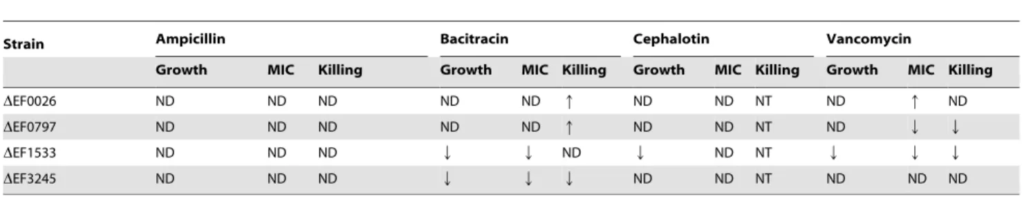
ml21, both DEF0797 and DEF1533) the MIC for vancomycin (Fig. 4D and Fig. S3). The summary of the antibiotic-resistant profile of each mutant strain is shown in Table 3.
Virulence ofDEF0026 andDEF0797 andDEF1533 was Altered in theG. mellonella Model
Larva of the LepidopteraGalleria mellonella has been shown to serve as a surrogate model to study bacterial virulence for a number of pathogens [25], including E. faecalis [16,26,27,28]. Here, we tested the virulence potential of theDEF0026,DEF0797,
DEF1533 andDEF3245 strains injected intoG. mellonella(Fig. 5). While theDEF3245 strain was able to killG. mellonellaas efficiently as the parent OG1RF strain, virulence ofDEF1533 was attenuated (p,0.05). Conversely, strains DEF0026 and DEF0797 were significantly more virulent than OG1RF (p,0.05).
Discussion
In this study, we used microarrays to obtain a snapshot of the transcriptional responses ofE. faecalisto four different antibiotics that target CW biosynthesis, namely ampicillin, bacitracin, cephalotin and vancomycin. Treatment with all four drugs, most Table 3.Summary of the antibiotic-resistance profile ofDEF0026,DEF0797,DEF1533 andDEF3245 in relation to that of OG1RF (wild-type strain).
Strain Ampicillin Bacitracin Cephalotin Vancomycin
Growth MIC Killing Growth MIC Killing Growth MIC Killing Growth MIC Killing
DEF0026 ND ND ND ND ND q ND ND NT ND q ND
DEF0797 ND ND ND ND ND q ND ND NT ND Q Q
DEF1533 ND ND ND Q Q ND Q ND NT Q Q Q
DEF3245 ND ND ND Q Q Q ND ND NT ND ND ND
ND, no differences in comparison to OG1RF. NT, not tested.
Q, sensitive in comparison to OG1RF. q, resistant in comparison to OG1RF. doi:10.1371/journal.pone.0064875.t003
Figure 4. Cell death kinetics of OG1RF,DEF3245, DEF0797, DEF1533 and DEF0026 in (A) chloramphenicol (160mg ml21), (B) ampicillin (80mg ml21), (C) bacitracin (320mg ml21), and (D) vancomycin (80mg ml21).Experiments were performed in triplicates with averages and standard deviations calculated for each time-point. Student’sttest was performed to verify significance.
notably bacitracin and vancomycin, resulted in an apparent metabolic downshift based on the repression of a number of genes involved in translation, energy metabolism, transport and binding. This may be an indication that cells are saving or redirecting energy resources to maintain CW integrity and survive the physiological stress imposed by the antibiotics. In fact, genes involved in CW metabolism were highly affected, and overwhelm-ingly up-regulated during antibiotic treatment. For example, several genes coding for penicillin binding proteins (PBPs), responsible for CW cross-linking, and genes from thedltoperon, responsible for D-alanylation of lipoteichoic acids (LTA), were highly induced by bacitracin, cephalotin and vancomycin. Notably, esterification of LTA with D-alanine has been directly linked to vancomycin tolerance in both S. aureus and E. faecium
[29,30].
Some attention has been devoted to investigate the possible association of oxidative stress with antibiotic-mediated cell death [31]. More specifically, these authors proposed that bactericidal drugs, irrespective of the cellular target, kill bacteria by triggering endogenous production of hydroxyl radical. In a separate study, a panel of strains deficient in the production of known antioxidant enzymes was used to suggest that accumulation of superoxide anion, but not peroxide, was responsible for the bactericidal effects of penicillin and vancomycin in E. faecalis [32]. However, two recent investigations using the Gram-negative paradigm E. coli
challenged the concept of the common ROS-mediated antibiotic killing pathway. Specifically, Liu and Imlay showed that different antibiotics targeting the cell wall, DNA replication or protein synthesis had little impact on respiration rates, did not promote H2O2 formation, caused no damage to ROS-sensitive metallo-enzymes and did not activate the OxyR regulon [33]. In the second study, Keren and co-workers observed no obvious differences in antibiotic survival ofE. colicells under aerobic or anaerobic conditions [34]. In our study, we found that the expression of a number of oxidative stress genes, such as ahpC
(alkyl hydroperoxide), katA (heme-dependent catalase), msrA
(methionine sulfoxide reductase A), nox (H2O-forming NADH oxidase),npr(NADH peroxidase) andohr(organic hydroperoxide resistance), was induced during bacitracin exposure. However, a few other typical antioxidant genes, such as sodA (superoxide dismutase) and tpx (thiol peroxidase), showed the opposite
transcriptional pattern (e.g. repressed by bacitracin). Thus, the linkage of oxidative stress gene expression with CW stress inE. faecalisis also not clear. Of note, previous transcriptional profiling studies of the envelope stress responses ofBacillus subtilis,S. aureus
and Streptococcus pneumoniae did not establish a firm correlation between the induction of oxidative stress genes and CW stress responses [35,36,37]. Given the recent controversy surrounding the common ROS-mediated antibiotic killing pathway model [31,33,34], further studies, perhaps by measuring the production of reactive oxygen species (ROS) as well as the activity of antioxidant enzymes during antibiotic treatment, are necessary to conclusively determine the relevance of oxidative stress damage in antibiotic-mediated killing of E. faecalis as well as other closely-related Gram-positive bacteria.
The expression of over 50 transcriptional regulators was affected by the different test conditions. Among those were 19 genes coding for two-component signal transduction systems (TCSTS) including members of the LiaRS, LytRS and VanRS families. The vanS and vanR TCSTS genes in OG1RF encode proteins with high homology with thevanGlocus responsible for vancomycin resistance in certain strains ofE. faecalis[38]. Multiple evidences derived from different Gram-positive bacteria indicate that the LiaRS and LytRS TCSTS are involved in regulation of CW metabolism. In Streptococcus gordonii, LiaSR was shown to control the expression of thedltoperon in response to cell envelope perturbations and extracellular pH [39]. InListeria monocytogenes, transcription of theliaRandliaSgenes was induced by bacitracin and vancomycin [40], and aliaSmutant strain exhibited increased sensitivity to cephalosporins [41]. Finally, treatment with vanco-mycin induced the expression of the LiaRS-encoding genes inS. pneumoniae [42]. In B. subtilis and S. aureus, lytR-like genes were induced by CW-active antibiotics [35,36]. The LytRS TCSTS of
S. aureushas been shown to play a role in the control of autolysis [43]. Notably, in a vancomycin intermediate S. aureus (VISA) strain, lytRS expression was repressed in comparison to the parental strain upon exposure to vancomycin, suggesting a role for LytRS in sensing envelope damage and controlling CW turn over and autolysis [8]. In addition to the putative LytRS TCSTS, transcription of 3 additional genes with putative LytR domains (EF0465, EF1212 and EF3245) was induced by at least 2 of the 4 tested antibiotics.
In an effort to discover new genes responsible for the intrinsic tolerance ofE. faecalisto CW-damaging agents, we selected 6 genes for mutational analysis based on three major criteria: (i) the genes were induced by all 4 antibiotics tested, (ii) the genes appeared to be uniquely found in enterococci or were restricted to Gram-positive bacteria, and (iii) the genes encode hypothetical proteins of unknown function or putative regulatory proteins based on the presence of a DNA-binding domain. Initially, we attempted to generate deletions in EF0026, EF0708, EF0797, EF1533, EF1753 and EF3245. However, we were unable to confirm mutations of EF0708 and EF1258, suggesting that these genes may perform an essential role for cell viability, or that we did not use appropriate conditions for mutant isolation. Further studies, such as the construction of conditional mutants, are necessary to confirm the essentiality of these genes. If proven to be essential, both EF0708 and EF1258 may be viewed as desirable targets for the development of novel therapeutic and prevention approaches.
The DEF0026 strain showed a two-fold higher MIC for vancomycin and enhanced survival during bacitracin killing (see Table 3). Given that EF0026 was induced under all tested conditions, these findings were unexpected. TheDEF0797 strain also showed enhanced survival during bacitracin killing but had a lower MIC and increased susceptibility to vancomycin. In
Figure 5. Galleria mellonella survival after injection with 56105CFU of OG1RF,DEF3245, DEF0797,DEF1533, DEF0026
and heat-killed (HK) OG1RF.Using the log-rank test to compare against the wild-type OG1RF strain, DEF1533 showed attenuated virulence whereas DEF0026 and DEF0797 were more virulent (p,0.05). Data presented is a representative of at least three independent experiments.
addition, bothDEF0026 andDEF0797 were more virulent inG. mellonella, an obviously undesirable trait in the drug discovery process. Based on these results, the EF0026 and EF0797 genes may not be desirable candidates for the development of new antibacterial therapies. In a recent study, a library of 177 insertion mutations in genes encoding putative surface or stress-response factors in E. faecalis V583 was screened for several phenotypes including resistance to antibiotics and virulence in G. mellonella
[27]. Two of the four genes investigated in our study, EF0797 and EF3245, were also characterized in the aforementioned large-scale study. In contrast with our results, the authors did not observe any important phenotypic differences in their EF0797 and EF3245 mutant strains [27]. However, both studies have relevant differences ranging from the different type wild-type strain used in the study (OG1RF vs. V583), mutant construction and general growth conditions.
EF1533 is a gene coding for a conserved hypothetical protein with 125 amino acid residues that is ubiquitously found in E. faecalis strains. Homologues of EF1533 can also be found inE. faecium and other enterococcal species as well as in lactobacilli, staphylococci and bacilli (<50% similarity). EF1533 appears to be
the last gene of a three-gene operon with the first gene (EF1531) coding for a putative TetR transcriptional regulator and the second gene (EF1532) coding for a hypothetical protein (see Fig. 3). The EF1533 mutant showed increased susceptibility to vancomy-cin and bacitravancomy-cin and grew more slowly under sub-MIC of cephalotin (see Table 3). Furthermore, virulence ofDEF1533 was significantly attenuated inG. mellonella. Collectively, these results indicate that EF1533 may play a role in CW homeostasis and support further characterization of EF1533 as a potential new drug target.
The DEF3245 strain showed attenuated virulence in the G. mellonellamodel and displayed increased sensitivity to bacitracin. Interestingly, the 538 amino acid residues long EF3245 is unique toE. faecalisand possess two distinct domains: an N-terminal type-2 phosphatidic acid phosphatase (PAPtype-2) superfamily domain and a C-terminal lytR-cpsA-psr superfamily domain of cell envelope-associated transcriptional attenuators. Members of the PAP2 superfamily have been proposed to serve as integral membrane proteins that catalyze the dephosphorylation of phospholipids [44,45,46]. InB. subtillis, overexpression of a PAP2 protein, named BcrC, is directly involved in bacitracin tolerance [47]. Thus, it is conceivable that EF3245 encodes a dual function protein that contributes to cell homeostasis and survival by modulating both membrane and CW composition. Members of the lytR-cpsA-psr superfamily are routinely found as part of the CW stress stimulon, and have been proposed to regulate CW homeostasis and biogenesis [48]. Noteworthy, LytR-CpsA-Psr proteins are present in all Gram-positive organisms, with the exception of the CW-deficientMollicutes, and rarely found in Gram-negative bacteria. In
S. aureusandS. mutans, inactivation of a member of the lytR-CpsA-Psr superfamily resulted in multiple phenotypes including increased susceptibility to CW-active antibiotics [49,50,51,52].
In this study, we unveiled the scope of the CW stress stimulon of
E. faecalis and conducted a preliminary characterization of four genes encoding putative hypothetical proteins that are unique to Gram-positive bacteria, or exclusively found in enterococci. In addition to the four isogenic mutants characterized here, several other genes with a potential role in CW homeostasis and cell survival were identified and may serve as the foundation for future studies. The identification of theE. faecalisCW antibiotic targeting stimulon is an important step for a better understanding of the intrinsic tolerance of enterococci towards CW-damaging agents.
Materials and Methods
Bacterial Strains and Growth Conditions
Bacterial strains used in this study are listed in Table 4. Strains were routinely maintained in Brain Heart Infusion (BHI) agar plates (BD, Franklin Lakes, NJ). For microarray analysis, a minimum of 4 independent cultures of E. faecalisOG1RF were grown in FMC medium [53] supplemented with 10 mM glucose to an optical density at 600 nm (OD600) of 0.3 and the cultures were divided in 5 aliquots. One aliquot was collected by centrifugation and immediately frozen (untreated control cells). The other aliquots were treated for 30 and 60 min with 1.256the minimum inhibitory concentration (MIC) of one of the following CW inhibitors: ampicillin (20mg ml21), bacitracin (80mg ml21),
cephalotin (80mg ml21), or vancomycin (10mg ml21). To assess
the ability ofE. faecalisOG1RF and its derivative mutant strains to grow in the presence of sub-inhibitory concentrations of antibi-otics, mid-exponential-phase (OD600,0.4) cultures were diluted
1:100 into conditioned BHI media and growth was monitored for 24 hours using a Bioscreen C growth monitor (Oy Growth Curves AB Ltd., Helsinki, Finland).
RNA Extraction
To isolate RNA from E. faecalis, cells were harvested by centrifugation at 4uC and then treated with the RNA Protect reagent (QIAGEN, Inc., Chatsworth, CA). Total RNA was isolated from homogenizedE. faecaliscells by the hot acid-phenol method as described previously [16]. RNA pellets were resuspended in nuclease-free H2O, and treated with DNase I (Ambion) at 37uC for 30 minutes. The RNA was purified again using the RNeasy mini kit (QIAGEN), including a second on-column DNase treatment that was performed as recommended by the supplier.
Microarray Experiments
Transcriptome analysis was performed using the E. faecalis
microarrays provided by the J. Craig Venter Institute Pathogen Functional Genomics Resource Center (PFGRC) (http://pfgrc.jcvi. org/index.php/microarray). The microarray slides consist of 6600, 70mer DNA probes encompassing all unique open reading frames (ORFs) from six sequencedE. faecalisstrains: OG1RF, ATCC 29200, TUSoD Ef11, HH22, TX0104, TX1322, and V583 including its three plasmids printed in duplicate. Additional details regarding the arrays can be found at http://pfgrc.jcvi.org/index.php/microarray/ array_description/enterococcus_faecalis/version1.html.
BRB-ArrayTools.html) with a cutoff Pvalue of 0.01. Additional details regarding array protocols are available at http://pfgrc.jcvi. org/index.php/microarray/protocols.html.
Microarray Data Accession Number
Microarray data have been deposited in the NCBI Gene Expression Omnibus (GEO) database (http://www.ncbi.nlm.nih. gov/geo) under GEO Series accession number GSE45306.
Real-time Quantitative PCR
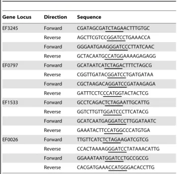
Microarrays were validated by real-time quantitative RT-PCR (qRT-PCR) by using primers specific for the six genes chosen for inactivation and three other randomly selected genes. Gene-specific primers (Table S3) were designed using Beacon Designer 2.0 software (Premier Biosoft International). Reverse transcription and real-time reverse transcriptase PCR were carried out according to protocols described elsewhere [55]. Student’s t test was performed to verify significance of the real-time PCR quantifications.
Construction of Mutant Strains
A markerless genetic exchange system [55] was used to obtain in-frame isogenic deletion strains inE. faecalisOG1RF. Briefly, two PCR products flanking the gene of interest were obtained with the primers listed in Table 5. The PCR products of the 59 and 39 flanking regions of each gene were digested with restriction enzymes (XbaI and BamHI or BamHI and NcoI) and individually ligated to the previously digested pCJK47 vector [56] usingE. coli
EC1000 [56] as the host strain. The resulting plasmids were electroporated into competent E. faecalis CK111/pCF10-101 (donor strain). Donor strains harboring the desired pCJK47 derivative plasmid were conjugated with E. faecalis OG1RF (recipient strain) and transconjugants were selected on BHI agar medium containing rifampin (200mg ml21), fusidic acid (25mg
ml21), and erythromycin (10mg ml21). The PheS* negative
counter-selection system was utilized to isolate double-crossover deletions [56]. Individual gene deletions were confirmed by PCR and further sequencing of the deletion site and flanking regions.
Minimum Inhibitory Concentration (MIC)
The MIC of selected CW inhibitors (ampicillin, bacitracin, cephalotin and vancomycin) to OG1RF and its derivative mutants was determined by a broth microdilution method in triplicates. Briefly, 3ml of exponentially-grown cultures (OD600 nm,0.7) was
added to the wells of microtiter plates containing two-fold serial dilutions of antibiotics in BHI. Plates were incubated aerobically at 37uC for 18 h. The lowest concentration of antibiotic that prevented growth was recorded as the MIC.
Antibiotic Time-kill Kinetics
Cultures were grown in BHI to exponential phase, serially diluted in fresh BHI to obtain 56106to 16107CFU ml21.
Time-kill studies were initiated by adding 5 or 10 times the MIC for each antibiotic for each mutant strain of ampicillin (80mg ml21),
bacitracin (80, 160 and 320mg ml21), chloramphenicol (80mg
ml21), or vancomycin (40, 80, 160mg ml21). Viable counts were
determined by plating cultures on BHI plates at time zero and then every 24 h. All experiments were performed at least in triplicates and Student’sttest was performed to verify significance at each time-point.
Table 4.Bacterial strains used in this study.
Strains Relevant Characteristics Source or reference
Enterococcus faecalis
OG1RF Laboratory strain; Rifampicin-R Fusisic Acid-R Lab stock
CK111/pCF10-101 OG1Spectomycinupp4::P23repA4 [55]
DEF3245 Deletion mutant of OGIRF This Study
DEF0797 Deletion mutant of OGIRF This Study
DEF1533 Deletion mutant of OGIRF This Study
DEF0026 Deletion mutant of OGIRF This Study
Escherichia coli
DH10B Cloning Host Lab stock
EC1000 Host for cloning RepA-dependent plasmid [56]
doi:10.1371/journal.pone.0064875.t004
Table 5.Primers used for gene deletion.
Gene Locus Direction Sequence
EF3245 Forward CGATAGCGATCTAGAACTTTGTGC
Reverse AGCTTCGTCCGGATCCTGAAACCA
Forward GGGAATGAAGGGATCCCTTATCAAC
Reverse GCTACAATGCCATGGAAAAGAGAGG
EF0797 Forward GCATAATCATCTAGACTTTCTAGCG
Reverse CGGTTGATACGGATCCTGATGATAA
Forward CGCTAAGACAGGATCCGATAAGAGA
Reverse GATTTCCTCCCATGGTACTACTCG
EF1533 Forward GCCTCAGACTCTAGAATTGCATTG
Reverse GGTCTTGTTGGATCCCTTCATACG
Forward GCATCAATGAGGATCCTTGGATAATC
Reverse GAAATACTTCCATGGCCCATGTGA
EF0026 Forward TTGTTCATCTCTAGAAGATCGTCG
Reverse CCACTAAAAGGGATCCTATAAACATTG
Forward GGAAATAATGGATCCTGCCGCCG
Reverse CACGATGAAACCATGGGACACCTTG
The underlined bases correspond to the restriction sites included to aid in the subsequent cloning of the PCR products.
Galleria Mellonella Infection
For theG. mellonellakilling assays, insects in the final larval stage were purchased from Vanderhorst Inc (St. Marys, OH). Groups of 20 larvae, ranging from 200 to 300 mg in weight with no signs of melanization were randomly chosen and used for subsequent infection. A Hamilton syringe (Hamilton, Reno, NV) was used to inject 5ml aliquots of bacterial inoculum (56105CFU) into the haemocoel of each larva via the last left proleg. Aliquots of the initial inocula were serially diluted and plated onto BHI agar plates to confirm initial infection dose. Control groups were injected with heat-inactivatedE. faecalisOG1RF (45 min at 75uC). After injection, larvae were kept at 37uC, and monitored for death at various time points during the course of 72 h. Kaplan–Meier killing curves were plotted and estimation of differences in survival compared by using the Mantel-Cox log-rank test. All data were analyzed with GraphPad Prism 4.0 software. Experiments were performed independently at least three times.
Supporting Information
Figure S1 Growth curve ofE. faecalisOG1RF in FMC. When cultures reached OD600of 0.3, ampicilin (20mg ml2
1
), bacitracin (80mg ml21), cephalotin (40mg ml21) or vancomycin (10mg ml21)
were added to culture aliquots. For microarray analysis, cells were harvested at 0, 30 and 60 min post antibiotic exposure.
(PDF)
Figure S2 Venn diagrams depicting overlaps in gene expression for each antibiotic between 30 and 60 minutes. Blue numbers indicate upregulated genes and numbers in red represent downregulated genes.
(PDF)
Figure S3 Cell death kinetics of the mutant strains with altered MICs. Experiments were performed using antibiotic concentra-tions ranging from 5 to 10X the MIC for each mutant strain. Experiments were performed in triplicates with average and standard deviations calculated for each time-point. Student’sttest was performed to verify significance (p,0.05) in comparison to the parent strain.
(PDF)
Table S1 Genes differentially expressed in Enterococcus faecalis
OG1RF cells treated with cell-wall inhibiting antibiotics for 30 and 60 min at ap-value,0.01.
(DOCX)
Table S2 Validation of microarrays by qRT-PCR. Data is presented in fold of change relative to control conditions measured by qRT-PCR, and by microarrays in parenthesis.
(DOCX)
Table S3 Quantitative RT-PCR primers used to validate microarray data.
(DOCX)
Acknowledgments
We thank the J. Craig Venter Institute for supplying the E. faecalis microarray slides.
Author Contributions
Conceived and designed the experiments: JA PT AAR AOG JKK JAL. Performed the experiments: JA PT AAR AOG JKK. Analyzed the data: JA PT AAR AOG JKK JAL. Contributed reagents/materials/analysis tools: JA PT AAR AOG JKK JAL. Wrote the paper: JA PT AAR AOG JKK JAL.
References
1. Arias CA, Murray BE (2012) The rise of theEnterococcus: beyond vancomycin resistance. Nat Rev Microbiol 10: 266–278.
2. Murray BE (1990) The life and times of theEnterococcus. Clin Microbiol Rev 3: 46–65.
3. Hollenbeck BL, Rice LB (2012) Intrinsic and acquired resistance mechanisms in Enterococcus. Virulence 3: 421–433.
4. Bearman GM, Wenzel RP (2005) Bacteremias: a leading cause of death. Arch Med Res 36: 646–659.
5. Weigel LM, Clewell DB, Gill SR, Clark NC, McDougal LK, et al. (2003) Genetic analysis of a high-level vancomycin-resistant isolate ofStaphylococcus aureus. Science 302: 1569–1571.
6. Pallares R, Pujol M, Pena C, Ariza J, Martin R, et al. (1993) Cephalosporins as risk factor for nosocomialEnterococcus faecalisbacteremia. A matched case-control study. Arch Intern Med 153: 1581–1586.
7. Fontana R, Ligozzi M, Pittaluga F, Satta G (1996) Intrinsic penicillin resistance in enterococci. Microb Drug Resist 2: 209–213.
8. McAleese F, Wu SW, Sieradzki K, Dunman P, Murphy E, et al. (2006) Overexpression of genes of the cell wall stimulon in clinical isolates of Staphylococcus aureusexhibiting vancomycin-intermediate-S. aureus-type resistance to vancomycin. J Bacteriol 188: 1120–1133.
9. Muthaiyan A, Silverman JA, Jayaswal RK, Wilkinson BJ (2008) Transcriptional profiling reveals that daptomycin induces theStaphylococcus aureuscell wall stress stimulon and genes responsive to membrane depolarization. Antimicrob Agents Chemother 52: 980–990.
10. Song Y, Lunde CS, Benton BM, Wilkinson BJ (2012) Further insights into the mode of action of the lipoglycopeptide telavancin through global gene expression studies. Antimicrob Agents Chemother 56: 3157–3164.
11. Breidenstein EB, Bains M, Hancock RE (2012) Involvement of the lon protease in the SOS response triggered by ciprofloxacin inPseudomonas aeruginosaPAO1. Antimicrob Agents Chemother 56: 2879–2887.
12. Aakra A, Vebo H, Snipen L, Hirt H, Aastveit A, et al. (2005) Transcriptional response of Enterococcus faecalis V583 to erythromycin. Antimicrob Agents Chemother 49: 2246–2259.
13. Vebo HC, Snipen L, Nes IF, Brede DA (2009) The transcriptome of the nosocomial pathogen Enterococcus faecalisV583 reveals adaptive responses to growth in blood. PLoS One 4: e7660.
14. Vebo HC, Solheim M, Snipen L, Nes IF, Brede DA (2010) Comparative genomic analysis of pathogenic and probioticEnterococcus faecalisisolates, and their transcriptional responses to growth in human urine. PLoS One 5: e12489.
15. Aakra A, Vebo H, Indahl U, Snipen L, Gjerstad O, et al. (2010) The Response of Enterococcus faecalisV583 to Chloramphenicol Treatment. Int J Microbiol 2010: 483048.
16. Gaca AO, Abranches J, Kajfasz JK, Lemos JA (2012) Global transcriptional analysis of the stringent response inEnterococcus faecalis. Microbiology 158: 1994– 2004.
17. Lopez G, Latorre M, Reyes-Jara A, Cambiazo V, Gonzalez M (2012) Transcriptomic response of Enterococcus faecalisto iron excess. Biometals 25: 737–747.
18. Solheim M, Aakra A, Vebo H, Snipen L, Nes IF (2007) Transcriptional responses ofEnterococcus faecalisV583 to bovine bile and sodium dodecyl sulfate. Appl Environ Microbiol 73: 5767–5774.
19. Zhang X, Paganelli FL, Bierschenk D, Kuipers A, Bonten MJ, et al. (2012) Genome-wide identification of ampicillin resistance determinants inEnterococcus faecium. PLoS Genet 8: e1002804.
20. Paulsen IT, Banerjei L, Myers GS, Nelson KE, Seshadri R, et al. (2003) Role of mobile DNA in the evolution of vancomycin-resistantEnterococcus faecalis. Science 299: 2071–2074.
21. Bourgogne A, Garsin DA, Qin X, Singh KV, Sillanpaa J, et al. (2008) Large scale variation inEnterococcus faecalisillustrated by the genome analysis of strain OG1RF. Genome Biol 9: R110.
22. Stone KJ, Strominger JL (1972) Inhibition of sterol biosynthesis by bacitracin. Proc Natl Acad Sci U S A 69: 1287–1289.
23. Doun SS, Burgner JW 2nd, Briggs SD, Rodwell VW (2005)Enterococcus faecalis phosphomevalonate kinase. Protein Sci 14: 1134–1139.
24. Heuston S, Begley M, Gahan CG, Hill C (2012) Isoprenoid biosynthesis in bacterial pathogens. Microbiology 158: 1389–1401.
25. Ramarao N, Nielsen-Leroux C, Lereclus D (2012) The InsectGalleria mellonellaas a Powerful Infection Model to Investigate Bacterial Pathogenesis. J Vis Exp. 26. de Oliveira NE, Abranches J, Gaca AO, Laport MS, Damaso CR, et al. (2011)
clpB, a class III heat-shock gene regulated by CtsR, is involved in thermotolerance and virulence ofEnterococcus faecalis. Microbiology 157: 656– 665.
27. Rigottier-Gois L, Alberti A, Houel A, Taly JF, Palcy P, et al. (2011) Large-scale screening of a targeted Enterococcus faecalismutant library identifies envelope fitness factors. PLoS One 6: e29023.
29. Peschel A, Vuong C, Otto M, Gotz F (2000) The D-alanine residues of Staphylococcus aureusteichoic acids alter the susceptibility to vancomycin and the activity of autolytic enzymes. Antimicrob Agents Chemother 44: 2845–2847. 30. Gutmann L, Al-Obeid S, Billot-Klein D, Ebnet E, Fischer W (1996) Penicillin
tolerance and modification of lipoteichoic acid associated with expression of vancomycin resistance in VanB-type Enterococcus faecium D366. Antimicrob Agents Chemother 40: 257–259.
31. Kohanski MA, Dwyer DJ, Hayete B, Lawrence CA, Collins JJ (2007) A common mechanism of cellular death induced by bactericidal antibiotics. Cell 130: 797– 810.
32. Bizzini A, Zhao C, Auffray Y, Hartke A (2009) The Enterococcus faecalis superoxide dismutase is essential for its tolerance to vancomycin and penicillin. J Antimicrob Chemother 64: 1196–1202.
33. Liu Y, Imlay JA (2013) Cell death from antibiotics without the involvement of reactive oxygen species. Science 339: 1210–1213.
34. Keren I, Wu Y, Inocencio J, Mulcahy LR, Lewis K (2013) Killing by bactericidal antibiotics does not depend on reactive oxygen species. Science 339: 1213–1216. 35. Cao M, Wang T, Ye R, Helmann JD (2002) Antibiotics that inhibit cell wall biosynthesis induce expression of theBacillus subtilissigma(W) and sigma(M) regulons. Mol Microbiol 45: 1267–1276.
36. Utaida S, Dunman PM, Macapagal D, Murphy E, Projan SJ, et al. (2003) Genome-wide transcriptional profiling of the response ofStaphylococcus aureusto cell-wall-active antibiotics reveals a cell-wall-stress stimulon. Microbiology 149: 2719–2732.
37. Rogers PD, Liu TT, Barker KS, Hilliard GM, English BK, et al. (2007) Gene expression profiling of the response of Streptococcus pneumoniae to penicillin. J Antimicrob Chemother 59: 616–626.
38. Reynolds PE, Courvalin P (2005) Vancomycin resistance in enterococci due to synthesis of precursors terminating in D-alanyl-D-serine. Antimicrob Agents Chemother 49: 21–25.
39. McCormick NE, Halperin SA, Lee SFm (2011) Regulation of D-alanylation of lipoteichoic acid inStreptococcus gordonii. Microbiology 157: 2248–2256. 40. Fritsch F, Mauder N, Williams T, Weiser J, Oberle M, et al. (2011) The cell
envelope stress response mediated by the LiaFSRLm three-component system of Listeria monocytogenesis controlled via the phosphatase activity of the bifunctional histidine kinase LiaSLm. Microbiology 157: 373–386.
41. Collins B, Guinane CM, Cotter PD, Hill C, Ross RP (2012) Assessing the contributions of the LiaS histidine kinase to the innate resistance ofListeria monocytogenesto nisin, cephalosporins, and disinfectants. Appl Environ Microbiol 78: 2923–2929.
42. Haas W, Kaushal D, Sublett J, Obert C, Tuomanen EI (2005) Vancomycin stress response in a sensitive and a tolerant strain ofStreptococcus pneumoniae. J Bacteriol 187: 8205–8210.
43. Brunskill EW, Bayles KW (1996) Identification of LytSR-regulated genes from Staphylococcus aureus.J Bacteriol 178: 5810–5812.
44. Zhang Y, Yang Z, Huang X, Peng J, Fei X, et al. (2008) Cloning, expression, and characterization of a thermostable PAP2L2, a new member of the type-2 phosphatidic acid phosphatase family from Geobacillus toebii T-85. Biosci Biotechnol Biochem 72: 3134–3141.
45. Waggoner DW, Gomez-Munoz A, Dewald J, Brindley DN (1996) Phosphatidate phosphohydrolase catalyzes the hydrolysis of ceramide 1-phosphate, lysopho-sphatidate, and sphingosine 1-phosphate. J Biol Chem 271: 16506–16509. 46. Sigal YJ, McDermott MI, Morris AJ (2005) Integral membrane lipid
phosphatases/phosphotransferases: common structure and diverse functions. Biochem J 387: 281–293.
47. Bernard R, El Ghachi M, Mengin-Lecreulx D, Chippaux M, Denizot F (2005) BcrC fromBacillus subtilisacts as an undecaprenyl pyrophosphate phosphatase in bacitracin resistance. J Biol Chem 280: 28852–28857.
48. Hubscher J, Luthy L, Berger-Bachi B, Stutzmann Meier P (2008) Phylogenetic distribution and membrane topology of the LytR-CpsA-Psr protein family. BMC Genomics 9: 617.
49. Wen ZT, Baker HV, Burne RA (2006) Influence of BrpA on critical virulence attributes ofStreptococcus mutans. J Bacteriol 188: 2983–2992.
50. Chatfield CH, Koo H, Quivey RG Jr (2005) The putative autolysin regulator LytR inStreptococcus mutansplays a role in cell division and is growth-phase regulated. Microbiology 151: 625–631.
51. Bitoun JP, Liao S, Yao X, Ahn SJ, Isoda R, et al. (2012) BrpA is involved in regulation of cell envelope stress responses inStreptococcus mutans. Appl Environ Microbiol 78: 2914–2922.
52. Rossi J, Bischoff M, Wada A, Berger-Bachi B (2003) MsrR, a putative cell envelope-associated element involved in Staphylococcus aureus sarAattenuation. Antimicrob Agents Chemother 47: 2558–2564.
53. Terleckyj B, Willett NP, Shockman GD (1975) Growth of several cariogenic strains of oral streptococci in a chemically defined medium. Infect Immun 11: 649–655.
54. Shi L, Reid LH, Jones WD, Shippy R, Warrington JA, et al. (2006) The MicroArray Quality Control (MAQC) project shows inter- and intraplatform reproducibility of gene expression measurements. Nat Biotechnol 24: 1151– 1161.
55. Kristich CJ, Chandler JR, Dunny GM (2007) Development of a host-genotype-independent counterselectable marker and a high-frequency conjugative delivery system and their use in genetic analysis ofEnterococcus faecalis. Plasmid 57: 131– 144.