PB 183
ABSTRACT: Recently, regular and white mineral trioxide aggregate (MTA) are being used in Dentistry as retrofilling materials. Genotoxicity and cytotoxicity tests form an important part of cancer research and risk assessment of potential carcinogens. Thus, the goal of this study was to examine the genotoxicity and cytotoxicity of regular and white MTA in vitro by the single cell gel (comet) assay and trypan blue exclusion test, respectively. Mouse lympho-ma cells were exposed to two presentation forms of MTA at final concentrations ranging from 1 to 1,000 µg/mL for 3 h at 37°C. The results showed that both compounds tested did not produce genotoxic effects at all concentrations evaluated. Likewise, no statistically significant differences (p > 0.05) were observed in cytotoxicity. Taken together, our results suggest that regular and white MTA are not genotoxins and are not able to interfere in cellular viability as assessed by single cell gel (comet) assay and trypan blue assay, respectively.
DESCRIPTORS: Mineral trioxide aggregate; Genotoxicity tests; Comet assay; Trypan blue; Mouse lymphoma cells.
RESUMO: Recentemente, o agregado de trióxido mineral (MTA) regular e branco estão sendo utilizados na Odon-tologia como materiais para obturação retrógrada de canais radiculares. Testes de genotoxicidade e citotoxicidade formam uma importante parte da pesquisa do câncer e da avaliação de risco de carcinógenos potenciais. Assim, o objetivo deste estudo foi examinar a genotoxicidade e citotoxicidade do MTA branco e regular in vitro pelo teste do cometa e teste de exclusão pelo azul de tripan, respectivamente. Células do linfoma murino foram expostas às duas formas de apresentação do MTA nas concentrações finais de 1 a 1.000 µg/mL por 3 horas a 37°C. Os resultados mostraram que ambos os compostos testados não produziram efeito genotóxico em todas as concentrações testa-das. Da mesma forma, nenhuma diferença estatisticamente significativa (p > 0,05) foi observada na citotoxicidade. Em suma, nossos resultados sugerem que o MTA regular e branco não são genotoxinas e não são capazes de inter-ferir na viabilidade celular conforme avaliado pelo teste do cometa e ensaio do azul de tripan, respectivamente. DESCRITORES: Mineral trióxido agregado; Testes de genotoxicidade; Teste de cometa; Azul tripano; Células do linfoma murino.
INTRODUCTION
Biocompatibility is the ability of a material to be used for a specific application without having toxic or injurious effects on biological function. In this context, such material should be easy to ma-nipulate, radiopaque, dimensionally stable, non-absorbable, and nontoxic4. In the 1990s, a new
material, mineral trioxide aggregate (MTA) (which is grey in colour) was developed as a retrofilling material. Herein, a number of biocompatibility studies have been conducted either in vitro or in vivo, and the results showed that MTA presents
good sealing ability and tissue healing7,9,10,12,13,24-27.
Recently, a new tooth-coloured form of MTA has been developed for use in endodontic practice in order to fulfill esthetic recommendations16.
How-ever, further biocompatibility data are needed to evaluate the risk of using these compounds2.
Taking into account the biocompatibility tests available in general, genotoxicity assays are of spe-cial concern since genotoxicity has gained wide-spread acceptance as an important and useful indicator of carcinogenicity1. A variety of assays
* Postdoctoral Researcher; ***Head Professor; ****Researcher, Center for Genotoxin and Carcinogen Evaluation, TOXICAN – De-partment of Pathology, School of Medicine of Botucatu, São Paulo State University.
** Assistant Professors, Department of Dental Clinics, University of Sacred Heart.
In vitro
biocompatibility tests of two commercial types of mineral
trioxide aggregate
Testes de biocompatibilidade
in vitro
de duas formas comerciais
do agregado de trióxido mineral
184 185
184 185
can assess genotoxicity, including those that as-sess metaphase chromosomal aberrations, micro-nuclei, sister chromatid exchanges and host cell reactivation. However, these methods are typically laborious and time-consuming or require highly trained technicians to accurately read and inter-pret slides. In the past decade, the single cell gel (comet) assay in alkaline version was developed. It is a rapid, simple, and reliable biochemical tech-nique for evaluating DNA damage in mammalian cells23. The basic principle of the single cell gel
(comet) assay is the migration of DNA fragments in an agarose matrix under electrophoresis. When viewed under a microscope, cells have the appear-ance of a comet, with a head (the nuclear region) and a tail containing DNA fragments or strands migrating towards the anode14. Previous studies
conducted by our group have proved that single cell gel (comet) assay is a suitable experimental model to test genotoxicity of compounds used in dental practice17,18,20,21.
Therefore, the purpose of this study was to investigate whether two commercial forms of MTA can induce DNA breakage in mouse lymphoma cells by the single cell gel (comet) assay. To moni-tor cytotoxic effects, the trypan blue exclusion test was used. These results will contribute to a better understanding of the mechanism of action of den-tal materials upon the cellular system.
MATERIALS AND METHODS
Cell culture
L5178Y mouse lymphoma cells were cultivat-ed in suspension in RPMI 1640 glutamax mcultivat-edium (Life Sciences, New York, USA) supplemented with 10% heat-inactivated horse serum and penicillin/ streptomycin (Life Technologies, New York, USA) at 37°C with 5% CO2 according to Rothfuss et al.22
(2000). Mouse lymphoma cells were first defrosted and subsequently sub-cultivated three times be-fore performing the experiment. Cell suspension was counted using a Neubauer® chamber (Kerka,
Germany) and seeded in 96-well microtitre plated (Corning, NY, USA) at a density of 1 × 10⁴ cells
per well (at a concentration of 1 × 10⁶/mL). All the
procedures in this study were in accordance with the ethical conducts described by the Committee of the School of Medicine of Botucatu, São Paulo State University.
Treatment of cells
The materials used were MTA (regular and white) (Angelus Soluções Odontológicas, Londrina, PR, Brazil). All materials tested were prepared in increasing final concentrations ranging from 1 to 1,000 µg/mL. The negative control group was treated with vehicle control (PBS) and the positive control group was treated with methyl methane-sulfonate (MMS at 10 µg/mL, Sigma Aldrich, St. Louis, USA). After incubation for 3 h at 37°C, the cells were centrifuged at 1,000 rpm (180 g) dur-ing 5 min and washed twice with fresh medium (Invitrogen Corporation, New York, USA) and re-suspended with fresh medium. Each individual treatment was repeated three times consecutively to ensure reproducibility.
Cytotoxicity assay
Cytotoxicity assay was performed using Try-pan blue (Sigma Aldrich, St. Louis, USA) staining after the treatment11. In brief, a freshly prepared
solution of 10 µl Trypan blue (0.05%) in distilled water was mixed to 10 µl of each cellular suspen-sion during 5 min, spread onto a microscope slide (Bioglass, Taubaté, Brazil) and covered with a cov-erslip. Non-viable cells appear blue-stained. At least 200 cells were counted per treatment.
Single cell gel (comet) assay
The protocol used for single cell gel (comet) assay followed the guidelines proposed by Tice et al.23 (2000). In brief, a volume of 10 µl of treated
or control cells (approximately 1 × 10⁴ cells) were
184 185
184 185
absolute ethanol (Merck, Darmstadt, Germany) and stored at room temperature until analysis. In order to minimize extraneous DNA damage from ambient ultraviolet radiation, all steps were per-formed with reduced illumination.
Comet capture and analysis
A total of 50 randomly captured comets from each slide6 were examined blindly at 400 X
mag-nification using a fluorescence microscope (Olym-pus, Orangeburg, USA) connected through a black and white camera to an image analysis system (Comet Assay II, Perceptive Instruments, Sufolk, Haverhill, UK). A computerized image analysis system acquires images, computes the integrated intensity profiles for each cell, estimates the comet cell components and then evaluates the range of derived parameters. Undamaged cells have an in-tact nucleus without a tail and damaged cells have the appearance of a comet. To quantify the DNA damage, tail moment was evaluated. Tail moment was calculated as the product of the tail length and the fraction of DNA in the comet tail. The comet tail moment is positively correlated with the level of DNA breakage in a cell. The mean value of the tail moment in a particular sample was taken as an index of DNA damage in this sample.
Statistical methods
Parameters from the comet assay and the cy-totoxicity assay were assessed by the Kruskal-Wal-lis non-parametric test, using SigmaStat software, version 1.0 (Jadel Scientific, Chicago, IL, USA). The level of statistical significance was set at 5%.
RESULTS
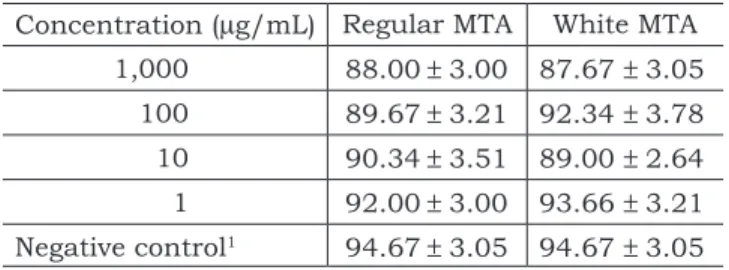
The cytotoxicity of MTA (grey and white) was measured in mouse lymphoma cells through try-pan blue assay in range-finding experiment prior to the determination of chemically induced geno-toxicity. The dose-response relationships of all compounds tested at concentrations ranging from 1-1,000 µg/mL on cell viability assessed by trypan blue assay are shown in Table 1. No significant statistically differences (p > 0.05) of viable cells were found for both endodontic materials tested and in all concentrations tested.
The single cell gel (comet) assay was used to measure DNA damage in mouse lymphoma cells
in vitro. DNA strand breaks were represented by the mean tail moment for 50 comets/sample. As
seen in Table 2, both materials did not induce DNA strand breaks at any concentration tested.
DISCUSSION
In this study, a cell culture technique was em-ployed in order to evaluate the biocompatibility for two forms of MTA. In vitro studies are simple, in-expensive to perform, provide a significant amount of information, can be conducted under controlled conditions, and may elucidate the mechanisms of cellular toxicity5. The results obtained from in vitro
assays might be indicative of the effects observed
in vivo. L5178Y, a continuous cell line, was used as the target cell in this experiment. Cell lines are easier to prepare and culture than primary cells (lymphocytes from peripheral blood). Primary cells are used in clinically simulated situations but are rather different between individuals. Our own most recent findings have shown that the two cell types do not differ much in sensitivity18.
Introduction of chemicals in the working envi-ronment requires the assessment of their harmful effects. The trypan blue exclusion test can be used
TABLE 1 - Effects of serial concentrations of regular and white MTA assessed by trypan blue exclusion test. Results are expressed as mean percentage of the con-trol (mean ± standard deviation).
Concentration (µg/mL) Regular MTA White MTA 1,000 88.00 ± 3.00 87.67 ± 3.05
100 89.67 ± 3.21 92.34 ± 3.78 10 90.34 ± 3.51 89.00 ± 2.64 1 92.00 ± 3.00 93.66 ± 3.21 Negative control1 94.67 ± 3.05 94.67 ± 3.05 1Phosphate buffer solution (pH 7.4).
TABLE 2 - Mean ± Standard deviation of DNA damage (tail moment) in mouse lymphoma cells exposed to regular and white MTA.
Concentration (µg/mL) Regular MTA White MTA 1,000 0.71 ± 0.36 0.58 ± 0.33
100 0.75 ± 0.25 0.83 ± 0.19 10 0.41 ± 0.17 0.77 ± 0.24 1 0.74 ± 0.18 0.61 ± 0.17 Negative control1 0.59 ± 0.16 0.59 ± 0.16
Positive control2 4.83 ± 1.20* 4.83 ± 1.20* 1Phosphate buffer solution (pH 7.4); 2Methyl methanesulfonate
186 187
186 187
to indicate cytotoxicity; dead cells take up the blue stain of trypan blue, whereas live cells have yellow nuclei. Cytotoxicity data obtained in our labora-tory on mouse lymphoma cultures demonstrated that either regular or white MTA were not able to interfere in cellular death at any concentration assessed. This is consistent with published data reporting that MTA has been found to be non-toxic
in vitro8,15,25.
The single cell gel (comet) assay is a sensitive method for the detection of DNA damage and re-pair induced by genotoxic compounds in individual level. The alkaline version, used in this study, is able to detect a variety of DNA lesions and incom-plete repair sites3,23. Therefore, and taking into
account the lack of data currently available, the assessment of the potential genotoxicity of MTA is justified. The results of this study indicated that the alkaline single cell gel (comet) assay, in the experimental conditions used, failed to detect the presence of DNA damage after treatment using both forms of MTA assessed at any concentration tested. These findings confirmed and extended the data already published, showing that MTA presents good biocompatibility7,12.
In the present study, as well as in all of our previous investigations using the single cell gel
(comet) assay, we have always excluded comets without clearly identifiable heads during the image analysis. Although it should be emphasized that it is still not completely understood what these ‘clouds’ actually represent, this type of comet was excluded based on the assumption that these cells represent dead cells, resulting from putative cy-totoxic effects of root-end filling materials rather than primary DNA-damage following direct interac-tion between DNA and a genotoxic agent19.
CONCLUSION
In summary, our results suggest that both regular and white MTA are not genotoxins and are not able to interfere in cellular death. Furthermore, the results presented here might be an additional argument to support the use of MTA in endodontic practice.
ACKNOWLEDGMENTS
CNPq (National Council for Scientific and Technological Development; grant number: 141501/2002-2) and TOXICAN (Núcleo de Ava-liação Toxicogenética e Cancerígena) supported this study.
REFERENCES
1. Auletta A, Ashby J. Workshop on the relationship between short-term information and carcinogenicity; Williamsburg, Virginia, January 20-23, 1987. Environ Mol Mutagen 1988;11:135-45.
2. Camilleri J, Montesin FE, Papaioannou S, McDonald F, Pitt Ford TR. Biocompatibility of two commercial forms of mineral trioxide aggregate. Int Endod J 2004;37:699-704. 3. Fairbairn DW, Olive PL, O’Neill KL. The comet assay: a
comprehensive review. Mutat Res 1995;339:37-59. 4. Gartner AH, Dom SO. Advances in endodontic therapy.
Dent Clin North Am 1992;36:357-78.
5. Geurtsen W. Substances released from dental resin composites and glass ionomer cements. Eur J Oral Sci 1998;106:687-95.
6. Hartmann A, Agurell E, Beevers C, Brendler-Schwaab S, Bur-linson B, Clay P, et al. Recommendations for conducting the
in vivo alkaline comet assay. Mutagenesis 2000;18:45-51. 7. Holland R, Souza V, Nery MJ, Otoboni JA, Filho Bernabe
PFE, Dezan E Jr. Reaction of rat connective tissue to im-planted dentin tubes field with mineral trioxide aggregate or calcium hydroxide. J Endod 1999;25:161-6.
8. Keiser K, Johnson CC, Tipton DA. Cytotoxicity of mineral trioxide aggregate using human periodontal ligament fi-broblasts. J Endod 2000;26:288-91.
9. Koh ET, McDonald F, Pitt Ford TR, Torabinejad M. Cel-lular response to mineral trioxide aggregate. J Endod 1998;24:543-7.
10. Koh ET, Torabinejad M, Pitt Ford TR, Brady K, Mc-Donald F. Mineral trioxide aggregate stimulates a biologi-cal response in human osteoblasts. J Biomed Mater Res 1997;37:432-9.
11. Mckelvey-Martin VJ, Green MHL, Schmezer P, Pool-Zobel BL, De Méo MP, Collins A. The single cell gel elec-trophoresis assay (comet assay): a european review. Mutat Res 1993;288:47-63.
12. Menezes R, Bramante CM, Letra A, Carvalho VGG, Garcia RB. Histologic evaluation of pulpotomies in dog using two types of mineral trioxide aggregate and regular and White Portland cements as wound dressings. Oral Surg Oral Med Oral Pathol Oral Radiol Endod 2004;98:376-9. 13. Mitchell PJC, Pitt Ford TR, Torabinejad M, McDonald
F. Osteoblast biocompatibility of mineral trioxide aggregate. Biomaterials 1999;20:167-73.
14. Olive PL, Banath JP, Durand RE. Heterogeneity in radiation-induced DNA damage and repair in tumor and normal cells measured using the comet assay. Radiat Res 1990;112:86-94.
15. Osório RM, Hefti A, Vertucci FJ, Shawley AL. Cyto-toxicity of endodontic materials. J Endod 1998;24:91-6. 16. Perez AL, Spears R, Gutmann JL, Opperman LA.
186 187
186 187
17. Ribeiro DA, Bazo AP, Franchi CAS, Marques MEA, Salvadori DMF. Chlorhexidine induces DNA damage in rat peripheral leukocytes and oral mucosal cells. J Periodontal Res 2004;39:358-61.
18. Ribeiro DA, Marques MEA, Salvadori DMF. Lack of genotoxicity of formocresol, paramonochlorophenol and calcium hydroxide on mammalian cells by comet assay. J Endod 2004;30:593-6.
19. Ribeiro DA, Pereira PC, Machado JM, Silva SB, Pessoa AW, Salvadori DM. Does toxoplasmosis cause DNA dam-age? An evaluation in isogenic mice under normal diet or dietary restriction. Mutat Res 2004;559:169-76.
20. Ribeiro DA, Salvadori DMF, Assis GF, Marques MEA. Does fluoride cause DNA damage? An in vitro evaluation us-ing rats oral mucosa cells. Braz J Oral Sci 2003;2:268-71. 21. Ribeiro DA, Scolastici C, Marques MEA, Salvadori DMF.
Fluoride does not induce DNA breakage in Chinese hamster ovary cells in vitro. Braz Oral Res 2004;18:192-6.
22. Rothfuss A, Merck O, Radermacher P, Speit G. Evalua-tion of mutagenic effects of hyperbaric oxygen (HBO) in vitro. II. Induction of oxidative DNA damage and mutations in the mouse lymphoma assay. Mutat Res 2000;471:87-94. 23. Tice RR, Agurell E, Anderson D, Burlinson B,
Hart-mann A, Kobayashi H, et al. Single cell gel/comet assay: guidelines for in vitro and in vivo genetic toxicology testing. Environ Mol Mutagen 2000;35:206-21.
24. Torabinejad M, Hong CU, Lee SF, Monsef M, Pitt Ford TR. Investigation of mineral trioxide aggregate for root-end-filling in dogs. J Endod 1995;21:603-8.
25. Torabinejad M, Hong CU, Pitt Ford TR. Cytotoxicity of four root end filling materials. J Endod 1995;21:489-92. 26. Torabinejad M, Rastegar AF, Keterring JD, Pitt Ford
TR. Bacterial leakage of mineral trioxide aggregate as a root-end filling material. J Endod 1995;21:109-12. 27. Wucherpfennig AL, Green DB. Mineral trioxide vs
Portland cement: two biocompatible filling materials [ab-stract]. J Endod 1999;25:308.