www.scielo.br/aabc
A reassessment of the role of serotonergic system
in the control of feeding behavior
MAGDA A. MEDEIROS, RICARDO H. COSTA-E-SOUSA, EMERSON L. OLIVARES, WELLINGTON S. CÔRTES and LUÍS C. REIS
Departamento de Ciências Fisiológicas, Instituto de Biologia
Universidade Federal Rural do Rio de Janeiro, BR 465, Km 07, 23890-000 Seropédica, RJ, Brasil Manuscript received on February 2nd, 2004; accepted for publication on September 9, 2004;
presented byFrederico G. Graeff
ABSTRACT
The role of serotonergic system in the feeding behavior was appraised by electrolytic lesions in the dorsal raphe nucleus (DRN) and administration of para-chlorophenylalanine (PCPA, 3 mg/5µl, icv). Chronic evaluations were accomplished through 120 and 360 days in PCPA-injected and DRN-lesioned rats, respectively. Acute food intake was evaluated in fasted rats and submitted to injection of PCPA and hydroxytryptophan (LHTP, 30 mg/kg, ip). DRN-lesioned rats exhibited 22-80% increase in food intake up to sixth month, whereas the obesity was evident and sustained by whole period. In PCPA-injected rats was observed an initial increase in the food intake followed by hypophagy from 25thto 30thday and a transitory increase of body weight from 5thto 60thday. In the acute study, the LHTP reverted partially the PCPA-induced increase in food intake of fasted rats suggesting a sustained capacity of decarboxylation of precursor by serotonergic neurons. Slow restoration of the levels of food intake in DRN-lesioned rats reveals a neuroplasticity in the systems that regulate feeding behavior. A plateau on the body weight curve in lesioned rats possibly represents the establishment of a new and higher set point of energetic balance.
Key words: food ingestion, serotonergic system, dorsal raphe nucleus, electrolytic lesion, para-chloro-phenylalanine, obesity.
INTRODUCTION
Evidences have implicated the serotonergic trans-mission in the feeding behavior. Basomedial nu-clei of hypothalamus and lateral hypothalamic area, admittedly involved with feeding behavior control, are innervated by serotonin neurons from the mid-brain raphe (Azmitia and Segal 1978, Parent et al. 1981). Studies with administration of serotonin precursors (Fernstrom and Wurtman 1971a, b, Fernstrom 1983), inhibitors of serotonin presynap-tic uptake, serotonin releasers, inhibitor of
trypto-Correspondence to: Luís Carlos Reis E-mail: [email protected]
after neurotoxic lesion of serotonin neurons, have reinforced the serotonergic hypothesis (Breisch et al. 1976, Saller and Stricker 1976).
Recent studies demonstrated the participation of other two systems of food intake control, the lep-tin, hormone secreted by adipocytes after increase of fat in the adipose tissue and, the orexin A, a neu-ropeptide which is expressed in the lateral hypotha-lamus after fasting and subsequent energy depletion (Janeckova 2001, Rodgers et al. 2002). Leptin reduces food ingestion and additionally stimulates mechanisms within basomedial hypothalamus con-cerned to fat oxidation, energetic balance and, there-fore, the body weight as well (Friedman and Halaas 1998, Grill and Kaplan 2002).
Subset of serotonin neurons within the raphe nuclei co-expresses serotonin transporter mRNA and leptin receptor mRNA (Finn et al. 2001). In this line of reasoning, Fernández-Galaz et al. (2002) evidenced the leptin uptake by serotonin neurons of the dorsal raphe nucleus (DRN) and Yamada et al. (2003) showed that the hypophagic effect of lep-tin is mediated by serotonergic activity and subse-quent 5HT2C receptor stimulation. On the other hand, orexin A receptors were identified in serotonin neurons of the DRN on which its excitation pos-sibly constitute a negative feedback loop for acute control of food ingestion, particularly carbohydrates (Brown et al. 2001).
However, evidences concerning the role of DRN ascending pathways in the regulation of feed-ing behavior are inconclusive. Observations con-cerning lesions are controversial in spite the above mentioned reports. Thus, Geyer et al. (1976) showed that electrolytic lesions of B7 area, cor-responding DRN, didn’t produce significant alter-ations in the food ingestion and gain of body weight, as well. When lesion was directed to B8 area, corre-sponding median raphe nucleus (MRN) the authors reported an increase of the food ingestion and body weight (Geyer et al. 1976, Blundell 1984). It is interesting to record that, these observations were made in 6 months old rats through 4 weeks. In opposing, Heym and Gladfelter (1982) don’t
evi-denced increase of food ingestion in young rats in same range of age and body weight between 150 and 200 g. In addition to those contradictory data, no evidence has been reported regarding body weight gain time course relatively to food ingestion in a longer observation period.
In the current study we reassessed the role of the serotonergic system in the control of food in-gestion. New approaches were now justified con-sidering the convergence of recent evidences for the role of serotonergic circuitry of DRN in the regu-lation of appetite and satiety mechanisms. Acute experiments were carried out in adult rats which re-ceived para-chlorophenylalanine (PCPA) into brain lateral ventricle and were submitted to fasting on the 4th day after microinjection and then treated with l-hydroxytryptophan (LHTP). In chronic exper-iments, adult rats were treated with PCPA or sub-mitted to DRN lesion and evaluated by 120 and 360 days, respectively.
MATERIALS AND METHODS
Animals and General Procedures
Wistar male rats from Fundação Oswaldo Cruz weighing 270-280 g were employed after previous adaptation to metabolic cage during one week. The animals were maintained underad libitumoffer of food and water. Experimental protocols were per-formed in laboratory with temperature control (25◦C) and lights on from 7:00 h to 19:00 h. Food ingestion was determined in the metabolic cages provided with chow container. Measurement of food ingestion was made by electronic precision scale in a cumulatively way, in the acute experiments and by 24-h interval in chronic evaluations. Experi-mental procedures were accomplished according to Brazilian College of Animal Experimentation and pertinent to Brazilian legislation.
Implantation of Canulae into Lateral Brain Ventricle and Microinjections
brain lateral ventricles in anesthetized rats (2.5% tri-bromoethanol, ip). Cannulae were placed by employing a stereotaxic device using the follow-ing coordinates: anterior-posterior, 0.9 mm poste-rior to bregma; lateral, 1.2-1.4 mm; vertical, 3.2-3.4 mm from skull calvaria (Paxinos and Watson 1986). Microinjections were made with a 10–µl Hamilton microsyringe.
Brain Serotonin Depletion
Brain serotonin depletion was produced by icv mi-croinjection of para-chlorophenylalanine methyl ester (PCPA, Sigma, St Louis, Mo, USA) an ir-reversible inhibitor of the tryptophan hydroxylase (TPO) at the dose of 3 mg/5µl, bilaterally, dur-ing 2 minutes under light anesthesia as described elsewhere (Breisch et al. 1976, Koe and Weissman 1966, Reis et al. 1994, Cooper et al. 1996). Con-trol group was treated with isotonic saline (5µl, icv, bilaterally). Acute experiments were fulfilled 4 days after icv microinjection. Chronic experiments were initiated after restoration of anesthesia effect.
Electrolytic Lesions of DRN
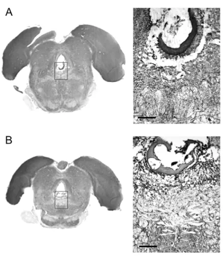
Electrolytic lesions of DRN were produced in rats anesthetized with tribromethanol (2.5%, ip), which were fixed in a Kopf stereotaxic device. DRN was placed through following coordinates (according to Paxinos and Watson atlas): anterior-posterior, 7.6– 7.8 mm posterior to bregma; lateral, 0.0 mm; ver-tical, 6.2–6.4 mm from skull calvaria (Paxinos and Watson 1986). Lesions were produced by passing an anodal current (2 mA, DC, for 10 sec) through nickel-chrome electrode guided into DRN. Control group received identical maneuver except current delivery (sham lesion). Isolated group of 12 rats were sacrificed 30 days after DRN electrolytic le-sion under profound anesthesia. Transcardiac infu-sion was made with saline and 10% formaldehyde and the brains were arrested for histological anal-ysis. Confirmation of lesions was made by histo-logical examination of coronal sections through the midbrain (10µm thickness) stained by cresyl violet.
Experimental Procedures
Investigations were carried out in three experi-mental sets:
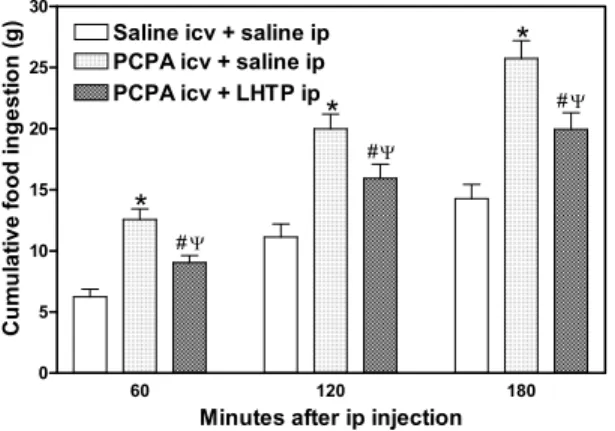
1) PCPA-treated rats were 24 hours fasted from the 4thday post-microinjection (N = 10). In fol-lowing day, fasted rats were given l-hydroxy-tryptophan, immediate precursor of serotonin synthesis (LHTP, Sigma, St Louis, Mo, USA) at the dose of 30 mg/kg, ip. Control group received isotonic saline (1ml/kg, ip) (N = 12). Both groups were further returned to metabolic cages. Food ingested was determined cumula-tively through 3 hours from 19:00 hours.
2) PCPA-treated rats recovered from anesthesia effect were returned to cages (N = 10) where chronic evaluation of food ingestion and body weight were made during 120 days. Control group was injected with saline (N = 12) and also transferred to cages.
3) DRN-lesioned (N = 10) and sham lesion rats were returned to cages after recovery of anes-thesia effect and observed through 360 days in which food ingested and body weight were de-termined.
Statistical analysis
Results were reported as means ±SE. Data were analyzed statistically by two-way analysis of vari-ance with repeated measures, and the significvari-ance between means was determined by the Newman-Keuls test. Differences between means were con-sidered to be significant whenP <0.05.
RESULTS
until the second day after central administration. However, from the 5th day serotonin-depleted rats presented an intense hyperphagia with gradual de-crease from 15th day (P < 0.05) (Figure 2). Af-ter transitory phase of hypophagy between 25thand 30thday PCPA-treated rats returned to control levels of food ingestion. At 40th day the mean values of food ingestion in PCPA-treated group were equiva-lent to controls. Body weight gain initiated on 5th day and maintained high in plateau way up to 60th day (P <0.05) and became comparable to controls at 90thday (P >0.05) post-injection (Figure 3).
60 120 180
0 5 10 15 20 25 30
Saline icv + saline ip PCPA icv + saline ip PCPA icv + LHTP ip
*
*
*
# # # < < <Minutes after ip injection
C u m u la ti ve f o od i ngest io n ( g )
Fig. 1 – Acute effect of the previous icv PCPA microinjectionvs ip LHTP injection on the food intake in nocturnal fasted rats. Data are presented as mean±standard error at 60, 120 and 180 min.
∗P <0.05 compared to control group. #P <0.05 compared
to PCPA, icv + saline, ip (ANOVA and Newman-Keuls test).
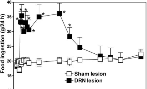
Typical electrolytic lesions of the DRN ex-tended in anterior-posterior direction (AP, 7.2-8.3 mm posterior to bregma) along rostral-dorsal and ventral-medial regions (Figure 4). DRN-lesioned rats displayed an intense hyperphagia from the 5th day post-surgery (P < 0.05) (Figure 5). This re-sponse maintained by 145 days on which the level of food ingestion remained higher than 30 g, whereas the sham lesioned rats continued in the 20 g level. From the 150th to 360th day the levels of food in-gested in DRN-lesioned group were comparable to controls. Body weight of the DRN-lesioned rats in-creased drastically from the 5thday in comparison to controls (P <0.05) (Figure 6). This feature
main--10 0 10 20 30 40 50 60 70 80 90 100 110 120 130
0 10 20 30 40 Saline icv PCPA icv
*
*
*
* *
*
* *
Days after icv PCPA injection
Fo od i n gest ion ( g )
Fig. 2 – Chronic effect of the icv injection of PCPA on food intake in rats. Data are presented as mean±standard error. *P <0.05 compared to sham lesion group (ANOVA and Newman-Keuls test).
-10 0 10 20 30 40 50 60 70 80 90 100 110 120 130
250 300 350 400 Saline icv PCPA icv
*
*
*
*
* * *
*
*
*
Days after icv PCPA injection
B o dy w e ig ht ( g)
Fig. 3 – Chronic effect of the icv injection of PCPA on the body weight in rats. Data are presented as mean±standard error. *P <0.05 compared to sham lesion group (ANOVA and Newman-Keuls test).
tained until the moment from which it developed a plateau significantly higher than controls ones, in spite the levels of food ingested between groups have been equivalent (P <0.05) (Figure 6).
DISCUSSION
Fig. 4 – Histological sections (10µm thickness), stained by cresyl violet, showing typical lesion of the DRN extending from 7.2 mm (panel A) to 8.3 mm posterior (panel B) to the bregma. Note topographical references around the delimited area. Aq: aqueduct; mlf: medial longitudinal fasciculus; pi: pineal gland; bar calibration, 200µm.
orexigenic response of the brain serotonin-depleted fasted rats. We presume that these conditions the serotonin neurons preserve the ability of decarboxy-late the LHTP for synthesis of serotonin. These sults possibly mean that serotonergic circuits are re-cruited during physiological ingestive process for the modulation of appetite intensity. In this context, the participation of the other systems of modulation must not be discarded. In chronic evaluation, the PCPA-treated rats expressed hyperphagic response concomitantly to a gradual increase of gain of body weight. These observations are similar to those re-ported by Breisch et al. (1976). However, we ev-idenced a gradual decrease of ingestive levels fol-lowed by hypophagy between 25thand 30thday
-15 10 35 60 85 110 135 160 185 210 235 260 285 310 335 360 10 15 20 25 30 35 40 Sham lesion DRN lesion
*
*
*
*
*
*
*
*
*
Days after DRN lesion
Fo od i n g e s ti o n ( g /24 h)
Fig. 5 – Chronic effect of the electrolytic lesion of DRN on the food intake in rats. Data are presented as mean±standard error. *P <0.05 compared to sham lesion group.
-15 10 35 60 85 110 135 160 185 210 235 260 285 310 335 360 200 250 300 350 400 450 500 550 600 650 Sham lesion DRN lesion
*
*
*
*
*
*
*
*
*
*
*
*
*
Days after DRN lesion
B od y w e ight ( g)
Fig. 6 – Chronic effect of electrolytic lesion of DRN on the body weight in rats. Data are presented as mean±standard error. *P <0.05 compared to sham lesion group.
the serotonin turnover, and possibly high compen-satory expression of other anorexigenic pathways (simultaneously to activation of control systems of the energetic balance), would restore the set point of body weight.
Data concerned to DRN-lesioned rats disclose that suppression of ascending circuits influenced the modulation mechanism of orexigenic activity as well as of energetic balance set point operation and therefore, of body weight adjustment. In this respect our observations parallel with findings regarding lesion of hypothalamic ventromedial nu-cleus (VMN) (Brobeck et al. 1943, Tepperman et al.
1943, Bray and York 1979, Hallonquist and Brandes 1983, Vilberg and Keesey 1984). These authors re-ferred an intense increase of the food ingestion asso-ciated with obesity for long time. Hallonquist and Brandes (1983) showed a gradual decrease of the ingestive levels after 12 weeks post-surgery, how-ever, with preservation of obesity. Data of the cur-rent work represent the former study of the feed-ing behavior in raphe-lesioned rats showfeed-ing similar feature to those achieved in VMN-lesioned rats. In other papers, the data regarded to hyper-phagiavsobesity are controversial possibly because the authors employed young rats and, in addition they performed the evaluations for a shorter period. Long time evaluation allows us to evidence either the restoring of food intake levels, 6 months after DRN lesion and the maintenance of high adipos-ity index. The recovery of the mean values of food ingestion suggests the achievement of a new home-ostatic status possibly consequent to orexigenic ac-tivity modulatory system plasticity. Future studies shall elucidate which neural circuit arranges that plasticity reaction. Is tempting to hypothesize that negative feedback loop from hypothalamus or origi-nating at peripheral sites would constitute one of the neural substrate disconnected by DRN lesion. This postulation is consonant with recent evidences which orexigenic neurons from lateral hypothala-mus project toward DRN where synapse with sero-tonergic neurons (Janeckova 2001, Rodgers et al. 2002). In this line of reasoning, leptin receptors were identified on DRN serotonergic neurons and, in addition, hypophagic effect of that hormone is partially mediated by serotonergic activity (Collin et al. 2000, Finn et al. 2001, Telles et al. 2003, Yamada et al. 2003).
ex-pression of neuropeptide Y (NPY). NPY represents the main convergence pathway of the orexigenic be-havior (Hillebrand et al. 2003, Kalra et al. 2003, Kalra and Kalra 2003). Briefly, daily rhythm of mRNA NPY demands the integrity of VMN (Dube et al. 1999). The VMN coordinately with hypota-lamic arcuate (AN) and dorsomedial (DMN) nuclei constitutes a circuitry responsive to leptin feedback which regulates the caloric ingestion and adjusts the energetic content of the adipose tissue (Bernardis and Berllinger 1998). Hassanain and Levin (2002) demonstrated in this context that fasted diet-induced obese (DIO) rats showed a 53% greater reduction in the ventromedial nucleus turnover than fasted diet-resistant rats. Thus, DIO-prone rats show abnor-malities in brain serotonin turnover which may pre-dispose them to become obese when dietary fat and caloric density are increased. These observations strengthen the findings of De Fanti et al. (2000) that showed a low capacity of serotonergic transmission from DRN toward VMN in Zucker rats, genetically obese. In this line, results of current study are ev-idences that serotonergic pathways are implicated with the acute control of food ingestion and chron-ically involved with the mechanisms of energetic balance set point adjustment and, therefore, with body weight regulation. These conclusions are based on (i) in the acute restoring (partially at least) of the appetite modulation in serotonin-depleted and –fasted rats induced by LHTP and, (ii) in the main-tenance of obesity for long time in DRN-lesioned rats despite of the normalization of food ingestion.
ACKNOWLEDGMENTS
We are grateful to Mr Ipojucan Pereira de Souza by animal care. This study was partially supported by Conselho Nacional de Desenvolvimento Científico e Tecnológico (CNPq).
RESUMO
O papel do sistema serotonérgico no comportamento ali-mentar foi avaliado através de lesões eletrolíticas do nú-cleo dorsal da rafe (L-NDR) e da administração de
para-clorofenilalanina (PCPA, 3 mg/5 µl, icv). Avaliações crônicas foram realizadas durante 120 e 360 dias em ratos injetados com PCPA e L-NDR, respectivamente. Avali-ações agudas foram realizadas em ratos em jejum e injeta-dos com PCPA e l-triptofano (LHTP, 30 mg/kg, ip). Ratos lesionados apresentaram um aumento de 22-80% na in-gestão de alimento até o sexto mês enquanto a obesidade foi evidenciada e mantida por todo o período. Ratos inje-tados com PCPA apresentaram um aumento da ingestão alimentar seguido de uma hipofagia do 25◦ao 30◦dia e um aumento transitório do peso corporal do 5◦ao 60◦. Agudamente, o LHTP reverteu parcialmente o aumento da ingestão de alimento em ratos tratados com PCPA e je-juados, sugerindo a preservação da capacidade de descar-boxilação do precursor pelos neurônios serotonérgicos. A lenta recuperação dos níveis de ingestão alimentar em ratos lesionados revela um mecanismo de neuroplastici-dade dos sistemas de regulação do comportamento ali-mentar. Estabelecimento de platô na curva de peso cor-poral dos ratos lesionados representaria o estabelecimento de um novo e mais elevado ponto de calibração do balanço energético.
Palavras-chave:ingestão de alimento, sistema serotonér-gico, núcleo dorsal da rafe, lesão eletrolítica, para-cloro-fenilalanina, obesidade.
REFERENCES
Azmitia EC and Segal M.1978. An autoradiographic analysis of the differential ascending projections of the dorsal and median raphe nuclei in the rat. J Comp Neurol 179: 641–668.
Bernardis LL and Berllinger LL. 1998. The dorso-medial hypothalamic revisited: 1998 update. Proc Soc Exp Biol Med 218: 284–306.
Blundell JE. 1984. Serotonin and appetite. Neurophar-macology. 23: 1537–1551.
Blundell JE. 1991. Pharmacological approaches to appetite suppression. TIPS 12: 147–157.
Bray GA and York DA. 1979. Hypothalamic and ge-netic obesity in experimental animals: An autonomic and endocrine hypothesis. Physiol Rev 59: 719–809.
Brobeck JR, Tepperman J and Long CNH. 1943. Ex-perimental hypothalamic hyperphagia in the albino rat. Yale J Biol Med 15: 831–853.
Brown RE, Sergeeva O, Eriksson KS and Haas HL. 2001. Orexin A excites neurons in dorsal raphe nu-cleus of the rat. Neuropharmacology 40: 457–459.
Chou-Green JM, Holscher TD, Dallman MF and
Akana SF. 2003. Repeated stress in young and old 5HT2C knockout mice. Physiol and Behav 70: 217– 226.
Collin M, Hakansson-Ovesjo ML, Misane I, Ogren
SO and Meister B. 2000. Decreased 5-HT trans-porte mRNA in neurons of the dorsal raphe nucleus and behavioral depression in the obese leptin-deficientob/obmice. Mol Brain Res 81: 51–61.
Cooper JR, Bloom FE and Roth RH. 1996. Serotonin (5-Hydroxytryptamine) and Histamine. In: Bloom FEet al. (Eds.), The Biochemical basis of neurophar-macology, 7th ed., New York: Oxford University Press, p. 352–409.
Curzon G. 1990. Serotonin and appetite. In: Neu-ropharmacology of serotonin. Ann N Y Acad Sci 600: 521–531.
Curzon G. 1991. Effects of tryptophan and of 5-hydro-xytryptamine receptor subtype agonists on feeding. Adv Exp Med Biol 294: 377–388.
De Fanti BA, Gavel DA, Hamilton JS and Horwitz
BA. 2000. Extracellular hypothalamic serotonin lev-els after dorsal nuclei stimulation of lean (Fa/Fa) and obese (fa/fa) Zucker rats. Brain Res 869: 6–14.
Dube MG, Xu B, Kalra PS, Sninsky CA and Kalra
SP. 1999. Disruption in neuropeptide Y and leptin signaling in obese ventromedial hypothalamic lesioned-rats. Brain Res 816: 38–46.
Fernández-Galaz MC, Diano S, Horvath TL and
Garcia-Segura LM. 2002. Leptin uptake by sero-tonergic neurones of the dorsal raphe. J Neuroen-docrinol 14: 429–434.
Fernstrom JD. 1983. Role of precursor availability in control of monoamine biosynthesis in brain. Physiol Rev 63: 484–546.
Fernstrom JD and Wurtman RJ. 1971a. Brain sero-tonin, content: Increase following ingestion of car-bohydrate diet. Science 174: 1023–1025.
Fernstrom JD and Wurtman RJ. 1971b. Brain
sero-tonin content: Physiological dependence on plasma tryptophan levels. Science 73: 149–152.
Finn PD, Cunningham MJ, Rickard DG, Clifton DK
and Steiner RA. 2001. Serotonergic neurons are targets for leptin in the monkey. J Clin Endocrinol Metab 86: 422–426.
Friedman JM and Halaas JL. 1998. Leptin and reg-ulation of body weight in mammals. Nature 395: 763–770.
Geyer MA, Puerto A, Menkes DB and Segal DS. 1976. Behavioral studies following lesions of the mesolimbic and mesostriatal serotonergic pathways. Brain Res 106: 257–270.
Grill HJ and Kaplan JM. 2002. The neuroanatomical axis for control of energy balance. Front Neuroen-docrinol 23: 2–40.
Hallonquist JD and Brandes JS. 1983. Ventromedial hypothalamic lesions in rats: Gradual elevation of body weight set-point. Physiol Behav 33: 831–836.
Hassanain M and Levin BE. 2002. Dysregulation of hypothalamic serotonin turnover in diet-induced obese rats. Brain Res 929: 175–180.
Heym J and Gladfelter WE. 1982. Locomotor activ-ity and ingestive behavior after damage to ascending serotonergic systems. Physiol Behav 29: 459–467.
Hillebrand JJG, De Wied D and Adan RAH. 2003. Neuropetides, food intake and body weight regula-tion: a hypothalamic focus. Peptides 23: 2283– 2306.
Janeckova R. 2001. The role of leptin in human physiol-ogy and pathophysiolphysiol-ogy. Physiol Res 50: 443–459.
Kalra SP and Kalra PS. 2003. Neuropetide Y: a phys-iological orexigen modulated by the feedback action of ghrelin and leptin. Endocrine 22: 49–56.
Kalra SP, Bagnasco M, Otukonyong EE, Dube MG
and Kalra PS. 2003. Rhythmic, reciprocal ghrelin and leptin signaling: new insight in the development of obesity. Regul Pept 111: 1–11.
Koe KB and Weissman A. 1966. p-Chlorophenyl-alanine: a specific depletor of brain serotonin. J Phar-macol Exp Ther 154: 499–516.
Lima HRC, Cavalcante-Lima HR, Cedraz-Mercez
PL, Costa-e-Sousa RH, Olivares EL,
Badauê-Passos Jr D, Medeiros MA, Côrtes WS and Reis
sodium appetite induced by sodium depletion or beta-adrenergic stimulation. An Acad Bras Cienc 76: 85– 92.
Nonogaki K, Strack AM, Dallman MF and Tecott
LH. 1998. Leptin-independent hyperphagia and type 2 diabetes in mice with a mutated serotonin 5-HT2C receptor gene. Nat Med 4: 1152–1156.
Parent A, Descarries L and Beaudet A. 1981. Orga-nization of ascending serotonin systems in the adult rat brain. A radioautographic study after intraven-tricular administration of [3H]5-hydroxytryptamine. Neuroscience 6: 115–138.
Paxinos G and Watson C. 1986. The Rat Brain in Stereotaxic Coordinates. 2ndedn. Academic Press, New York, NY, USA.
Reis LC, Ramalho MJ, Favaretto AL, Gutkowska
J, McCann SM and Antunes-Rodrigues J. 1994. Participation of the ascending serotonergic system in the stimulation of atrial natriuretic peptide release. Proc Natl Acad Sci USA 91: 12022–12026.
Richard F, Sanne JL, Bourde O, Weissman D, Ehret
M, Casch C, Maitre M and Pujol JF. 1990. Vari-ation of tryptophan-5-hydroxylase concentrVari-ation in the rat raphe dorsalis nucleus after p-chloro-phenylalanine administration. I. A model to study the turnover of the enzymatic protein. Brain Res 536: 41–45.
Rodgers RJ, Ishii Y, Halford JCG and Blundell JE. 2002. Orexins and appetite regulation. Neuropep-tides 36: 303–325.
Saller CF and Stricker EM. 1976. Hyperphagia and increased growth in rats after intraventricular injec-tion of 5,7-dihydroxytryptamine. Science 192: 385– 387.
Tecott LH, Sun LM, Akana SF, Strack AM,
Lowest-ein DH, Dallman MF and Julius D. 1995. Eating disorder and epilepsy in mice lacking 5-HT2C sero-tonin receptors. Nature 374: 542–546.
Telles MM, Guimarães RB and Ribeiro EB. 2003. Ef-fect of leptin on the acute feeding-induced hypotha-lamic serotonergic stimulation in normal rats. Regul Pep 115: 11–18.
Tepperman J, Brobeck JR and Long CNH. 1943. The effect of hypothalamic hyperphagia and of alterations in feeding habits on the metabolism of the albino rat. Yale J Biol Med 15: 855–874.
Vilberg TR and Keesey RE. 1984. Reduced energy expenditure after ventromedial lesions in female rats. Am J Physiol 247: R183–R188.