(Annals of the Brazilian Academy of Sciences)
Printed version ISSN 0001-3765 / Online version ISSN 1678-2690 www.scielo.br/aabc
Juvenile development of Callinectes danae Smith, 1869 (Crustacea, Decapoda, Brachyura, Portunidae) under laboratory conditions
EDUARDO A. BOLLA Jr1, VIVIAN FRANSOZO2 and MARIA LUCIA NEGREIROS-FRANSOZO1
1NEBECC, Departamento de Zoologia, Instituto de Biociências de Botucatu, Universidade Estadual Paulista/ UNESP,
Distrito de Rubião Júnior, s/n, Caixa Postal 510, 18618-970 Botucatu, SP, Brasil
2
Departamento de Ciências Naturais – Zoologia, Universidade Estadual do Sudoeste da Bahia/ UESB, Estrada do Bem Querer, Km 04, Campus de Vitória da Conquista, Caixa Postal 95, Bairro Universidade, 45031-900 Vitória da Conquista, BA, Brasil
Manuscript received on November 23, 2012; accepted for publication on March 28, 2013
ABSTRACT
The juvenile development of Callinectes danae was investigated from megalopae obtained in neuston samples at Ubatuba, São Paulo, Brazil. The individuals were raised in the laboratory under constant
temperature (25 ± 1°C), filtered sea water from the collection location (35‰), and natural photoperiod.
Newly hatched Artemia sp. nauplii were offered as food on a daily basis and ornamental-fish food was also provided for the juveniles from the 4th stage on. Twelve stages of the juvenile phase were obtained. The main
morphological features that allowed recognition of the first juvenile stage were drawn and described. All the subsequent stages obtained were examined and measured, and the main changes in relation to the first
stage were recorded. Sexual dimorphism becomes apparent from the fourth juvenile stage onwards. Some
appendages and morphological features proved to be of great importance in the identification of species, including the number of segments of the antennal flagellum and the number of setae on the maxilla and on the
1st, 2nd and 3rd maxillipeds. These can probably be used for future comparisons and species identifications.
Key words: juvenile stages, morphology, portunid, post-larval, swimming crab.
Correspondence to: Eduardo Antonio Bolla Jr E-mail: [email protected]
INTRODUCTION
The life cycle of marine brachyurans, in general, has two distinct periods, the planktonic and the benthic. Each phase has its own particularities, which can be reflected in the animal’s development, mainly during the change from the larval to the juvenile phase (Kurata 1962). The planktonic period comprises the larval development, which includes a zoea phase with several zoeal stages, and a final megalopa phase with a unique megalopa stage, whose settlement marks the transition between the planktonic and the benthic
period (Anger 2001). In the benthic period, early development up to the pubertal molt is characterized as the juvenile phase. This phase comprises a few early “undifferentiated” stages (characterized by a non-external sexual differentiation of the individual) and “immature” stages, during which juveniles can be sexed by observing the number of pleopods on their abdomen. Only in the later immature stages the gonads begin to differentiate, and usually the relative growth of certain body parts starts to differ slightly between the sexes (Hartnoll 1982).
studied by Flores et al. (1998), to eleven stages in
Sesarma rectum Randall, 1840, described by Fransozo
(1986/87). The undifferentiated stages end with the pre-pubertal molt followed by the first immature stage. The immature juveniles can show a highly variable number of stages and when the individual reaches sexual maturity at the puberty molt, the adult phase begins. As the study of juvenile development in decapods is still incipient, the nomenclature of the terms “phase” and/or “stages” is also variable. Some authors use different nomenclatures for the stages in juvenile phase in several species. In most cases they do not use the separation between undifferentiated and immature stages. However, we adopted here the concept described above.
Studies of the early life cycle of crabs amplify taxonomic knowledge and also provide information on their ecology, physiology, phylogeny, and so on (Felder et al. 1985, Anger 2001). A pioneer complete study on juvenile development in Brachyura was accomplished by Shen (1935), who described
Carcinus maenas (Linnaeus, 1758) (Portunidae)
up to the ninth stage of the juvenile phase. Studies in the following decades dealt mostly with the description of the first stage of the juvenile phase. More complete descriptions appeared again during the 1980s. The species for which the juvenile development has been studied and which occur on the Brazilian coast are listed in Table I.
The family Portunidae Rafinesque, 1815 is represented on the Brazilian coast by 21 species (Melo 1996, Calado 1996, Carqueija and Gouvêa 1996, Tavares and Mendonça Jr 1996) of which 6 belong to the genus Callinectes. The geographical distribution of Callinectes danae Smith, 1869 includes much of the Western Atlantic: Bermuda, Florida, Gulf of Mexico, Antilles, Colombia, Venezuela and Brazil (from the states of Paraíba to Rio Grande do Sul). This crab occurs from brackish to hypersaline waters, in mangroves and muddy estuaries, on sandy beaches to the open sea, and from the intertidal zone to 75 m depth (Melo 1996).
In the family Portunidae, juvenile development has been described for a relatively small number of species. For the genus Callinectes, only C.
sapidus Rathbun, 1896 and C. ornatus Ordway,
1863 have been studied, both up to the 11th stages
of the juvenile phase, by Barutot et al. (2001) and Bolla Jr et al. (2008), respectively. Ogburn et al. (2011) also studied some species of portunids, but only superficial descriptions of the carapace of
C. sapidus and C. similis were provided. Due to
the scarcity of investigations on this aspect, it is difficult to define characteristics that are peculiar in the juvenile phase for portunids. Only detailed studies from laboratory rearing will allow the comparison and the establishment of features for portunid species.
This study presents morphological details and the most significant features that identify the juvenile phase of C. danae. We also provide information to recognize this species in natural environments, during its juvenile phase. This information will contribute to species identification and culture for economic purposes.
MATERIALS AND METHODS
The juvenile development of C. danae was studied using material from the collection of larvae and juveniles of the “Núcleo de Estudos em Biologia, Ecologia e Cultivo de Crustáceos (NEBECC)”, Departamento de Zoologia, Instituto de Biociências, UNESP, Botucatu, São Paulo, Brazil. This material consisted of juveniles (exuviae and individuals) obtained from raising of megalopae captured in the neuston of Ubatuba Bay (23°26’S; 45°03’W), Ubatuba/SP. The climate was tropical/subtropical, with mean temperatures of water surface around 18°C during winter and 29°C during summer (Negreiros-Fransozo and Fransozo 2003).
megalopae were separated into covered acrylic containers (30 ml) with filtered sea water (salinity 35) from the sampling area, and transported to the NEBECC laboratory. The megalopae were individually raised at 24 ± 1°C in labeled and numbered containers with filtered and aerated sea water. The containers were inspected daily, and debris and exuviae or dead individuals were removed. The water in the containers was partially renewed over one day, and completely replaced
every second day with filtered and aerated sea water. After inspection, individuals were fed with newly hatched Artemia sp. nauplii ad libitum and, for juveniles from 4th stage on, ornamental-fish food was also provided. Dead individuals and their respective exuviae were fixed in 80% ethanol and glycerin at 2:1, and stage changes and deaths were recorded. These techniques are similar to those used by Guimarães and Negreiros-Fransozo (2005) and Negreiros-Fransozo et al. (2007).
Super Families Species Juvenile stages described Stage of the sexual differentiation (based on pleopods) Authors
Aethroidea Hepatus pudibundus (Herbst, 1785) 1st to 8th 3rd Hebling and Rieger 2003
Grapsoidea
Armases rubripes (Rathbun, 1897) 1st to 10th 5th Negreiros-Fransozo et al. 2011
Cyrtograpsus angulatus Dana, 1851 1st to 13th 4th Rieger and Beltrão 2000
Neohelice granulata (Dana, 1851) (described as Chasmagnathus granulata) 1
st
to 8th 3rd Rieger and Nakagawa 1995
Pachygrapsus transversus (Gibbes, 1850) 1st to 8th 3rd Flores et al. 1998
Percnon gibbesi (H. Milne-Edwards, 1853) 1st ? Paula and Hartnoll 1989
Sesarma rectum Randall, 1840 1st to 15th 12th Fransozo 1986/87 Majoidea Pyromaia tuberculata (Lockington, 1877) 1st to 9th 2nd Flores et al. 2002 Ocypodoidea Uca (Leptuca) cumulanta Crane, 1943 1
st
to 8th 3rd Hirose et al. 2010
Uca (Minuca) burgersi Holthuis, 1967 1st to 10th 4th Vieira et al. 2010
Portunoidea
Arenaeus cribrarius (Lamarck, 1818) 1st to 3rd ? Stuck and Truesdale 1988
Callinectes ornatus Ordway, 1863 1st to 11th 4th Bolla Jr et al. 2008
Callinectes sapidus Rathbun, 1896 1st to 11th 4th Barutot et al. 2001
Charybdis hellerii (A. Milne-Edwards, 1867) 1st to 3rd ? Dineen et al. 2001
Xanthoidea
Acantholobulus bermudensis (Stimpson, 1871) (described as Hexapanopeus heblingi) 1
st
to 14th 6th Martin et al. 1984, M.D. Rodrigues, unpublished data
Eriphia gonagra (Fabricius, 1871) 1st to 10th 4th Fransozo and Negreiros-Fransozo 1987
Eurypanopeus abbreviatus (Stimpson, 1860) 1st to 7th 4th Fransozo and Negreiros-Fransozo 1987
Eurytium limosum (Say, 1818) 1st to 10th 4th Guimarães and Negreiros-Fransozo 2005
Hexapanopeus caribbaeus Stimpson, 1871 1st to 11th 5th R.R.R.Vieira, unpublished data
Menippe nodifrons Stimpson, 1859 1st to 8th 4th Fransozo et al. 1988
Panopeus austrobesus Williams, 1983
(described as Panopeus herbstii) 1 st
to 15th 4th Hebling et al. 1982
Panopeus occidentalis Saussure, 1857 1st to 14th 6th M.D. Rodrigues, unpublished data
Panopeus rugosus A. Milne-Edwards, 1880 1st to 14th 6th M.D. Rodrigues, unpublished data
TABLE I
List of species with occurrence in the Brazilian coast, with some stages of the juvenile phase known to date.
All individuals in the different stages were dissected, measured, drawn, and described based on fixed exuviae and dead individuals, using a stereoscopic microscope (Zeiss SV6) or a compound optical microscope (Zeiss Axioskop 2, with Nomarski optics), both equipped with a drawing tube, ocular micrometer, and Axiovision image system.
At least ten individuals of the first stage of the juvenile phase (J1) were dissected, drawn, and analyzed with regard to their structure and the number and types of setae. In addition, at least three specimens of each of the following stages were dissected and analyzed for evidence of morphological changes over the developmental period. These changes were recorded in tables, and the most noticeable changes were illustrated with line drawings.
The terminology adopted for the descriptions was based on Clark et al. (1998) and Pohle et al. (1999). Setae types were named according to Pohle (1989) and Garm (2004). The setal sequence was presented from the proximal to the distal part of the appendages. Systematic classifications were based on Ng et al. (2008).
RESULTS
During the rearing experiments of C. danae, the juvenile development was followed until the 12th stage, when the last specimen died.
MORPHOLOGY OF THE FIRST STAGE OF C. danae
The general shape of the first stage (Fig. 1a) was similar to that of the adult: carapace flattened dorso-ventrally, slightly convex, wider than long, and two small frontal teeth. It also presented: eight finely serrate lateral spines plus the pair of spines common to portunids; small plumose setae on the orbital margin and small granules and sparse simple setae over the carapace surface, in addition to well-developed stalked eyes.
Antennule (Fig. 2b) had a developed basal segment bearing several plumose and simple setae;
numerous small marginal granules; bisegmented peduncle with sparse simple setae; bisegmented endopod (ventral flagellum) with, respectively, 3 and 10 simple setae on the proximal and distal segments; exopod (dorsal flagellum) constituted by 7 segments with 0, 14, 10, 8, 5, 0 and 0 long aesthetascs from the proximal to the distal segment, respectively. Exopod with 0, 1, 2, 3, 1, 4 and 3 simple setae from the proximal to the distal segment.
Antenna (Fig. 2a) bore a 3-segmented antennal peduncle, provided with simple and plumose setae. Antennal flagellum 10-segmented with 0, 2, 4, 2, 2, 2, 3, 2, 4 and 3 simple setae, all of them terminal.
Mandible (Fig. 2c) possessed an incised, well chitinized blade; palp 3-segmented with 2 serrate and 14 plumose setae on the distal segment and 1 long plumose seta on the middle segment.
Maxillule (Fig. 2e) exhibited a coxal endite with 2 plumose setae, 15 simple setae of several sizes and 2 serrate setae; basial endite with 4 small simple setae and 1 long seta on the proximal margin, 6 serrate setae, 9 simple, 7 cuspidate and 3 plumose on distal margin; bisegmented endopod with 3 plumose setae and 1 simple seta on proximal segment, and 3 plumose setae and 2 simple setae on the distal segment. Two simple setae on protopod margin.
Maxilla (Fig. 2d) had a bilobed coxal endite with 6 plumose setae and 1 simple seta on the proximal segment, 2 simple setae and 6 plumose setae on the distal segment; basial endite, bilobed, with 3 plumose setae and 7 simple setae on the proximal segment, 3 plumose setae and 13 simple setae on the distal segment; endopod with 1 plumose and 4 simple marginal setae; exopod (= scaphognathite) with 85 plumose marginal setae and 63 simple surface setae.
1 small seta on the shaft of this region; bisegmented exopod with 13 plumose, 2 simple and 5 cuspidate setae on the proximal segment, 7 plumose and 5 simple setae on the distal segment; epipod well developed, with 58 simple setae.
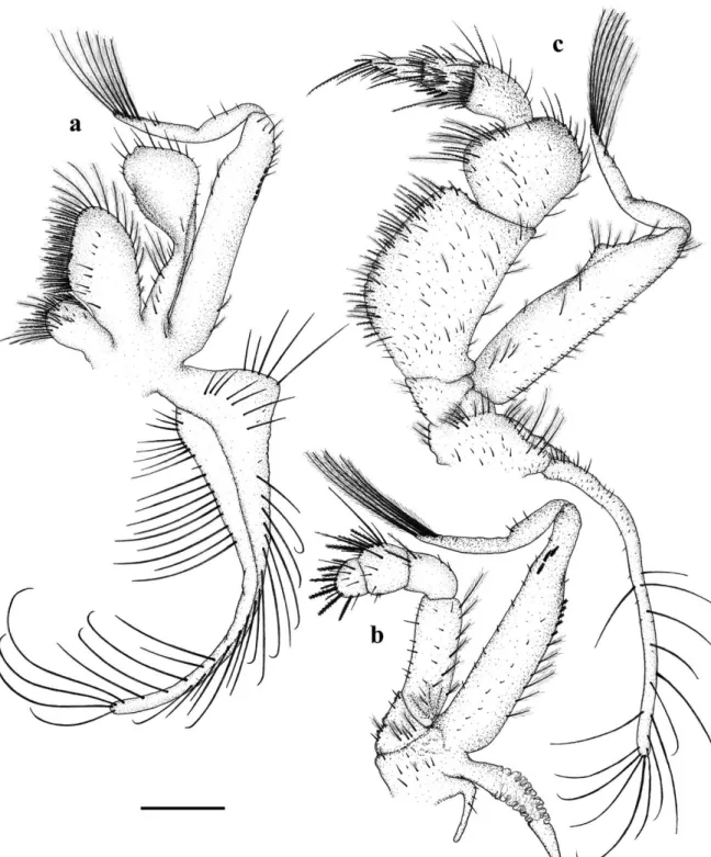
Second Maxilliped (Fig. 3b) possessed a 5-segmented endopod with 6, 8, 2, 1 and 0 plumose setae from the proximal to the distal segment, 3 and 4 serrate setae and 0 and 5 cuspidate setae, on the 4th and 5th segments, respectively; and 5,
16, 4, 11 and 5 simple setae from the proximal to the distal segment; bisegmented exopod with 9 plumose setae, 18 simple setae and 8 cuspidate setae on the proximal segment, 3 simple setae and 6 plumose setae on the distal segment; protopod with 3 plumose setae and 6 simple setae; rudimentary epipod provided with 3 simple setae.
Third Maxilliped (Fig. 3c) exhibited a 5-segmented endopod with several marginal spines on the first and second segments, several
Figure 3 -Callinectes danae. 1st undifferentiated stage: A, 1st maxilliped; B, 2nd maxilliped; C, 3rd maxilliped. Scale: 0.2mm.
setae on each segment: 17, 11, 12, 19 and 6 serrate setae, 35, 11, 1, 0, 0 plumose setae, 0, 0, 5, 4, 0 plumodenticulate setae and 63, 21, 9, 5, 7 simple setae from the proximal to distal segment;
simple setae; epipod with around 53 small simple setae on the proximal portion and 15 long simple setae on the distal portion.
Chelipeds (Fig. 4a) were symmetrical, with marginal spines and three strong inner ones on merus; granules and small spines on external margin of the carpus and propodus; simple and plumose setae sparsely distributed over the appendage surface, plus long plumose setae on the external margin of the merus. Second, third (Fig. 4b) and fourth pereiopods similar, sparse simple setae and several plumose setae present, mainly on the inner and external margins of the propodus and dactyls.
Last pair of pereiopods (Fig. 4c) – a spine on the coxa; propodus and dactyl flattened and paddle-shaped with several marginal plumose setae.
Thoracic sternites (Fig. 1c) had simple and plumose setae sparsely distributed over surface; concentric and semicircular wrinkle on the 7th somite (near the coxa).
Abdomen (Fig. 1b) bore 6 somites, wider than long, several sparse simple and plumose setae; the first 5 somites with horizontal ridges; telson smooth with small simple and plumose setae. Rudimentary pleopods visible ventrally on the 2nd to 5th somites (Fig. 1, PL2–PL5).
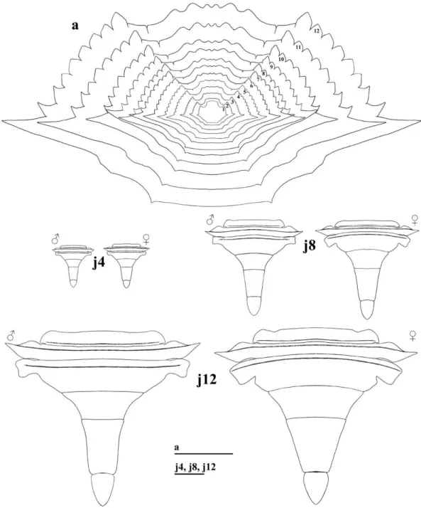
MORPHOLOGY OF THE 2nd TO 12th STAGES OF C. danae As the individuals grew in the following stages, in each successive ecdysis the proportion between carapace width and carapace length varied very little. This proportion reached the same as observed in the adult phase, from the 12th stage on (Fig. 5a).
The abdomen increased in size during the growth period, but it changed slightly in shape; the sex of individuals could be distinguished only at the 12th stage. As observed in adult swimming crabs, the
male abdomen had an inverted “T” shape and the female abdomen was semicircular (Fig. 5, J4–J12).
The most remarkable morphological changes, observed as of the 2nd stage, were related to the secondary sexual characteristics. The existing rudimentary pleopods in the 1st stage disappeared in the 2nd stage. In the 4th stage the pleopods
reappeared, but in different numbers and locations, according to each sex, as follows:
Male: two pairs of pleopods on the first (PL1)
and second (PL2) somite of the abdomen, PL2
with one reduced rami (Fig. 6, J4). In the 5th and
6th stages (Fig. 6, J
5and J6) no significant changes,
PL1 only increased in size. In the 7th and 8th stages (Fig. 6, J7and J8), PL2 exhibited one reduced rami
and PL1 had plumose setae on its base and simple
setae along its length. From the 9th stage on (Fig. 7,
J9–J12), PL2 was uniramous and both PL1 and PL2
were gradually larger, with more setae. From the 10th stage on, there was presence of bulges on PL1
distally. Morphology of 12thstage pleopods was the
same as seen in adults.
Figure 7 -Callinectes danae. pl1 and pl2: pleopods of the 1st and 2nd male abdominal somites, respectively, from 9th (j9), 10th (j10), 11th (j11) and 12th (j12) stages on. Scale: j9, 0.2mm; j10, j11 and j12, 0.4mm.
Female: four biramous pleopods from the 2nd
to the 5th abdominal somite (PL2, PL3, PL4 and
PL5); simple setae on endopod of PL2 and PL3 only
(Fig. 8, J4). 5th stage pleopods (Fig. 8, J5) were
larger, with more setae. In the 6th stage (Fig. 8, J 6),
PL2 endopod segmentation begun, a few
brush-shaped setae on PL2 and PL3. In the 7th and 8th
stages (Fig. 8, J7 and J8), number of brush-shaped
setae increased, endopod of all pleopods gradually was larger and more segmented. In the 9th stage (Fig. 9, J9), there were simple articulated setae
(instead of brush-shaped setae) on the endopod of all pleopods. From the 10th stage on, pleopods
In the remaining body structures, some signi-ficant changes occurred in the following: 1) Endopod of first maxilliped had a hollow in the apical region and a foliaceous shape on the internal margin from the 2nd stage on; 2) First segment of the antennal peduncle had a projection similar to the antennal scale, mainly from the most advanced stages on.
Other appendages did not show remarkable morphological changes, but the number of setae increased on each segment of the appendages. On the epipod of the third maxilliped, some
denticle-serrulate harpoon-shaped setae appeared from the 3rd stage on. Such setae were also found on the epipod of the 2nd maxilliped from the 4th stage on, and on the epipod of the 1st maxilliped in the 5th stage.
The main morphological characteristics which allow the identification of the first 12 stages of C.
danae are shown in Table II.
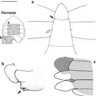
The vestigial aperture of the female gonopores appeared from the 4th stage on (Fig. 10a, white arrow). In males, it was not possible to verify in what stage the gonopores appeared.
Figure 9 -Callinectes danae. pl2 to pl5: pleopods from the 2nd to 5th female abdominal somites, respectively, from 9th (j9), 10th (j10), 11th (j11) and 12th (j12) stages on. Scale: j9, 0.2mm; j10, j11 and j12, 0.4mm.
The abdomen remained sealed throughout the stages observed. Detailed inspection revealed two pairs of “closing mechanisms” or “sternal buttons” on the sternal plates: an anterior one (located on the 5th sternite, at the level of the distal region of the
6th abdominal somite) (Fig. 10a, black arrow); and another, posterior one (located on the 8th sternite, at
the level of the lateral region of the 2nd abdominal
somite) (Fig. 10b, black arrow), which did not allow the extension of the abdomen.
DISCUSSION
Characteristics J1 J2 J3 J4 J5 J6 J7 J8 J9 J10 J11 J12 Number of segments
on antennule endopod 2 2 3 3 3 4 4 4 5 5 5 6
Number of segments
on antennule exopod 7 8 9 10 11 12 13 14 15 16 18 (19) 20
Number of segments
of antennal flagellum 10 10 12 13 18 20 22 24 (23) 29 (30) 32 34 (33) 39
Antenna length 1.29 1.42 1.9 2.27 2.96 3.62 4.51 4.98 6.07 6.86 8.4 9.58
Number of setae on the
basis of maxillule protopod 1 2 2 4 4 4 6 7 (8) 11 (10) 12 12 (13) 13 (14) Number of marginal setae
on maxilla exopod ±85 ±102 ±148 ±161 ±183 ±229 ±270 ±298 ±325 ±359 ±406 ±425 Length of maxilla exopod 0.67 0.77 1.06 1.37 1.59 2.04 2.28 2.64 3.25 3.74 4.39 4.98 Number of apical setae
on 1st maxilliped 11 14 23 ±33 ±38 ±49 ±65 ±73 ±81 ±89 ±101 ±112
Length of basal segment
of 1st maxilliped exopod 0.62 0.74 0.92 1.16 1.36 1.69 1.98 2.18 2.57 2.92 3.55 4.14 Length of basal segment
of 2nd maxilliped exopod 0.64 0.74 0.96 1.23 1.42 1.82 2.12 2.29 2.73 3.2 3.89 4.46 Length of basal segment
of 3rd maxilliped exopod 0.63 0.75 0.92 1.18 1.35 1.71 1.96 2.15 2.62 3.02 3.59 4.11 Length of 2nd pereiopod merus 0.95 1.14 1.64 2.13 2.56 3.37 3.82 4.33 5.38 6.12 7.42 8.91
TABLE II
Callinectes danae Smith, 1869. The main morphological characteristics for the identification of
the first 12 stages of the juvenile phase. The dimensions were taken in millimeters and represent mean values. Numbers in parenthesis mean alternative values with low occurrences.
± indicates “approximately”.
Figure 10 -Callinectes danae, 4th stage of the juvenile phase:
A, white arrow, the rudimentary female gonopores; black arrow, anterior “closing mechanism” position; B, posterior “closing mechanism” position, pointed by the arrow; C, schematic drawing of the position of the abdomen over the posterior “closing mechanism”.
(Saussure 1858), and found 8 lateral spines in addition to the pair that is common to portunid crabs. Yatsuzuka and Sakai (1980) also described the first undifferentiated stage of P. pelagicus, which showed the same characteristics as P. anceps and also a serrate rostrum. Only C. ornatus (studied by Bolla Jr et al. 2008), C. similis (studied by Ogburn et al. 2011) and C. danae (present study) have been reported bearing a similar serrate rostrum starting from the first undifferentiated stage. In C. sapidus,
according to Barutot et al. (2001) and Ogburn et al. (2011), the rostrum is smooth in the first undifferentiated stage, and the spines appear only as of the 6th stage. This feature might be useful as a diagnostic of this species on the Brazilian coast, as it is easy to distinguish. Nevertheless, further studies on other species of the genus Callinectes
are needed.
setae on the body appendages. However, because of the difficulty in discerning characteristics in the morphology of different setae types with only optical microscopy, the study of setae in the juvenile phases has been limited to counting them and noting their location on the animal’s body (Rieger and Beltrão 2000).
Certain appendages and morphological features have proven to be highly useful in discriminating species, due to their particularities, which can be observed with minimal manipulation of the specimens. These will likely prove to be useful for future comparisons and species identifications. Among these are the following features: number of segments of the antennal flagellum (i.e., C. sapidus,
C. danae and C. ornatus, which have 8, 10 and 10(11)
segments, respectively), and the number of setae on the maxilla and on the 1st, 2nd and 3rd maxillipeds.
With regards to the sternal region, the presence of wrinkles in concentric semicircles on the 7th somite of the first undifferentiated stage [present
in C. danae and C. ornatus, but not noted by
Yatsuzuka and Sakai (1980) and Barutot et al. (2001) in P. pelagicus (Linnaeus, 1758) and C.
sapidus, respectively], could be interpreted as a
remnant of the sternal spine that is conspicuous in the portunid megalopa (Kurata 1975). Such wrinkles are not found in the next stages, reinforcing this supposition. Nevertheless, further studies are needed in order to confirm the origin of this feature.
As mentioned by Guinot and Bouchard (1998), the anterior abdomen sealing mechanism, located on the 5th thoracic sternite of C. danae, is in accordance
with the typical system of sealing found in the Eubrachyura. However, those authors did not mention the closing mechanism (8th sternite). This mechanism may be exclusive to portunids, but only through additional studies of species within and outside the family would this hypothesis be able to be tested.
The sexual dimorphism in the abdominal shape of males and females is not evident throughout the juvenile phase observed for C. danae. For C.
sapidus, C. ornatus and P. pelagicus, studied up to
the 11th, 11th and 3rd juvenile stages, respectively, such dimorphism is also not observed. This feature probably develops in later immature stages, in contrast to representatives of other families, in which the sexual differentiation observed in the abdomen is most pronounced in the early immature stages. Examples include Inachus
dorsettensis (Pennant 1777), a majid studied by
Ingle (1977); P. transversus, a grapsid studied by Flores et al. (1998); and C. angulatus, a varunid studied by Rieger and Beltrão (2000); in which this differentiation occurs from the 3rd, 6th, and 4th
immature stages on, respectively.
The presence of the rudimentary pleopods in the first undifferentiated stage can also differ among species; this feature is observed in C. danae, C. ornatus
and P. pelagicus, but not in C. sapidus. Nevertheless,
except for P. pelagicus which shows rudimentary pleopods up to the 3rd undifferentiated stage, in all other species studied, these pleopods totally disappear by the 2nd undifferentiated stage. This feature might be interpreted as a remnant characteristic from the megalopa stage, since they occupy the same location on somites of the megalopa pleopods. In the following stages, the pleopods appear in accordance with each sex (two pairs for males, and four pairs for females), and the juvenile stage in which such appearance can be observed varies for each species, as shown in Table I. In portunids, the pleopods commonly appear in the 4th stage of the juvenile phase, as in
C. danae, C. ornatus and C. sapidus.
A distinctive and interesting feature with regards to the 2nd pair of pleopods of males (PL2) is
the fact that this appendage is biramous until the 8th
stage in C. danae and until the 6th stage in C. ornatus,
and, at least, until the 7th stage in C. sapidus (as can
be seen in Barutot et al. 2001). More interesting yet is the fact that only C. sapidus has a biramous first
pleopod (PL1) in males. In the remaining portunid
species studied, the PL1 remains uniramous as of its
Table III presents a comparative analysis, using the main morphological characteristics, among the species of the genus Callinectes which occur on the Brazilian coast and for which their juvenile development is known to date.
It is of great importance to consider further studies on the early phases of decapod crustacean
life cycles, for a better understanding of their life history, taxonomy or ecological and functional issues, since these subjects are interconnected. Such studies are also important to define characteristics that are peculiar to the juvenile phase, and they could allow us to identify species for various purposes, notably aquaculture and fisheries management.
JUVENILE I C. sapidus C. ornatus C. danae
Barutot et al. (2001) Bolla Jr et al. (2008) This study
Antennule endopod segments 2 2 2
Antennule exopod segments 7 7 7
Antenna segments 3 + 8 3 + 11 3 + 10 *
Mandible palp segments 3 3 3
Maxillule endopod segments 2 2 2
Maxillule endopod (E), basial endite (EB), coxal endite (EC), protopod (P) setae
4, 2 (E); 23 to 25 (EB);
15 to 17 (EC); 2 (P) 3, 4 (E); 29 (EB);20 (EC); 3 (P) 4, 5 (E); 30 (EB);19 (EC); 2 (P) Maxilla exopod (Ex), endopod (E), basial
endite (EB), coxal endite (EC) marginal setae
60 to 70 (Ex); 0 (E); 20 to
23 (EB); 10 to 13 (EC) 22 (EB); 13 (EC)88 (Ex); 4 (E); 26 (EB); 15 (EC)85 (Ex); 5 (E); *
1st maxilliped exopod segments 2 2 2
1st maxilliped endopod segments 1 1 1
1st maxilliped exopod (Ex), endopod (E), basial endite (EB), coxal endite (EC), epipod (Ep) setae
16 to 19 (Ex); 17 (E); 38 to 44 (EB); 17 to
21 (EC); 39(Ep)
29 (Ex); 35 (E); 67 (EB); 26 (EC);
49(Ep)
32 (Ex); 35 (E); 58 (EB); 33 (EC); 58 (Ep) *
2nd maxilliped exopod segments 2 2 2
2nd maxilliped exopod (Ex), endopod (E), epipod (Ep) setae
25 to 27 (Ex); 12, 2, 3, 12 to 14 (E)
52 (Ex); 27, 8, 14,
14 (E); 6 (Ep) 44 (Ex); 11, 24, 6,15, 14 (E); 3 (Ep) *
3rd maxilliped exopod segments 2 2 2
3rd maxilliped exopod (Ex), protopod (P), epipod (Ep) setae
18 to 20 (Ex); 14 to
17 (P); 22 to 24 (Ep) 56 (Ex); 16 (P);28 (Ep)
79 (Ex); 66 (P);
68 (Ep) *
Number of pleopods 0 4 rudimentary pairs 4 rudimentary pairs *
* = most important characteristics for comparison among species.
TABLE III
Diagnostic characters which allow the differentiation and identification of the first undifferentiated stage of the known species of the genus Callinectes with regards to juvenile phase.
ACKNOWLEDGMENTS
The authors are grateful to Conselho Nacional de Desenvolvimento Científico e Tecnológico (CNPq) for the first author’s Master of Science scholarship (# 553174/2008-8), to Fundação de Amparo à Pesquisa do Estado de São Paulo (FAPESP) for providing the financial support during neuston collections (#2004/15194-6), to all the members of NEBECC for their assistance during this study, and to Dr. Janet W. Reid for her help with the English review. All sampling in this
investigation has been conducted in accordance with applicable state and federal Brazilian laws.
RESUMO
O desenvolvimento juvenil de Callinectes danae foi investigado a partir de megalopas obtidas em amostras de neuston em Ubatuba, São Paulo, Brasil. Os indivíduos foram criados em laboratório sob temperatura constante (25 ± 1°C), água do mar filtrada proveniente do local de coleta (35‰) e fotoperíodo natural. Nauplii recém-eclodidos de Artemia
e também ração de peixes ornamentais foi fornecida a juvenis a partir do 4º estágio. Foram obtidos 12 estágios da fase juvenil. As principais características morfológicas que permitiram o reconhecimento do primeiro estágio juvenil foram desenhadas e descritas. Todos os estágios subsequentes obtidos foram analisados e medidos, e as principais alterações em relação ao primeiro estágio foram registradas. O dimorfismo sexual torna-se evidente a partir do 4º estágio juvenil. Alguns apêndices e características morfológicas provaram ser de grande importância para identificação das espécies, incluindo o número de segmentos do flagelo antenal e o número de cerdas na maxila e no 1º, 2º e 3º maxilípedes. Estes, provavelmente, podem ser usados para futuras comparações e identificações de espécies.
Palavras-chave: estágios juvenis, morfologia, portunídeos, pós-larva, siri.
REFERENCES
ANGER K. 2001. The Biology of Decapod Crustacean Larvae (Crustacean Issues 14). Lisse: A. A. Balkema Publishers, 419 p.
BARUTOT RA, VIEIRA RRR AND RIEGER PJ. 2001. Desenvolvimento juvenil de Callinectes sapidus Rathbun, 1896 (Crustacea: Decapoda: Portunidae), em laboratório, a partir de megalopas coletadas no plâncton. Comun Mus Cienc Tecnol PUCRS, Ser Zool 14: 23-42.
BOLLA JR EA, NEGREIROS-FRANSOZO ML AND FRANSOZO A. 2008. Juvenile development of Callinectes ornatus
Ordway, 1863 (Crustacea: Decapoda: Portunidae), from megalopae obtained from neuston samples. Zootaxa 1788: 1-20.
CALADO TCS. 1996. Registro de Charybdis hellerii (Milne Edwards, 1867) em águas de litoral brasileiro (Decapoda: Portunidae). Bol Estud Cienc Mar 9: 175-180.
CARQUEIJA CRG AND GOUVêA EP. 1996. A ocorrência, na costa brasileira, de um Portunidae (Crustacea, Decapoda) originário do Indo-Pacífico e Mediterrâneo. Nauplius 4: 165-168.
CLARK PF, CALAZANS DD AND POHLE GW. 1998. Accuracy and standardization of brachyuran larval descriptions. Invertebr Reprod Dev 33: 127-144.
DINEEN JF, CLARK PF, HINES AH, REED SA AND WALTON HP. 2001. Life history, larval description, and natural history of Charybdis hellerii (Decapoda, Brachyura, Portunidae), an invasive crab in the Western Atlantic. J Crust Biol 21: 774-805.
FELDER DL, MARTIN JW AND GOY JW. 1985. Patterns in early post-larval development of decapods. In: WENNER AM (Ed), Larval Growth, Crustacean Issues, vol. 2, Rotterdam: Balkema Press, Rotterdam, Netherlands, p. 163-225.
FLORES AAV, MARQUES FPL AND NEGREIROS-FRANSOZO ML. 2002. Postlarval stages and growth patterns of the spider crab Pyromaia tuberculata (Brachyura, Majidae) from laboratory-reared material. J Crust Biol 22: 314-327. FLORES AAV, NEGREIROS-FRANSOZO ML AND FRANSOZO
A. 1998. The megalopa and juvenile development of
Pachygrapsus transversus (Gibbes, 1850) (Decapoda, Brachyura), compared with other grapsid crabs. Crustaceana 71: 197-222.
FRANSOZO A. 1986/87. Desenvolvimento dos estágios juvenis de Sesarma (Holometopus) rectum Randall, 1840 (Decapoda, Grapsidae) obtidos em laboratório. Naturalia 12: 77-87.
FRANSOZO A AND NEGREIROS-FRANSOZO ML. 1987. Morfo-logia dos primeiros estágios juvenis de Eriphia gonagra
(Fabricius, 1781) e Eurypanopeus abbreviatus (Stimpson, 1860) (Crustacea, Decapoda, Xanthidae), obtidos em laboratório. Pap Avulsos Zool 36: 257-277.
FRANSOZO A, NEGREIROS-FRANSOZO ML AND HIYODO CM. 1988. Développement juvénile de Menippe nodifrons
Stimpson, 1859 (Crustacea, Decapoda, Xanthidae) au laboratoire. Rev Hydrobiol Trop 21: 297-308.
GARM A. 2004. Revising the definition of the crustacean seta and setal classification systems based on examinations of the mouthpart setae of seven species of decapods. Zool J Linn Soc 142: 233-252.
GUIMARãES FJ AND NEGREIROS-FRANSOZO ML. 2005. Juvenile development and growth patterns in the mud crab
Eurytium limosum (Say, 1818) (Decapoda, Brachyura, Xanthidae) under laboratory conditions. J Nat Hist 39: 2145-2161.
GUINOT D AND BOUCHARD J-M. 1998. Evolution of the abdominal holding systems of brachyuran crabs (Crustacea, Decapoda, Brachyura). Zoosystema 20: 613-694.
HARTNOLL RG. 1982. Growth. In: BLISS DE (Ed), The biology of Crustacea: embryology, morphology and genetics, vol. 2, New York: Academic Press Inc, New York, USA, p. 111-185.
HEBLING NJ, FRANSOZO A AND NEGREIROS-FRANSOZO ML. 1982. Desenvolvimento dos primeiros estágios juvenis de
Panopeus herbstii H. Milne-Edwards. 1834 (Crustacea, Decapoda, Xanthidae), criadas em laboratório. Naturalia 7: 177-188.
HEBLING NJ AND RIEGER PJ. 2003. Desenvolvimento juvenil de Hepatus pudibundus (Herbst) (Crustacea, Decapoda, Calappidae), em laboratório. Rev Bras Zool 20: 531-539. HIROSE GL, BOLLA JR EA AND NEGREIROS-FRANSOZO ML.
2010. Post-larval morphology, growth, and development of Uca cumulanta Crane, 1943 (Crustacea, Decapoda, Ocypodidae) under laboratory conditions. Invertebr Reprod Dev 54: 95-109.
INGLE RW. 1977. The larval and post-larval development of the scorpion spider crab, Inachus dorsettensis (Pennant) (Family: Majidae), reared in the laboratory. Bull Br Mus Nat Hist Zool 3: 331-348.
KURATA H. 1975. Larvae of Decapoda Brachyura of Arasaki, Sagami Bay-V. The swimming crabs of subfamily Portuninae. Bull Nansei Reg Fish Res Lab 8: 39-65. LEBOUR MV. 1944. Larval Crabs from Bermuda. Zoologica
29: 113-128.
MARTIN JW, FELDER DL AND TRUESDALE FM. 1984. A comparative study of morphology and ontogeny in juvenile stages of four western Atlantic xanthoid crabs (Crustacea: Decapoda: Brachyura). Phil Trans R Soc Lond B 303: 537-604.
MELO GAS. 1996. Manual de Identificação dos Brachyura (Caranguejos e Siris) do Litoral Brasileiro. São Paulo: Plêiade/FAPESP, 603 p.
NEGREIROS-FRANSOZO ML, FERNANDES CS, SILVA SMJ AND FRANSOZO A. 2011. Early juvenile development of
Armases rubripes (Rathbun 1897) (Crustacea, Brachyura). Invertebr Reprod Dev 2011: 1-12.
NEGREIROS-FRANSOZO ML AND FRANSOZO V. 2003. Morphometric study of the mud crab, Panopeus austrobesus
Williams, 1983 (Decapoda, Brachyura) from a subtropical mangrove in South America. Crustaceana 76: 281-294. NEGREIROS-FRANSOZO ML, WENNER EL, KNOTT D AND
FRANSOZO A. 2007. The megalopa and early juvenile stages of Calappa tortugae Rathbun, 1933 (Crustacea, Brachyura) reared in laboratory from neuston samples. Proc Biol Soc Wash 120: 469-485.
NG PKL, GUINOT D AND DAVIE PJF. 2008. Systema Brachyurorum: Part I. An annotated checklist of extant brachyuran crabs of the world. Raffles Bull Zool Suppl 17: 1-286.
OGBURN MB, STUCK KC, HEARD RW, WANG SY AND FORWARD JR RB. 2011. Seasonal variability in morphology of blue crab, Callinectes sapidus, megalopae and early juvenile stage crabs, and distinguishing characteristics among co-occurring Portunidae. J Crust Biol 31: 106-113. PAULA J AND HARTNOLL RG. 1989. The larval and post-larval
development of Percnon gibbesi (Crustacea, Brachyura, Grapsidae) and the identity of the larval genus Pluteocaris. J Zool 218: 17-37.
POHLE G. 1989. Structure, function, and development of setae on gill-grooming appendages and associated mouthparts of pinnotherid crabs (Decapoda: Brachyura). Can J Zool 67: 1690-1707.
POHLE G, MANTELATTO FLM, NEGREIROS-FRANSOZO ML AND FRANSOZO A. 1999. Decapod larvae. In: BOLTOVSKOY D (Ed), South Atlantic Zooplankton, vol. 2, Rotterdam: Balkema Books, Rotterdam, Netherlands, p. 1281-1351. RIEGER PJ AND BELTRãO R. 2000. Desenvolvimento juvenil
de Cyrtograpsus angulatus Dana (Crustacea, Decapoda, Grapsidae), em laboratório. Rev Bras Zool 17: 405-420. RIEGER PJ AND NAKAGAWA C. 1995. Desenvolvimento juvenil
de Chasmagnathus granulata Dana, 1851 (Crustacea, Decapoda, Grapsidae) em laboratório. Nauplius 3: 59-74. SHEN CJ. 1935. An Investigation of the Post-Larval
Development of the Shore-Crab Carcinus maenas, with Special Reference to the External Secondary Sexual Characters. Proc Zool Soc Lond 1935: 1-33.
STUCK KC AND TRUESDALE FM. 1988. Larval development of the speckled swimming crab, Arenaeus cribrarius
(Decapoda: Brachyura: Portunidae) reared in the laboratory. Bull Mar Sci 42: 101-132.
TAVARES M AND MENDONçA JR JB. 1996. Charybdis hellerii
(A. Milne Edwards, 1867) (Brachyura: Portunidae), eighth nonindigenous marine decapod recorded from Brazil. Crustaceana Res 25: 151-207.
VIEIRA RRR, PINHO GLL AND RIEGER PJ. 2010. Juvenile development of Uca (Minuca) burgersi Holthuis, 1967 (Crustacea, Brachyura, Ocypodidae) in the laboratory. Atlântica 32: 59-70.
YATSUZUKA K AND SAKAI K. 1980. The larvae and juvenile crabs of Japanese Portunidae (Crustacea, Brachyura). I