UNIVERSIDADE DE LISBOA
FACULDADE DE MEDICINA DA UNIVERSIDADE DE LISBOA
HOST-PARASITE INTERACTIONS MEDIATED BY
TOLL-LIKE RECEPTORS IN MALARIA LIVER INFECTION
Ana Rita Serra da Costa França
DOUTORAMENTO EM CIÊNCIAS BIOMÉDICAS
ESPECIALIDADE EM CIÊNCIAS BIOPATOLÓGICAS
UNIVERSIDADE DE LISBOA
FACULDADE DE MEDICINA DA UNIVERSIDADE DE LISBOA
HOST-PARASITE INTERACTIONS MEDIATED BY
TOLL-LIKE RECEPTORS IN MALARIA LIVER INFECTION
Ana Rita Serra da Costa França
Tese orientada pelo Prof. Doutor António Coutinho
e pela Prof. Doutora Maria Manuel Mota
Faculdade de Medicina da Universidade de Lisboa
Instituto Gulbenkian de Ciência
Instituto de Medicina Molecular
DOUTORAMENTO EM CIÊNCIAS BIOMÉDICAS
ESPECIALIDADE EM CIÊNCIAS BIOPATOLÓGICAS
A impressão desta dissertação foi aprovada pela Comissão Coordenadora
do Conselho Científico da Faculdade de Medicina de Lisboa em reunião de
22 de Setembro de 2009.
A malária constitui uma das doenças infecciosas mais devastadoras, afectando actualmente 5 a 10% da população mundial. O audacioso plano de erradicação da malária é um objectivo em curso que requer o desenvolvimento de novas estratégias de combate à infecção e que é limitado pelo conhecimento insuficiente da biologia do Plasmodium, o parasita que provoca a malária, e das complexas interacções que mantém com o hospedeiro.
O Plasmodium é um organismo protozoário com um ciclo de vida complexo que envolve um mosquito vector do género Anopheles e um hospedeiro vertebrado. A fase hepática da malária é a primeira etapa do ciclo de vida do parasita no hospedeiro mamífero, durante a qual não é revelado nenhum sintoma da doença. É, no entanto, nos hepatócitos que o Plasmodium se desenvolve e replica originando milhares de novos parasitas que são depois libertados na corrente sanguínea. A passagem do parasita para o sangue dará lugar à fase sintomática da doença. Assim, apesar de ser clinicamente silenciosa, a infecção do fígado pelo Plasmodium é determinante no estabelecimento de qualquer infecção de malária. A fase hepática da malária constitui um excelente alvo para a criação de uma vaccina ou de novas abordagens terapeúticas uma vez que conferirá protecção completa ao hospedeiro humano, impedindo a manifestação clínica de sintomas e, mais importante, a transmissão da doença para outros indivíduos. A compreensão dos mecanismos fundamentais de controlo da infecção no fígado irá certamente contribuir para o desenvolvimento de novas estratégias de combate à infecção. Considerando que as terapias existentes actualmente tenderão a tornar-se insuficientes ou obsoletas, devido em grande parte ao desenvolvimento de resistências pelo parasita, será crucial produzir vacinas eficazes contra a infecção por Plasmodium.
O principal desafio para qualquer hospedeiro é o de detectar o parasita e de induzir uma resposta defensiva rápida. Os Toll e os Toll-like receptors (TLRs) constituem uma família de receptores do sistema imune inato, que desempenham esse papel essencial de reconhecimento e defesa contra agentes infecciosos. Estão presentes tanto em organismos invertebrados como vertebrados, reflectindo uma notável conservação de
estes reconhecem moléculas ou famílias moleculares específicas conservadas evolutivamente que se encontram presentes em microrganismos. Estas moléculas não são encontradas em mamíferos, excepto em condições de stress celular ou inflamação, tornando-as alvos importantes no desenvolvimento de novas estratégias de intervenção imunológica. Os diferentes membros desta família de receptores reconhecem lípidos, proteínas, lipoproteínas, carbohidratos, péptidos, lipopeptídeos e estruturas de ácidos nucleicos que são na generalidade expressas por grupos específicos de organismos patogénicos. Quando reconhecidas pelos TLRs, induzem a activação da imunidade inata, assegurando a geração de uma resposta imune adaptativa contra os parasitas. Actualmente, embora seja consensual a noção de que os TLRs estão envolvidos na detecção de parasitas, e tendo sido previamente descrito que os hepatócitos expressam os TLRs 1 a 9, a nossa compreensão acerca da sua função na resposta imune inata contra a infecção do fígado pelo Plasmodium ainda é limitada.
O trabalho apresentado nesta tese focou-se essencialmente na determinação do papel de alguns destes receptores e de uma molécula adaptadora envolvida na via de sinalização dos mesmos, MyD88, durante a fase hepática da malária.
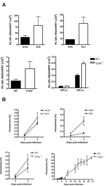
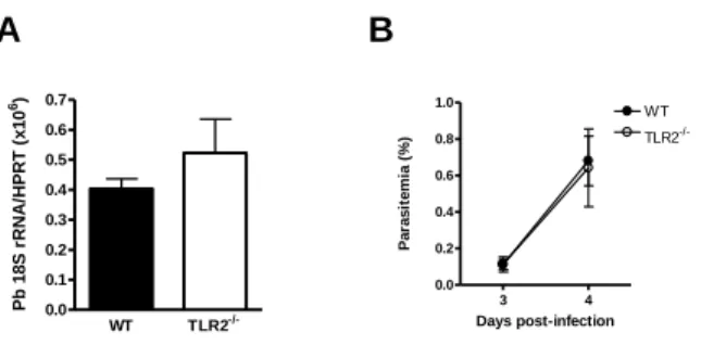
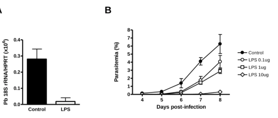
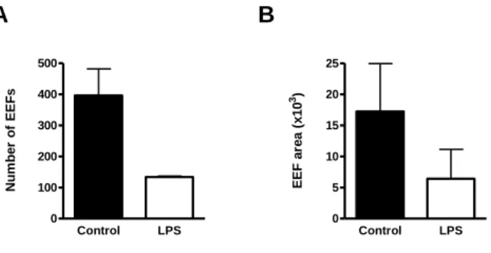
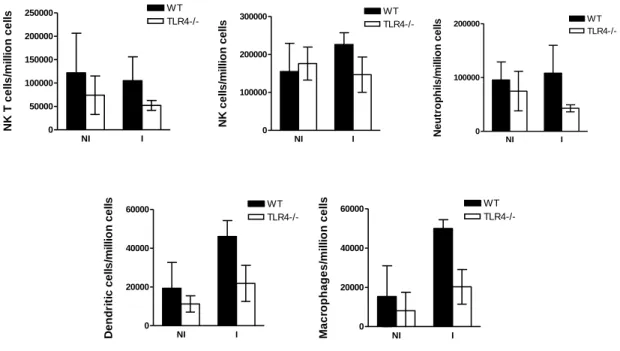
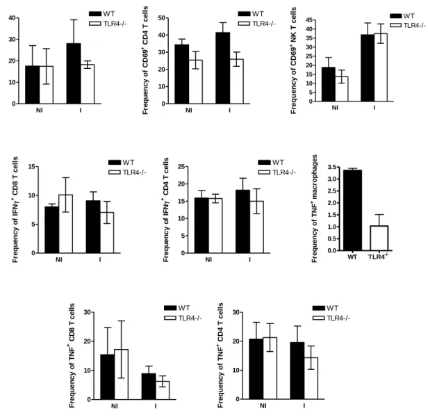
Enquanto que a infecção de ratinhos mutantes para TLR2 por esporozoítos de P. berghei – a forma invasiva transmitida por mosquitos Anopheles e que infecta hepatócitos de roedores – é semelhante à dos ratinhos controlo wild-type, a infecção de ratinhos que não expressam os receptores TLR4 e TLR9 apresenta diferenças significativas importantes relativamente aos controlos. Ratinhos com deficiências na expressão de TLR4 e TLR9 revelam uma maior susceptibilidade e resistência, respectivamente, à infecção do fígado pelo parasita. A expressão reduzida ou ausência de TLR4 reduz a intensidade da resposta imune, permitindo uma maior proliferação do parasita no fígado. Pelo contrário, a ausência de expressão de TLR9 parece ser prejudicial para o parasita, uma vez que os níveis de infecção pelo parasita são reduzidos em mutantes para TLR9. Estas observações sugerem que estes dois receptores desempenham papéis importantes na infecção pela forma hepática de P. berghei. De facto, o tratamento com LPS e CpG, ligandos de TLR4 e TLR9 respectivamente, em simultâneo com a infecção, provoca uma diminuição nos níveis de infecção no fígado. O receptor TLR9 poderá ser importante na infecção e desenvolvimento do parasita durante a primeira fase do seu ciclo de vida no
hospedeiro. Adicionalmente, a molécula recrutada por todos os TLRs na sua via de sinalização, MyD88, é aqui descrita como mediadora da imunidade protectora induzida por esporozoítos de P. berghei atenuados por irradiação, a única forma até agora descrita de imunização efectiva contra a forma hepática do parasita. Embora a molécula adaptadora MyD88 seja relevante no processo de imunização com esporozoítos irradiados, os ratinhos que não expressam MyD88, quando injectados com parasitas viáveis, revelam níveis de infecção muito semelhantes aos ratinhos controlo wild-type. Os resultados apresentados nesta tese sugerem que os TLRs são mediadores de interacções entre o parasita e o hospedeiro durante a fase hepática da malária e poderão ainda estar envolvidos no desenvolvimento de imunidade conferida pela imunização com esporozoítos irradiados. A activação destes receptores na fase inicial da infecção poderá ainda ditar o grau de desenvolvimento da patologia em etapas mais tardias da doença, e consequentemente, determinar o aparecimento ou a ausência de malária cerebral, a manifestação mais letal desta doença.
Abstract
Malaria is one of the most severe human infectious diseases, affecting 5 to 10% of the world's population every year. Although malaria eradication has emerged as a desirable if audacious goal, it is consensual that the development of novel intervention strategies is limited by our current understanding of the biology of Plasmodium, the causative agent of malaria, and of the complex relationships that the parasite maintains with its hosts.
Plasmodium has a complex life cycle that oscillates between a mosquito vector and a vertebrate host. Upon infection of its mammalian host, each Plasmodium sporozoite establishes itself in liver hepatocytes where it replicates into thousands of new parasites (merozoites) that are subsequently released into the bloodstream, infecting red blood cells and causing malaria. Liver infection by Plasmodium is an ideal target for the development of anti-malaria strategies, as it is the first step of infection and it is clinically silent. Indeed, Plasmodium liver stage is the epitome of a perfect malaria vaccine or drug target since complete protection from infection of the treated human host abrogates clinical manifestation and, importantly, transmission of the disease. Thus, understanding the key events during liver infection will certainly facilitate our progress towards novel intervention strategies against malaria.
The prime challenge for any invaded host is to detect the pathogen and orchestrate a rapid defensive response. A set of essential surface and endosomal molecules that comprise the Toll or Toll-like family of receptors perform this role in invertebrate and vertebrate organisms, reflecting a remarkable conservation of function. Functional analysis of mammalian Toll-like receptors (TLRs) has revealed that they recognize specific evolutionarily conserved microbial molecules or molecular families that are present among pathogens and are usually critical to the pathogen's function. These molecules are not found in mammals except in cell stress and inflammation, making them crucial targets for immune intervention. The members of the TLR family recognize lipids, proteins, lipoproteins, carbohydrates, peptides, lipopeptides and nucleic acid structures that are broadly expressed by specific groups of pathogens, providing the
basis for innate immunity and ensuring multiple mechanisms of the adaptive immune response against parasites.
Although it is clear that pathogen detection involves members of the TLR family and that hepatocytes express the majority of all known TLRs, our current understanding about their function in the innate response to Plasmodium liver infection remains elusive. The work presented in this thesis aimed to determine the role of some of these innate receptors and their adaptor molecule, MyD88, not only in the establishment of Plasmodium in the liver but also during an immunization process with attenuated forms of Plasmodium sporozoites.
Liver stage infection by Plasmodium is strongly affected by manipulation of TLR4 and TLR9. While TLR2-deficient mice are infected as wild-type control mice, TLR4 and TLR9-deficient mice show increased susceptibility and resistance to liver stage infection, respectively. While TLR4 absence dampens the inflammatory response, increasing the parasite load in the liver, the lack of TLR9 seems to be detrimental for the parasite. On the other hand, administration of LPS and CpG (TLR4 and TLR9 ligands, respectively), at the time of sporozoite injection, strongly reduces the levels of Plasmodium liver load. In humans, immunization with large numbers of radiation-attenuated sporozoites (RAS) remains the only protocol that leads to the induction of sterile immunity. MyD88 is a mediator of protective immunity induced by P. berghei RAS, despite the fact that when injected with viable sporozoites, MyD88-deficient mice are infected as normal wild-type control mice.
Altogether, the findings presented herewith suggest that TLRs not only mediate major events in the establishment of Plasmodium liver stage infection but are also key players during the establishment of sterile immunity.
Palavras-chave
Malária Plasmodium Fígado Hepatócito Imunidade inata Toll-like receptors Interacções hospedeiro-parasitaKeywords
Malaria Plasmodium Liver Hepatocyte Innate immunity Toll-like receptors Host-pathogen interactionsAbbreviations
cDNA complementary deoxyribonucleic acid
CM cerebral malaria CSP circumsporozoite protein CTL cytotoxic T-cell DAPI 4'6-diamidino-2-phenylindole DCs dendritic cells DDT dichloro-diphenyl-trichloroethane
DMEM Dulbecco's modified eagle medium
EEF exoerythrocytic form
ERK 1/2 extracellular signal-related kinase ½
FACS fluorescence activated cell sorting
FCS foetal calf serum
GAS genetically attenuated sporozoites
GFP green fluorescence protein
HPRT hypoxanthine guanine phosphoribosyltransferase
Hsp60 heat shock protein 60
Hsp70 heat shock protein 70
HSPGs heparan sulphate proteoglycans
i.p. intraperitoneal
i.v. intravenous
IL-6 interleukin-6
IRAK IL-1 receptor associated kinase
iRBC infected RBC
JNK jun NH2-terminal kinase
KO knockout
LRR leucin-rich repeat
MALP2 dipalmitoylated mycoplasm lipoprotein-2
MMTV mouse mammary tumor virus
MyD88 myeloid differentiation factor 88
NF-κB nuclear factor kappa-B
NLRs Nod-like receptors
P. Plasmodium
PAMP pathogen-associated molecular pattern
PbA Plasmodium berghei ANKA
PBS phosphate buffered saline solution
PCR polymerase chain reaction
pen/strep penicillin/streptomycin
PFA paraformaldehyde
PfEMP1 P. falciparum-erythrocyte membrane protein 1
PRR pattern recognition receptor
PV parasitophorous vacuole
qRT-PCR quantitative real-time polimerase chain reaction
RAS radiation attenuated sporozoites
RLRs RIG-I-like receptors
RNA ribonucleic acid
RPMI Roswell Park Memorial Institute (medium)
rRNA ribosomal RNA
RSV respiratory syncytical virus
RT-PCR reverse transcriptase PCR
s.d. standard deviation
TIR Toll/interleukin-1 receptor
TLR Toll-like receptor
TNF-α tumour necrosis factor α
TRAF6 tumor necrosis factor receptor-associated factor 6
Contents
Resumo
Abstract
Palavras-chave
/Keywords
Abbreviations
Contents
Chapter 1
Chapter 2
Chapter 3
Chapter 4
Chapter 5
Chapter 6
References
Acknowledgments
General IntroductionMaterials and Methods
TLR4 deficiency is associated with impaired immune response to malaria liver stage infection
TLR9 is required for Plasmodium berghei liver infection
MyD88 is a mediator of protective immunity induced by Plasmodium radiation attenuated sporozoites
General Discussion iii vii ix xi xiii 3 35 43 65 79 93 123 103
Plasmodium, the causative agent of malaria, is a protozoan parasite that has been discovered more than 125 years ago. Still, malaria remains a major global-health problem today. According to the World Health Organization (WHO) World Malaria Report 2008 the most recent numbers shows that malaria still kills approximately one million people each year, essentially young children and pregnant women. This public health burden creates a significant barrier to economic and social development of the most affected countries (Snow, Trape et al. 2001). So far, due to the complexity of the Plasmodium parasite and its life cycle, the available options for preventing malaria remain limited to vector control and chemoprophylaxis. While drug-resistant strains of the parasite are emerging and insecticide-resistant strains of the mosquito vector are spreading around, the efforts to design an effective vaccine or to develop new drugs capable of reducing malaria morbidity, mortality or transmission remain unsuccessful.
Malaria – Past, present and future
The term malaria is derived from the Italian mala aria, which means “bad air”, from the early association of the disease with marshy areas. Towards the end of the 19th century, Charles Laveran, a French army surgeon, observed parasites in the blood of a patient suffering from malaria, and Dr Ronald Ross, a British medical officer in Hyderabad, India, discovered that mosquitoes were responsible for transmitting malaria. The Italian professor Giovanni Grassi subsequently showed that human malaria could only be transmitted by Anopheles mosquitoes (reviewed inTuteja 2007).
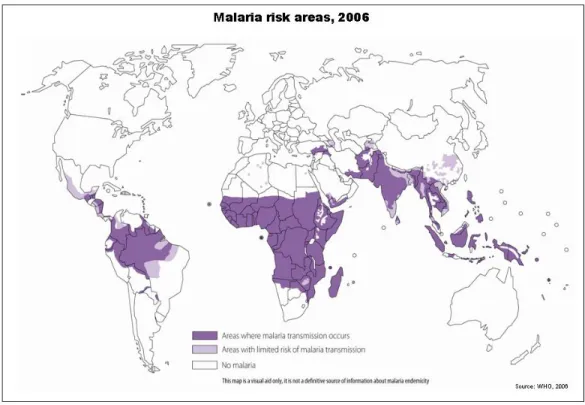
Currently, over two billion people, representing more than 40% of the world’s population, are at risk of malaria (Snow, Guerra et al. 2005). The most affected populations are from developing countries in both the subtropical and tropical regions, many of which are endemic for the disease. Despite its broad distribution, most of the malaria cases and deaths occur in sub-Saharan Africa. Nevertheless, Asia, Latin America, the Middle East and parts of Europe are also affected (World Health Organization, 2007) (see Figure 1). Malaria is widespread where the temperature and rainfall are most suitable for the development of Anopheles mosquitoes, the transmission vector of the malaria parasite, Plasmodium. In the past, malaria also occurred widely in other regions with temperate
climate conditions such as Western Europe and the United States. However, public health measures and economic development have been successful in achieving complete eradication of the disease, except for cases imported via international travel.
Figure 1 Malaria risk areas, 2006 (World Health Organization, 2007).
To counteract this major public health problem The Global Malaria Eradication Programme was created by the WHO in 1955 and was based on two different kinds of approaches. First, chloroquine was used for treatment and prevention of malaria infection, and second, the insecticide dichloro-diphenyl-trichloroethane (DDT) was applied as a vector control strategy. The implementation of these two types of measures led to some beneficial effects, especially in areas with lower transmission rates, like Sri Lanka and India (WHO, The World Health Report 1999). However, global eradication was officially abandoned in 1972, mainly due to the emergence of chloroquine-resistant
Plasmodium parasites and DDT-resistant Anopheles mosquitoes (Brito 2001), but also due to lack of political motivation. Still, in certain countries like Thailand, where better health infrastructure and sustained anti-vector measures, associated to economic development, were implemented, parasite transmission has actually declined. In other countries, like Sri Lanka and Madagascar, the resurgence of malaria caused devastating epidemics, which have affected entire populations (Roberts, Manguin et al. 2000). Then, the emergence of chloroquine and sulfadoxine-pyrimethamine-resistant Plasmodium parasites in Africa (Snow, Trape et al. 2001) became a major issue that required a serious intervention from the international community, which led to new vector controlling measures and new drug combinations with artemisinin derivatives. These measures were very successful in some areas, reinvigorating the program for global eradication (Greenwood, Fidock et al. 2008).
Currently, the WHO’s Global Malaria Programme (GMP), as well as other international initiatives that have been launched to challenge malaria, postulates that malaria control requires an integrated approach that comprises prevention and treatment with effective antimalarials. Several other organizations support the implementation of prevention and treatment programs. In fact, there are several ways of decreasing malaria prevalence but none currently offers an absolute blockade (Greenwood and Mutabingwa 2002). Thus, it is urgent to develop new tools to prevent and treat malaria and to overcome the increasing parasite drug resistance observed over the past few decades. Many believe that the three combined strategies of drug treatment, vaccination and vector control will ultimately be a requisite to significantly decrease malaria infection and transmission (Miller and Greenwood 2002; Ballou, Arevalo-Herrera et al. 2004).
Vaccines, drugs and anti-vector measures are being developed to prevent infection, disease, and transmission. Interestingly, the most widely used old compounds are both derived from ancient herbal therapies. Quinine, isolated from cinchona bark in 1820, and artemisinin, which was obtained from the plant Artemisia annua in 1972 and currently constitutes the most used drug for treatment, being an extremely effective antimalarial. To overcome parasite drug resistance and to extend the useful life of current drugs, combination therapy is being progressively more employed. Further, progress towards developing a vaccine is incomplete. There is no clinically approved malaria vaccine available thus far, even though some are currently in the clinical development and
testing phases (Girard, Reed et al. 2007; Todryk and Hill 2007). Another prospective option for reducing malaria transmission is by using genetically modified mosquitoes that are refractory to parasite transmission (Christophides 2005). Nevertheless, to be effective, these mosquitoes need to be successful in the wild, competing with the already evolutionary and ecologically established mosquitoes.
The availability of genome sequences for humans, Anopheles mosquitoes and Plasmodium parasites has created new expectations for future research achievements. As part of a major and global effort to eradicate malaria, research must be directed to a deeper knowledge of Plasmodium biology but also to the mammalian host and vector response to the parasite. In that respect, a better understanding of the host immune mechanisms operating against Plasmodium infection can also give rise to more powerful interventions. These would be critical to counteract antimalarial drug resistance and the requirement of an effective vaccine against Plasmodium, which hinders malaria control.
Plasmodium
Malaria parasites are eukaryotic unicellular microorganisms that belong to the genus Plasmodium. More than 100 species of Plasmodium can infect several animal species such as birds, reptiles, and a variety of mammals, although only five different species of Plasmodium infect humans under natural conditions: P. falciparum, P. vivax, P. malariae, P. ovale and more recently P. knowlesi. These five species differ in their morphology, geographical distribution, relapse patterns and drug responses. Host immunity against them is also distinct. Among these five species, P. falciparum is the most virulent and the major cause of mortality. It is also distinguished by its ability to adhere to the endothelium during the blood stage of the infection and to sequester in several organs, including the brain. The sequestration of P. falciparum-infected erythrocytes in the brain has been thought to be a major cause of severe malaria-associated pathology including cerebral malaria, a complication that is often fatal. However, this view is today contested by proposals of alternative pathogenic mechanisms. Plasmodium vivax is usually found in tropical areas outside Africa because most Africans lack the Duffy blood group antigen, essential for P. vivax invasion, which
is expressed on the surface of erythrocytes. Both P. vivax (the most widespread species) and P. ovale (the least common malaria parasite, restricted to West Africa) are capable of remaining latent in the liver for weeks to many years as quiescent liver stage forms – hypnozoites, which turns infection with these parasites difficult to eliminate. The beginning of a new round of pre-erythrocytic schizogony results in relapses of malaria infection. Despite not being able to form hypnozoites, P. malariae can persist for decades as a long-lasting asymptomatic blood stage infection. It is found worldwide, but with relatively low frequency (reviewed in Collins and Jeffery 2007). A fifth species, P. knowlesi, which was originally described as a malaria parasite of non-human primates, has been recently described in naturally infected humans in specific regions, such as Malaysia, where the monkey population is highly prevalent (Singh, Kim Sung et al. 2004).
Plasmodium life cycle
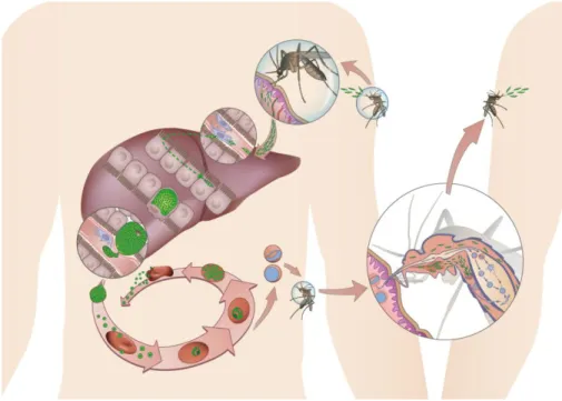
Plasmodium parasites have a complex life cycle in their mosquito vector and vertebrate hosts (see Figure 2). Vertebrate infection begins with the bite of an infected female Anopheles mosquito. Sexual stage differentiation and development of sporozoites, the liver infective form of the parasite, take place in the mosquito vector and its transfer occurs during a blood meal. It was thought that sporozoites moved rapidly away from the site of injection. However it was recently described that most of the infective sporozoites remain at the injection site for hours, with only slow release into the bloodstream (Yamauchi, Coppi et al. 2007). Once in circulation, sporozoites travel to the liver and enter parenchymal cells to initiate the hepatic phase of the disease. Prior to invasion of a final hepatocyte where the parasite will develop and replicate, sporozoites traverse several hepatocytes before invading a final one (Mota, Pradel et al. 2001). The structures on sporozoites surface responsible for hepatocyte invasion are mainly the thrombospondin domains on the circumsporozoite protein (CSP) and on thrombospondin-related adhesive protein (TRAP). These domains specifically bind to heparan sulfate proteoglycans (HSPGs) on the hepatocytes (Frevert, Sinnis et al. 1993). Once inside the hepatocyte, each sporozoite gives rise to tens of thousands of
merozoites through a process termed schizogony. The time for asexual replication in the liver differs, depending on the infecting parasite and infected host. The prepatent period – the time between sporozoite inoculation and the appearance of parasites in the blood – takes 6 to 18 days for human species, and 42 to 48 hours for murine malaria parasites. Using a rodent malaria parasite, P. berghei, it has been shown that liver stage parasites “manipulate” their host cells to ensure the safe release of merozoites into the blood. Hepatocyte-derived merosomes appear to act as shuttles that ensure the protection of parasites from the host immune system and the release of viable merozoites directly into the bloodstream (Sturm, Amino et al. 2006). The release of many thousands of merozoites into the blood, initiates the erythrocytic stage of malaria, a pathogenic cyclic phase in which merozoites develop into mature schizonts within erythrocytes, which rupture and release a new wave of merozoites that invade a new batch of red blood cells (reviewed in Prudencio, Rodriguez et al. 2006).
Each merozoite invades an erythrocyte and divides by schizogony to form an erythrocytic schizont, containing up to 20 daughter merozoites. These merozoites can reinfect other erythrocytes, giving rise to a cyclical blood-stage infection with a periodicity of 48–72 hours, depending on the Plasmodium species.
During the blood stage of infection, unknown factors trigger a subset of developing merozoites to differentiate into male and female gametocytes, which, when taken up by a feeding mosquito, give rise to extracellular gametes, carrying on the Plasmodium’s life cycle. In the mosquito mid-gut, the gametes fuse to form a motile zygote (ookinete), which penetrates the mid-gut wall and forms an oocyst, within which meiosis takes place and genetically distinct haploid sporozoites develop (Aravind, Iyer et al. 2003). The mosquito becomes infectious to its next blood meal donor around two weeks after ingesting gametocytes.
To complete its life cycle, Plasmodium must pass through the mosquito midgut and salivary glands, penetrate skin vessels, traverse macrophages and several hepatocytes prior to enveloping itself in a vacuole and finally, to attach to the surface of erythrocytes before invasion. The unicellular malaria parasite exploits a pool of around 5000 genes (Gardner, Hall et al. 2002) to undergo remarkable changes and face distinct environments and barriers to parasite infection and development. Since all these processes require a series of specific molecular interactions, they are considered as potential targets for the development of new tools against malaria.
The pre-liver infection
The liver stage of Plasmodium infection appears as a good target for developing anti-malaria vaccines or prophylactic drugs. Given that it is the first obligatory step of infection and that Plasmodium remains in the liver for about a week in human malaria infections, the immune system has enough time to mount a response. However, the immune response is usually unsuccessful as the blood stage of infection will follow. Even so, it can be improved to make it more effective. Furthermore, the liver stage is a relevant target to prevent the infection at an early stage, once it is clinically silent and either an effective vaccine or prophylaxis would avoid the development of disease. In
addition, the infection is achieved with only a small number of infected hepatocytes and so, there are only small numbers to eradicate (Rosenberg, Wirtz et al. 1990). In recent years, despite some experimental constraints, particularly in humans, some advances have been made to understand both parasite and host mechanisms of infection and response, respectively. The use of malaria rodent models to study the liver stage of infection has been crucial to allow manipulation of liver infection, intravital imaging, gene knockouts and a sort of scientific approaches that would never be possible in humans. These progresses are having important implications in the development of potential tools for novel clinical approaches and for vaccine design.
During a blood meal, infected Anopheles mosquitoes inoculate Plasmodium sporozoites into the dermal and subdermal tissue of the host. Over time, the sporozoites migrate through the skin until they reach a blood vessel, then traversing the endothelium, sporozoites move into the blood circulation, from where they reach the liver to initiate a malaria infection. Using intravital microscopy of the skin during a mosquito bite, Vanderberg and colleagues, directly evidenced that mosquito probing introduces sporozoites into dermal tissue and these migrate through the dermis and into blood vessels. Furthermore, they also pointed out the role of anti-sporozoite antibodies in blocking sporozoite invasion of these dermal blood vessels (Vanderberg and Frevert 2004).
Interestingly, a quantitative real-time analysis of the fate of sporozoites was used after a mosquito bite in rodents, to demonstrate that only a portion of the parasites entered circulation, whereas some are drained by lymphatics and stop at the proximal lymph node (Amino, Thiberge et al. 2006). Once there, the authors postulate that most are degraded by dendritic cells, but a few sporozoites are capable of partially differentiating into exoerythrocytic stages. This event occurs during sporozoite migration after mosquito probing, and the fact that parasites can partially develop in lymph nodes might have a major influence in host immunity against Plasmodium infection and therefore must be considered in vaccine development against the preerythrocytic stages of the parasite. It was also proposed that host cell traversal is important for progression of the malaria parasite through the dermis to the liver. Using a murine parasite, P. berghei, it has been recently shown that cell traversal is important in the host dermis for preventing sporozoite destruction by phagocytes and arrest by nonphagocytic cells (Amino,
Giovannini et al. 2008). In addition, during the migration in the skin before entering the liver, sporozoites are very susceptible to neutralization by antibodies. These bind sporozoite surface proteins, mainly the CSP (Yoshida, Nussenzweig et al. 1980) that uniformly covers the external surface of sporozoites, and effectively opsonize the parasite (Vanderberg and Frevert 2004). Still, when sporozoites are able to rapidly reach a blood vessel, they are then carried to the liver in a very short period of time, entering the sinusoids through the portal fields. Sinusoids display a very specialized endothelium, essentially consisted by fenestrated endothelial cells and resident liver macrophages, known as Kupffer cells (Bouwens, De Bleser et al. 1992; Prudencio, Rodriguez et al. 2006).
The liver infection
Sporozoites attach to the endothelial inner layer of liver sinusoids by interactions between parasite surface proteins, both CSP and TRAP (Robson, Hall et al. 1988), with host extracellular matrix proteoglycans (reviewed in Kappe, Buscaglia et al. 2004). These parasite surface proteins, CSP and TRAP, are also thought to play essential roles in sporozoite gliding motility and host cell infection (Sultan, Thathy et al. 1997). Since sporozoites do not adhere to sinusoidal endothelia in vitro (Pradel and Frevert 2001), the gliding along the sinusoid lining is thought to be mediated by the interaction of CSP and TRAP with the unique highly sulphated liver HSPGs protruding from the extracellular matrix in the space of Disse and through fenestrae of the endothelial cells (Robson, Frevert et al. 1995; Pradel, Garapaty et al. 2002).
After attaching to sinusoids endothelium, sporozoites must cross the sinusoidal barrier to reach hepatocytes. Surprisingly, Frevert and colleagues suggested that instead of taking what would seem a safer route through endothelia, the parasites traverse Kupffer cells without suffering any harm. The authors proposed a model in which these liver macrophages function as the sporozoite gate to the liver. Thus, according to this model, sporozoites glide along the endothelium, then penetrate and traverse Kupffer cells to reach hepatocytes. Notably, the CSP from sporozoites, by binding to ribosomes and inhibiting protein synthesis (Frevert, Galinski et al. 1998), seems to suppress the
respiratory burst in Kupffer cells (Usynin, Klotz et al. 2007) and to inhibit antigen presentation and cytokine release, thereby disabling the Kupffer cells’ potential anti-parasitic function (Steers, Schwenk et al. 2005). However, it has been shown that Kupffer cells can phagocytize sporozoites (Meis, Verhave et al. 1983) and interfere with the development of the exo-erythrocytic forms (Vreden, Sauerwein et al. 1993) presumably by the production of IL-6 (Vreden, van den Broek et al. 1992). In addition, in vivo depletion of Kupffer cells prior to infection with P. berghei sporozoites results in increased parasitemia (Vreden, Sauerwein et al. 1993).
Once inside the liver parenchyma, sporozoites traverse several hepatocytes before infecting a final one (Mota, Pradel et al. 2001). Mota and colleagues described that sporozoite host cell traversal caused breaching of the plasma membrane of the hepatocyte, followed by rapid repair. These observations emphasized that sporozoite migration through several cells in the mammalian host seems to be required for liver stage development and maintenance of the life cycle. The same authors proposed that sporozoites must cross the cytosol of several hepatocytes before invading a final one by the formation of a parasitophorous vacuole (PV), a specialized membrane compartment. Cell traversal seems to depend on at least two sporozoite secretory proteins – SPECT (sporozoite microneme protein essential for cell traversal) and SPECT2 (Ishino, Yano et al. 2004; Ishino, Chinzei et al. 2005). Inside the PV, in which the parasite is confined from the host cell cytoplasm (Mikolajczak and Kappe 2006), the parasite proliferates and differentiates into thousands of erythrocyte-invasive forms, the merozoites (Hollingdale 1985). The initial formation of the vacuole is dependent on secretory proteins that are characterized by cysteine domains. Indeed, sporozoites lacking 6-cysteine proteins enter hepatocytes but cannot form a PV (Labaied, Harupa et al. 2007) and do not undergo the subsequent liver stage development.
Several other parasite proteins have been described as being important in nutrient uptake from the host infected hepatocyte. UIS3 protein (Mikolajczak, Jacobs-Lorena et al. 2007) is one these proteins that seem to be inserted in the PV membrane. Genetically modified parasites that lack the expression of these proteins, display arrested development early in infection (Mueller, Camargo et al. 2005; Tarun, Dumpit et al. 2007). In fact, these proteins are critical for parasite development, since both 6-Cysteine protein-deficient (Labaied, Harupa et al. 2007) and PV protein-knockout
parasites are consequently attenuated and cannot give rise to a blood stage infection (Mueller, Camargo et al. 2005; Tarun, Dumpit et al. 2007).
Liver innate immunity
Vertebrate immunity can be divided into innate and adaptive, both of which are responsible for the recognition and elimination of pathogens. The innate immune system, also present in invertebrates, constitutes the first line of defense against parasites. The innate immune response is mediated largely by polymorphonuclear leukocytes (PMNs) such as neutrophils, macrophages, dendritic cells (DCs), mast cells and NK cells. The functions of these innate cellular components comprise opsonization, activation of complement and phagocytosis that kills the pathogens and concurrently coordinate additional host responses by synthesizing a wide range of inflammatory mediators and cytokines (Medzhitov 2001).
In general, the infectious agent is killed and degraded in antigen-presenting cells within the maturing phagossome, and components of the pathogen are presented to T cells, resulting in the activation of the adaptive immune response and in the establishment of protective immunity (Aderem and Ulevitch 2000). The recognition by the adaptive immune system and the efficiency of the response relies on the generation of random and highly diverse repertoires of B and T cell antigen receptors, clonal selection and expansion of lymphocytes with receptors of relevant specificity, followed by subsequent memory acquisition in most cases.
The liver is an organ that stands between the gastrointestinal tract and the systemic circulation. Blood from the intestines, rich in bacterial products and in food-derived antigens, encounters the resident population of immune cells. The vast majority of the non-parenchymal cells found in the liver are specialized macrophages called Kupffer cells (KCs). Additionally, CD4 and CD8 T cells, DCs, natural killer (NK) cells and NKT cells are also found in the liver (reviewed in Crispe 2003).
The primary antigen-presenting cells (APCs) in the liver are DCs, which migrate to lymph nodes in order to present antigens to antigen-specific lymphocytes, which in turn migrate to the site of infection. Not only DCs but also KCs as well as liver sinusoidal
endothelial cells (LSECs), hepatic stellate cells (HSCs) and hepatocytes are able to present antigens in the liver. These are resident cells and do not enter the draining lymph nodes.
KCs constitute the liver resident macrophages and their interaction with leucocytes has been found to be relevant for host defense, liver regeneration and establishment of liver immune tolerance (reviewed in Bilzer, Roggel et al. 2006). By residing in liver sinusoids, KCs are among the first immune cells to come in contact with bacteria or bacterial components derived from the gastrointestinal tract. Additionally, KCs are thought to be responsible for the rapid clearance of bacteria from the blood stream (Klein, Zhadkewich et al. 1994).
The most relevant factors in KC activation are endogenous complement factors C3a and C5a, β-glucans from bacteria and fungi, and LPS. In particular, KC activation by LPS, involving the LPS-binding protein CD14 and TLR4, activates the NF-κB pathway that ultimately will lead to production of pro-inflammatory cytokines, tumor necrosis factor-α (TNF-α) and IL-1, as well as large amounts of nitric oxide (NO). The release of pro-inflammatory cytokines, such as IL-1, IL-6, and TNF-α, promotes the infiltration of neutrophils to eliminate bacteria. TNF-α also induces apoptosis in hepatocytes under pathological conditions (Schumann and Tiegs 1999). Additionally, KCs produce IL-12 and IL-18 which activate NK cells to produce anti-viral interferon-γ (IFN-γ). However, following initial activation to produce pro-inflammatory cytokines, KCs release IL-10 which down-regulates the production of TNF-α, IL-6 and other cytokines (Knolle, Schlaak et al. 1995) and thereby probably contributes to the intrahepatic cell populations capability to induce tolerance.
LSECs can also be efficient APCs in the liver (Crispe 2009). They constitutively express MHC class I and II, as well as the co-stimulatory molecules CD40, CD80, and CD86. LSECs take up the antigen and present it to CD4 and CD8 T cells.
Physiological concentrations of endotoxin, which are continuously present in portal venous blood (Crispe 2009), seem to induce the release of IL-10 from LSECs and KCs (Knolle, Schlaak et al. 1995) and to downregulate CD4 T cell activation by LSECs through down-modulation of the expression of MHC class II, CD80 and CD86 (Knolle, Germann et al. 1999). In contrast, TLR4 activation of APCs by endotoxin induces T cell activation (Pasare and Medzhitov 2005). These observations indicate that the liver
tolerogenic effect might be related to the continuous exposure of sinusoidal cells to bacterial products from the gut.
Nevertheless, in contrast to TLR4 activation, activation of TLR3 by polyI:C or activation of TLR9 by CpG oligonucleotides, has been shown to induce CD8 T cell-mediated hepatitis in transgenic mouse models and has been suggested to induce autoimmunity by breaking immune tolerance in the liver (Sacher, Knolle et al. 2002; Lang, Georgiev et al. 2006). As well, TLR9 activation has been described to exacerbate CD4 T cell induced hepatitis (Jiang, Sun et al. 2009).
HSCs are well known for their involvement in hepatic fibrosis and storage of vitamin A, and were recently shown to function as APCs (Winau, Hegasy et al. 2007). They are able to present protein or lipid antigens to MHC class I and II or to CD8 and CD4 T cells as well as to NKT cells. Upon bacterial infection, HSC elicited a protective antigen-specific T cell response that eliminated the pathogens (Winau, Hegasy et al. 2007). Hepatocytes constitute the parenchymal cell population in the liver, which primarily perform metabolic functions. Still, they are also involved in immunoregulation by their ability to function as APCs. It has been shown recently that direct interactions occur between lymphocytes and hepatocytes through cytoplasmic extensions penetrating the liver endothelial fenestrations (Warren, Le Couteur et al. 2006). Apart from their constitutive MHC class I expression, hepatocytes also express MHC class II under inflammatory conditions, present the antigen and activate CD4 T cells (Herkel, Jagemann et al. 2003).
T lymphocytes in the liver display an activated phenotype, constituting a source of both IFN-γ and IL-4 (Klugewitz, Blumenthal-Barby et al. 2002). However, the primary consequence of T cell priming in the liver seems to be tolerance (Knolle, Schmitt et al. 1999; Limmer, Ohl et al. 2000). Indeed, as mentioned above, antigen presentation by LSECs induces apoptosis in CD8 T cells (Bertolino, Trescol-Biemont et al. 1998), a mechanism which might frustrate efforts to prime an effective local immune response. NK cells are present at an unusual high frequency among liver lymphocytes. By synthesizing IFN-γ, which promotes secretion of chemokine CXCL9 by hepatocytes, NK cells are thought to have a crucial role in T cell recruitment, and thus in promoting T cell-mediated immunity (Itoh, Morita et al. 2001). As for NKT cells, even though having a limited T cell receptor (TCR) diversity, they were shown to play a role in the immune
response to the liver stage of malaria infection (Gonzalez-Aseguinolaza, de Oliveira et al. 2000) and to be crucial for the establishment of anti-tumour immunity (Cui, Shin et al. 1997).
Immune responses to malaria
The life cycle of Plasmodium includes several stages, which can be targeted by host immune responses, either by blocking the intermediary critical steps between the different infection stages, or by directly killing the parasite.
Following the injection of Plasmodium parasites by a female Anopheles mosquito into the human dermis, sporozoites have to evade antibodies and other opsonizing molecules such as complement, when accessing blood vessels in the skin and then to pass through liver macrophages and hepatocytes to initiate liver stage infection.
Liver developing parasites are a target of cytotoxic T lymphocytes (CTLs) and at the time of merosome release, merozoites have to evade circulating antibodies to invade erythrocytes. Blood stage parasites are susceptible to opsonizing antibodies and macrophages, and cytokine responses have been related to both protection and disease during this stage of infection. In fact, antibodies that block binding of P. falciparum– infected erythrocytes to endothelium might prevent disease and control parasitemia. Human antibodies specific for gametocytes can block transmission to mosquitoes, although these might require complement for parasite killing. Parasites can also be killed by Anopheles mosquito innate immune responses during early or late sporogonic stages and lead to refractoriness to infection (reviewed in Greenwood, Fidock et al. 2008). It has been demonstrated that the main anti-parasite effector mechanism in the liver is IFN-γ produced mainly by CS-specific CD8 and CD4 T cells that inhibit parasite development within hepatocytes (Schofield, Ferreira et al. 1987; Weiss, Sedegah et al. 1988; Hoffman, Isenbarger et al. 1989; Romero, Maryanski et al. 1989; Rodrigues, Cordey et al. 1991). CD8 CTLs that are capable of recognizing Plasmodium antigens presented by MHC class I molecules on the surface of infected hepatocytes seem also to play a role. CTLs kill the hepatocytes through pore-forming perforin proteins, which allow apoptosis-inducing granzymes to enter and kill the target cells. However, immunity
can also be achieved in perforin knockout mice immunized with irradiated sporozoites (Renggli, Hahne et al. 1997). Moreover, IFN-γ production may enhance cytolytic mechanisms by increasing class I molecules on the surface of hepatocytes, making those hepatocytes better targets for CTL lysis, and activating supplementary effector cells such as natural killer cells and macrophages, which are also able to kill the target cells through CTL-like mechanisms, TNF-α induction and NO production.
In the erythrocytic stage, the potential targets for an immune response are merozoites in the blood stream or intraerythrocytic parasites. Since HLA class I or II molecules are absent from the surface of the parasite or the infected red blood cell (RBC), it is generally agreed that humoral responses are critical in blood-stage immunity. It was shown in mouse models that B cells and antibodies are important in parasite elimination (Langhorne, Cross et al. 1998). The mechanisms by which antibodies are effective consist of blockade of the invasion of RBCs by merozoites (Blackman, Heidrich et al. 1990), antibody-dependent cellular killing mediated by cytophilic antibodies (Bouharoun-Tayoun, Oeuvray et al. 1995) and binding of antibody to parasite-induced molecules on the RBC surface, leading to greater clearance of infected RBCs (Bull, Lowe et al. 1998). Several studies suggest that responses to many antigens are involved in protection (Gray, Corran et al. 2007). Many presumed target antigens on the infected RBC surface and antigens either on the merozoite surface or released during merozoite invasion have been identified as being potentially protective.
The immune response to malaria is extremely complex, and is both species- and stage-specific. The generation and maintenance of clinically protective immune responses requires repeated infections over the lifetime of the individual. There are several mechanisms playing a role in adaptive immunity against malaria: antibodies that prevent invasion of sporozoites into liver cells, CD8 T cells and IFN-γ that inhibit parasite development in infected hepatocytes, antibodies that block invasion of merozoites into erythrocytes, antibodies that prevent sequestration of infected erythrocytes by preventing binding to adhesion molecules on the vascular endothelium, IFN-γ and CD4 T cells that activate macrophages to phagocytose intra-erythrocytic parasites and free merozoites, antibodies that neutralize parasite glycosylphosphatidylinositol and inhibit induction of the inflammatory cytokine cascade and, finally, antibodies that mediate
complement-dependent lysis of extracellular gametes, prevent fertilization of gametes and the development of zygotes (reviewed in Stevenson and Riley 2004).
As described above, the adaptive immune responses known to be protective at each stage of the parasite’s life cycle have been the subject of many studies in mice models of malaria infection and are quite well known. However, unlike other infections with intracellular pathogens, including viruses, bacteria and some protozoan parasites (Janeway and Medzhitov 2002; Akira, Uematsu et al. 2006; Beutler, Jiang et al. 2006), the role of the innate immune mechanisms against Plasmodium infection has not been often addressed.
Vaccine development against malaria
It is known that older children and adults with exposure to malaria infection can develop complete protection from severe illness and death, even though sterile immunity is probably never achieved. However, the viability of malaria vaccination is supported by a number of observations. Both epidemiological data and mathematical models demonstrate that immunity to malaria might be acquired with age, since only a few adults die of malaria in endemic regions (Gupta and Day 1994). This might be due either to the acquisition of tolerance – the infection proceeds at the same rates, but there are no clinical symptoms (clinical immunity) – or to the development of immunity to the parasite infection, in individuals that are continuously exposed to the disease throughout their life time. Some epidemiological data correlate specific immune responses with reduced malaria incidence, which indicates that it might be possible to develop immunity against the parasite. For instance, a considerable number of associations were done with immune responses to the main highly variable protein on the surface of infected erythrocytes, P. falciparum erythrocyte membrane protein 1 (PfEMP1), which constitutes a difficult target for immunization due to its high rates of antigenic switching and considerable diversity (Bull, Lowe et al. 1998; Dodoo, Staalsoe et al. 2001). Additionally, in the 1970s, it was shown that both humans and mice could be immunized with RAS, meaning that immunity against the pre-erythrocytic malarial stages by itself could induce
sterile protection for several months (Nussenzweig, Vanderberg et al. 1967; Clyde, Most et al. 1973).
Despite it being possible to culture sporozoites in vitro (Al-Olayan, Beetsma et al. 2002), an extensive production is not a viable process, while immunization by thousands of infected mosquitoes is unfeasible as well. Thus, a huge effort has been made in the past 25 years seeking to understand the immune mechanisms responsible for this protection and to identify the parasite proteins that are the targets of these protective immune responses.
Thus far, sterile protective immunity against challenge with Plasmodium spp. sporozoites has been induced in several model systems and in humans by immunization with RAS. The primary target of this RAS-induced protection is the infected hepatocyte and several immune mechanisms have been identified as critical mediators of protection against sporozoite infection. Studies in mice indicate that CD8 effector T cells producing IFN-γ are the main mechanism by which these vaccines may act (Schofield, Villaquiran et al. 1987).
T cells specific for parasite-derived peptide/class I MHC molecule complexes on the surface of infected hepatocytes are the primary immune effectors, which kill parasites in infected hepatocytes, while antibodies against sporozoite surface proteins, such as CSP, are thought to have a minor function. CD4 T cells that recognize parasite-derived peptides presented by class II MHC molecules on the surface of infected hepatocytes as well as other factors, including NO, are also important effectors in protection against sporozoite infection (reviewed in Doolan and Martinez-Alier 2006).
It was recently shown in mice that parasites deficient in certain proteins, which induce arrested development in the liver, were also able to induce sterile protection. Indeed, a single-dose immunization with specific genetically attenuated sporozoites (GAS) can induce full protection against subsequent sporozoite infection. Moreover, prime-boost immunizations are capable of inducing protection for at least 6 months. Several GAS that infect hepatocytes but are unable to establish blood-stage infections in vivo have been described: UIS3-deficient sporozoites (Mueller, Labaied et al. 2005), UIS4-deficient sporozoites (Mueller, Camargo et al. 2005), P36p-UIS4-deficient sporozoites (van Dijk, Douradinha et al. 2005; Douradinha, van Dijk et al. 2007) and P52-deficient sporozoites, in P. falciparum, an ortholog of the rodent parasite gene P36p (van Schaijk,
Janse et al. 2008). The protection conferred by GAS is, to some extent, achieved by antibodies that prevent sporozoite invasion of hepatocytes. However, CD8 T cells are essential to eliminate the remaining infected hepatocytes both by direct cytotoxicity and by IFN-mediated mechanisms (Jobe, Lumsden et al. 2007; Mueller, Deckert et al. 2007), similar to those conferring protection induced by RAS (Doolan and Hoffman 2000). Because many do not believe a live-attenuated vaccine will ever be feasible, research has also been directed towards identifying parasite antigens that should be included in a vaccine. Despite extensive work on this subject, the number or the identity of the antigenic determinants involved in the protection against liver infection conferred by immunization with RAS remain unclear. The genes that need to be targeted in a designed subunit vaccine in order to elicit similar levels of immunity are still undefined. There may be numerous antigens that contribute to the sterile protective immunity elicited by the immunization with RAS. This means that subunit vaccines that comprise only one or a few antigens might need to induce immune responses that are significantly stronger than those that are induced by years of natural exposure to the parasite, in order to be able to confer worthwhile protection (Hill 2006).
Although hundreds of other Plasmodium genes are expressed in sporozoites and exoerythrocytic forms (EEFs), the CSP seems to be an immunodominant protective antigen in RAS (Kumar, Sano et al. 2006). The CSP is a surface protein that covers Plasmodium sporozoites and the plasma membrane of EEFs and has been detected in the cytoplasm of the infected hepatocytes (Hollingdale, Leland et al. 1983). Recently, CSP was shown to influence the expression of over one thousand host genes involved in several metabolic processes in order to create an appropriate niche for the parasite growth and development in liver hepatocytes (Singh, Buscaglia et al. 2007). The presence of CSP in the hepatocyte seems to enhance parasite growth of the liver stages both in vitro and in vivo (Singh, Buscaglia et al. 2007). Nevertheless, regardless of being considered as a major candidate antigen for vaccines targeting the pre-erythrocytic stages of Plasmodium infection, sterile immunity can be induced despite the absence of specific immune responses to the CSP expressed by the parasite used for challenge (Gruner, Mauduit et al. 2007).
The sequencing of the genome of P. falciparum (Gardner, Hall et al. 2002) provided a most useful tool for vaccine research. However, this might become a double-edged
sword: the approximately 5300 open reading frames that were identified, many of completely unknown function, may scatter the enormous efforts that are being developed thus far. It is not obvious whether the identification of thousands of putative candidate antigens to generate a vaccine would be helpful or not in a field that is already struggling with a small number of putative vaccine candidates. Still, malaria research is already gaining from the genome sequencing and related proteome project (Florens, Washburn et al. 2002; Hall, Karras et al. 2005). For instance, the stage-specificity of expression of many antigens in the Plasmodium life cycle is being better defined, revealing the unique expression of liver and blood stage antigens.
Four antigens expressed by P. falciparum RAS in hepatocytes have been extensively investigated as putative vaccine targets: PfCSP, Pf Sporozoite Surface Protein 2/Thrombospondin related anonymous protein (PfSSP2/TRAP), Pf Exported Protein-1 (PfEXP-1), and Pf Liver Stage Antigen-1 (PfLSA1). Immunizing mice with the rodent malaria orthologues of PfCSP, PfSSP2/TRAP, and PfEXP-1 protects them from sporozoites infection. Moreover, immunization of people with PfCSP partially protects them against experimental and natural challenge (reviewed in Hoffman and Doolan 2000). So, currently, despite a long history of disappointing results with other candidates, the most success has been achieved using a PfCSP-based recombinant protein, called RTS,S, one subunit vaccine which has shown promise in phase IIb clinical trials (Bojang, Milligan et al. 2001; Alonso, Sacarlal et al. 2004; Alonso, Sacarlal et al. 2005; Aponte, Aide et al. 2007). This leading malaria vaccine candidate incorporates a fusion protein, which comprises CSP and the HBV surface antigen and aggregates as virus-like particles, together with the adjuvant AS02, which is based on monophosphoryl lipid A and QS-21 (Stoute, Slaoui et al. 1997). However, the RTS,S immunized volunteers were not protected after six months post−immunization and the vaccine only induced antibody and CD4 T cell responses, while CD8 T cell responses were not detectable (Lalvani, Moris et al. 1999).
A second approach has been to elicit CD8 T cell responses, against the known Pf proteins expressed by irradiated sporozoites in hepatocytes. A PfCSP DNA vaccine has been reported to be safe (Le, Coonan et al. 2000) to induce CD8 CTL in malaria-naïve volunteers (Wang, Doolan et al. 1998) and to elicit CD8 T cell-dependent IFN-γ production (Wang, Epstein et al. 2001).
These promising results are also stimulating the search for additional vaccines. However, the field must be aware that natural immunity to malaria might consist of a complex mixture of diverse immune responses, some of probably no protective value and some potentially counter-protective, such as pro-inflammatory responses that have been implicated in the pathology of cerebral malaria (CM).
Likewise, ongoing large efforts are testing P. falciparum attenuated parasites, by deletion of essential genes, as vaccine candidates in humans and developing a mean to deliver attenuated vaccines.
Toll-like receptors
For a long time a dogma persisted according to which the innate branch of immunity was considered to be entirely unspecific and somewhat less evolved then the adaptive immunity branch. With the discovery of a class of recognition receptors, which bind highly conserved specific molecules among pathogens, the unforeseen specificity of innate immunity was recognized, despite at a much lower level then the specificity observed in adaptive immunity.
TLRs are one subset of a group known as pattern recognition receptors (PRRs). This set of recognition receptors of the innate immune system recognizes conserved molecular structures known as pathogen-associated molecular patterns (PAMPs), which are present in large groups of pathogens (Medzhitov and Janeway 1997). Mannose receptors, such as C-type lectins, scavenger receptors, opsonins (acute phase proteins, complement proteins), NOD (nucleotide-binding oligomerization domain) receptors and CARD (caspase activating and recruitment domain)-containing proteins, such as RIG-1 (retinoic acid-inducible 1) and MDA-5 (melanoma differentiation-associated gene-5) are also other members of this functionally-defined family.
The Toll receptor was originally identified in Drosophila as an essential receptor for the establishment of the dorso-ventral pattern in developing embryos (Hashimoto, Hudson et al. 1988). In 1995, it was demonstrated that the Toll pathway in Drosophila embryos is crucial for immune responses (Lemaitre, Meister et al. 1995), and that Toll-mutant flies were highly susceptible to fungal infection (Lemaitre, Nicolas et al. 1996). The
involvement of Toll in flies’ immune defense drove the research to other animal models. In 1997, the first mammalian homologue of the Drosophila Toll receptor was identified. A mammalian homologue of Drosophila Toll receptor, currently designated as TLR4, was shown to induce the expression of genes involved in inflammatory responses (Medzhitov, Preston-Hurlburt et al. 1997). Afterwards, several other proteins that are structurally similar to TLR4 were also identified and designated as TLRs (Rock, Hardiman et al. 1998). Since then, mammalian TLRs have been a major focus in the immunology field. They participate in the first line of defense against invading pathogens and play a significant role in inflammation, immune cell regulation, survival and proliferation. TLRs are Type I transmembrane proteins characterized structurally by the presence of a Leucin-Rich Repeat (LRR) domain in the extracellular part and a TIR (Toll IL-1 receptor) domain in the intracellular portion.
Thus far, 11 human TLRs (TLR1–TLR11) and 13 (TLR1–TLR13) mouse TLRs have been identified. Toll homologues can also be found in plants (O'Neill and Greene 1998) and in worms (Rich, Allen et al. 2000). In Drosophila, nine Toll receptors were identified to date, but only Toll 1, was shown to play a role in host defense (Hoffmann and Reichhart 2002).
Mammalian TLRs recognize a wide spectrum of ligands that comprise modified lipids, proteins and nucleic acids. TLR2 recognizes an extensive array of microbial compounds, such as bacterial lipopeptides (Aliprantis, Yang et al. 1999), peptidoglycan and lipoteicoic acid from Gram-positive bacteria (Schwandner, Dziarski et al. 1999). TLR2 can form heterodimers with both TLR1 and TLR6 (Ozinsky, Underhill et al. 2000) recognizing several other PAMPs including triacylated lipoproteins (Alexopoulou, Thomas et al. 2002), the dipalmitoylated mycoplasm lipoprotein 2 (MALP2) (Takeuchi, Kawai et al. 2001) and fungal zymosan (Ozinsky, Underhill et al. 2000).
TLR4 recognizes the Gram-negative bacterial lipopolysaccharide (LPS). Already in the early 1970’s it was reported that antibody production by B cells could be induced by the mitogen LPS (Coutinho, Moller et al. 1973). Despite there being no specificity associated to LPS stimulation alone, B cells stimulated with this microbial component together with antigen give rise to an antigen-specific response (Coutinho, Gronowicz et al. 1974). Following these findings, the mouse strain C3H/HeJ was reported to be hyporresponsive to LPS, and the authors speculated that the observed defect in B cell
activation could be due to the absence of the receptor for LPS (Coutinho 1976). This was later confirmed by showing that using an antibody which bound the surface of B cells inhibited their LPS-mediated activation and immunoglobulin production, competing with LPS for the receptor (Coutinho, Forni et al. 1978).
Later, two independent studies identified a mutation in the gene responsible for the LPS hyporesponsiveness in two mouse strains that were hyporesponsive to LPS, the C57Bl10/ScCr and the C3H/HeJ (Poltorak, He et al. 1998; Qureshi, Lariviere et al. 1999).
Afterwards, other TLR4 agonists with similar immunostimulatory properties to LPS in mice were identified: Taxol (Kawasaki, Akashi et al. 2000), the envelope proteins of mouse mammary tumour virus (MMTV) (Rassa, Meyers et al. 2002), the fusion protein of respiratory syncytical virus (RSV) (Kurt-Jones, Popova et al. 2000) and a few fungal structural components (Shoham, Huang et al. 2001; Wang, Warris et al. 2001). More recently, the heat shock protein 60 (Hsp60) (Ohashi, Burkart et al. 2000), Hsp70 (Asea, Rehli et al. 2002; Correia, V., personal communication; Vabulas, Ahmad-Nejad et al. 2002), gp96 (Vabulas, Braedel et al. 2002) and β-defensin (Biragyn, Ruffini et al. 2002) have been described as endogenous ligands for TLR4. Fibronectin, heparan sulfate and hyaluronic acid are extracellular matrix components that are produced in response to tissue injury during inflammation. The extra domain A of fibronectin (Okamura, Watari et al. 2001), polysaccharide fragments of heparan sulphate (Johnson, Brunn et al. 2002) and oligosaccharides of hyaluronic acid (Termeer, Benedix et al. 2002) have also been reported as powerful agonists of TLR4. In addition, host fibrinogen is able of triggering chemokine secretion by macrophages, through recognition by TLR4 (Smiley, King et al. 2001).
TLR9 recognizes bacterial DNA and synthetic oligodeoxynucleotides containing CpG dinucleotides (Hemmi, Takeuchi et al. 2000). TLR9 has also been reported to be required in the recognition of host chromatin-IgG complexes (Leadbetter, Rifkin et al. 2002). Recently, hemozoin purified from P. falciparum was described as a new non-DNA ligand for TLR9 (Coban, Ishii et al. 2005).
TLRs can be expressed in two different cellular localizations. TLRs 3, 7, 8, and 9, are expressed in intracellular compartments, the endosomes. Intracellular TLRs sense viral