Characterization of HCV Interactions with Toll-Like
Receptors and RIG-I in Liver Cells
Erika A. Eksioglu1, Haizhen Zhu2, Lilly Bayouth1, Jennifer Bess1, Hong-yan Liu1, David R. Nelson3, Chen Liu1,3*
1Department of Pathology, Immunology and Laboratory Medicine, University of Florida College of Medicine, Gainesville, Florida, United States of America,2State Key Laboratory of Chemo/Biosensing and Chemometrics, School of Biology and Research Center of Cancer Prevention and Treatment of Hunan University & Hunan Tumor Hospital, Changsha, Hunan Province, China,3Department of Medicine, Division of Hepatobiliary Diseases, University of Florida College of Medicine, Gainesville, Florida, United States of America
Abstract
Background and Aim:The aim of this study was to examine the mechanisms of IFN induction and viral escape. In order to accomplish the goal we compared our new hepatoma cell line LH86, which has intact TLR3 and RIG-I expression and responds to HCV by inducing IFN, with Huh7.5 cells which lack those features.
Methods:The initial interaction of LH86 cells, Huh7.5 cells or their transfected counter parts (LH86 siRIG-I, siTLR3 or siTLR7 and Huh7.5 RIG-I, TLR3 or TLR7) after infection with HCV (strain JFH-1) was studied by measuring the expression levels of IFNb, TRAIL, DR4, DR5 and their correlation to viral replication.
Results:HCV replicating RNA induces IFN in LH86 cells. The IFN induction system is functional in LH86, and the expression of the RIG-I and TLR3 in LH86 is comparable to the primary hepatocytes. Both proteins appear to play important roles in suppression of viral replication. We found that innate immunity against HCV is associated with the induction of apoptosis by RIG-I through the TRAIL pathway and the establishment of an antiviral state by TLR3. HCV envelope proteins interfere with the expression of TLR3 and RIG-I.
Conclusion:These findings correlate with the lower expression level of PRRs in HCV chronic patients and highlight the importance of the PRRs in the initial interaction of the virus and its host cells. This work represents a novel mechanism of viral pathogenesis for HCV and demonstrates the role of PRRs in viral infection.
Citation:Eksioglu EA, Zhu H, Bayouth L, Bess J, Liu H-y, et al. (2011) Characterization of HCV Interactions with Toll-Like Receptors and RIG-I in Liver Cells. PLoS ONE 6(6): e21186. doi:10.1371/journal.pone.0021186
Editor:Jean-Luc E. P. H. Darlix, Institut National de la Sante´ et de la Recherche Me´dicale, France
ReceivedDecember 1, 2010;AcceptedMay 23, 2011;PublishedJune 17, 2011
Copyright:ß2011 Eksioglu et al. This is an open-access article distributed under the terms of the Creative Commons Attribution License, which permits unrestricted use, distribution, and reproduction in any medium, provided the original author and source are credited.
Funding:This work was supported by NIH-DK002958 and NIH-A1061158. EAE was a Ruth Kiershtein NRSA pre-doctoral fellow (F31) while engaged in this project: NIH-F31AI071353. The funders had no role in study design, data collection and analysis, decision to publish, or preparation of the manuscript.
Competing Interests:The authors have declared that no competing interests exist.
* E-mail: [email protected]
Introduction
Hepatitis C virus (HCV), a member of the family flaviviridae, infects approximately 170 million people worldwide leading to chronic liver disease in up to 80% of them [1,2]. Failure to induce type I IFN, has been correlated with the lack of an appropriate innate response [3,4]. In particular, chronically infected hepato-cytes have a reduced expression of the pathogen recognition receptors (PRRs) and levels of interferona(IFNa) which correlate with poor responses to standard treatment of care (Ribavirin and pegylated IFN) [5,6]. How this down regulation occurs has not yet been fully elucidated.
An important mechanism of initial immune response is through the recognition of the virus’ genetic material by PRRs, such as Toll-like receptors (TLR3 and TLR7 for RNA) and the cytosolic receptor DexH(D) RNA helicase retinoic acid inducible gene-I (RIG-I), followed by the induction of cytokines including type I IFN [7,8,9,10]. This cytokine cascade starts with the binding of foreign RNA to specific sites inside the receptor followed by the coupling to adaptor molecules specific for each receptor (TRIF
and Cardif respectively) that converge at transcription factors (IRF-3, NF-kB and ATF2/c-jun) in charge of genes related to the amplification of the signal and initiation of adaptive responses [9,11,12]. Both TLR3 and RIG-I share most of these pathways’ molecules and can in theory have complementary effects on most cells. While these pathways are normally associated with the induction of cytokines it was recently discovered that RIP-1, a molecule normally associated with apoptosis by functioning downstream of TNFR by TRADD interaction, is also associated with TRIF or Cardif. This binding helps modulate the type of response to either pro-inflammatory or pro-apoptotic effects in an IRF-3-dependent manner [13,14,15]. Therefore, as long as both pathways are active (before pathogenic disruption) type I IFNs, pro-inflammatory cytokines and apoptosis can work together to induce clearance.
indirect link between the actions of PRRs in viral infection which represents a novel mechanism of viral pathogenesis for HCV.
Materials and Methods
Cells culture, reagents and plasmids
LH86 cells have been developed by our group from a resected, well-differentiated, hepatocellular carcinoma and Huh-7.5 cells were kindly provided by Dr. Charles M. Rice (Rockefeller University, New York, NY)[16,17]. All cell lines were propagated in DMEM supplemented with 10% FBS, 200mM L-glutamine, non-essential amino acids, penicillin, and streptomycin (complete DMEM or cDMEM). The expression vector pTOPO was from Invitrogen (Carlsbad, CA) and the negative control siRNA was produced by T7 polymerase in vitro with the Ambion Silencer siRNA construction Kit or purchased (both control and kit from Ambion, Austin, TX). The plasmids for TLR3 and TLR7 (pUNO-hTLR3-HA and pUNO-hTLR7-HA) were purchased from Invivogen (San Diego, CA) and the siRNAs targeting those genes were purchased from Santa Cruz Biotechnology (Santa Cruz, CA). Core, Envelope E1/E2 and NS3/4A were amplified from pJFH-1 plasmid and cloned using TOPO TA Cloning kit from Invitrogen (Table 1 shows PCR primers used for amplifica-tion).
Cells were transfected using Lipofectamine following the manufacturer’s recommendations (Invitrogen). Briefly, in a 6-well tissue culture plate (Fisherbrand), 16105Huh7.5 or LH86 cells were seeded in 2 mL of cDMEM and incubated at 37uC overnight. The next day 2mg of DNA diluted in a total of
20mL serum free media and 20mL of lipofectamine diluted to a 100mL were incubated for 45 minutes at room temperature before mixing. The mixture was incubated for another 15 minutes under the same conditions. After this incubation the cells were mixed with serum free media to a total 1 ml volume and layered on top of prewashed adherent cells. The transfected cells were incubated for another 24 hours before changing into complete DMEM. Stable cell clones were selected using antibiotics for a minimum of 4 weeks. All experiments were observed daily by light microscopy and cells collected for total RNA isolation with Trizol reagent (Invitrogen, Carlsbad, CA). Poly (I:C) was obtained from InvivoGen (San Diego, CA).
HCV constructs and viral particle generation
pJFH-1 plasmid and pJFH-1/GND plasmid (negative control) were gifts from Dr. Takaji Wakita (Department of Virology II, National Institute of Infectious Diseases, Tokyo, Japan) [18]. The linearized DNA was purified and used as a template for in vitro
transcription using MEGAscript kit (Ambion, Austin, TX).In vitro
transcribed genomic JFH-1 or JFH-1/GND RNA was delivered into Huh-7.5 cells by electroporation. The transfected cells were transferred to complete DMEM medium and cultured for the
indicated period. Cells were passaged every 3–5 days and corresponding supernatants were collected and filtered with a 0.45mm filter device before freezing at280uC. Viral titers were
expressed as focus-forming units per milliliter (ffu/mL) and determined by the average number of NS5A-positive foci detected at the highest dilution of a serial dilution culture using Huh-7.5 cell line as host cells.
Reverse Transcription and Polymerase Chain Reaction (RT-PCR)
RT-PCR of total RNA to obtain cDNA was performed using the Superscript II (50 U reverse transcriptase per reaction) first-strand synthesis for RT-PCR kit (Invitrogen) primed with oligo (dT)12–18(Invitrogen) according to the manufacturer’s instructions.
After reverse transcription, cDNA was used for quantitative real-time RT-PCR using fluorophore-labeled LUX primers from Invitrogen, or SYBR (Table 2) some of which we have published before [19,20,21]. Reactions were conducted in a 96-well spectrofluorometric thermal cycler (StepOne Plus Sequence detector system, Applied Biosystems). Fluorescence was monitored during every PCR cycle at the annealing step. Results were analyzed with StepOne Plus software version 2.1 from Applied Biosystems. The PCR conditions were as follows: 50uC, 2 min; 95uC, 2 min (Super UDG master mix, Invitrogen) or 10 min (SYBR Green Master Mix, Applied Biosystems) and 40 cycles of 95uC, 15 s; 60uC–62uC (depending on the primer set), 30 s and
Table 1.Primer sets used for cloning.
59-TTGGATCCATGAGCACAAATCCTAAACC-39
Core 59-CGTCTAGATCAAGCAGAGACCGGAACGGTGAT-39
59-GCTCTAGAGCCCAGGTGAAGAATACCAG-39
E1/E2 59-CGGGATCCTCATGCTTCGGCCTGGCCCAACAAG-39
59-TTGGATCCATGGCTCCCATCACTGCTTATGCC -39
NS3/4A 59-CGTCTAGATCAGCATTCCTCCATCTCATCAAAAGCC-39
doi:10.1371/journal.pone.0021186.t001
Table 2.Primers sets used for Real time RT-PCR(D-Lux primers show fluorochrome in the sequence).
59-CCTTCTTTAATGGTGGCTCCAT-39
HCV 39UTR 59-GGCTCACGGACCTTTCACA-39
Probe: 59-TTAGCCCTAGTCACGGCT-39
59-CAGCCGATTCATCGAGCACTCGC[FAM]G-39
IFNb 59-TTCCAGGACTGTCTTCAGATGG-39
I-8U 59CACGGTCATAGCATTCGCCTACTCCG[FAM]G-39
59GTCACGTCGCCAACCATCTT-39
59-GACTGCTCGGAGGAGGACTCGCAG[FAM[C-39
G1P3 59CAGGATCGCAGACCAGCTCA-39
59-ATGGTACCTCATGGCTATGATGGAGGTC-39
TRAIL 59-AAGCGGCCGCTCATAGTGTATCATCCTGAAAACTGA-39
59-GGGTCCACAAGACCTTCAAGT-39
DR4 59-TGGTGTAACCCACACCCTCT-39
59-AGAGGGATTGTGTCCACCTG-39
DR5 59AATCACCGACCTTGACCATC-39
59-CATCTGCCTCCCCATATTCCT-39
RANTES (CCL5) 59-GCGGGCAATGTAGGCAAA-39
59-ATTATTCCTGCAAGCCAATTTTG-39
IP-10 (CXCL10) 59-TCACCCTTCTTTTTCATTGTAGCA-39
59-ACAACTTAGCACGGCTCTGGA-39
TLR3 59-ACCTCAACTGGGATCTCGTCA-39
59-GACTGGACGTGGCAAAACAA-39
RIG-I 59-TTGAATGCATCCAATATACACTTCTG-39
59-CGACCGGAGTCAACGGATTTGGT[JOE]G-39
GADPH 59-GGCAACAATATCCAGTTTAGCA-39
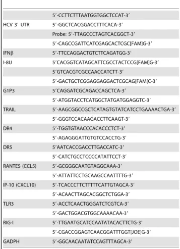
Figure 1. IFN response is dependent on viral replication.A) IFNbmRNA expression was measured daily from the total RNA of LH86 cells
treated with an MOI of 0.1 of HCV/JFH-1. The expression was calculated by theDDCt method where uninfected cells were the experimental control
and the housekeeping gene GAPDH was the internal control. Error bars represent the SEM of three separate experiments. The ‘‘No virus’’ control indicates cells that were cultured with uninfected Huh7.5 supernatant, ‘‘HCV’’ is the supernatant from infected Huh7.5 cells as described in the
methods section (MOI = 0.1), ‘‘heated HCV’’ is the same supernatant as ‘‘HCV’’ but heated for 15 minutes at 72uC and ‘‘UV-treated HCV’’ was exposed
95uC, 1 min. All genes were analyzed through the 2-DDCtmethod following previously described calculations [22].
HCV copies were determined using a standard curve of JFH-1 full-length RNA transcribedin vitro. PCRs were run in duplicates and overall data represents triplicate experiments and are represented as means 6SEM. The Taqman FAM-labeled primer-probe set used in our experiments was originally designed for HCV genotype 1. Compared to genotype 2a there is a mismatch in the forward primer in the ninth nucleotide (59to 39). However, this primer set was demonstrated in our laboratory to be highly efficient, even more so than our previously published 59UTR primer when used with in vitro transcribed JFH-1. Furthermore, most of the experiments carried here were
performed initially with both primers and similar results were obtained.
Immunofluorescence
Cells were grown on glass coverslips later to be washed and fixed on ice with 5% acetic acid in 100% ethanol (Fisherbrand). Cells were washed with 1X PBS and incubated with either goat anti-human TLR3 (Santa Cruz Biotechnology), goat anti-human TLR7 (Santa Cruz Biotechnology), rabbit anti-human RIG-I (ProSci Inc, Poway, CA) or mouse anti-HCV NS5A monoclonal antibody (established at our institution) for 1 hour. The respective secondary antibody (donkey anti-goat IgG FITC conjugated for TLR3 and TLR7, goat anti-rabbit IgG FITC conjugated, goat
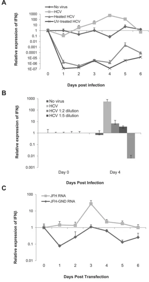
Figure 2. Both TLRs and RIG-I prevent viral replication and are needed for the induction of IFN.A) IFNbmRNA expression at day 4
post-infection in LH86 cells after transfecting with a control siRNA, or siRNA against TLR3 or TLR7. Expression was calculated by theDDCt method where
uninfected cells were the experimental control and the housekeeping gene GAPDH was the internal control. Error bars represent the SEM of three separate experiments. No virus indicates cells were cultured with the same volume in uninfected Huh7. 5 supernatant, HCV is the supernatant from
infected Huh7.5 cells as described in the methods section (MOI = 0.1). B) IFNbmRNA expression at day 4 post-infection in Huh7.5 cells expressing
TLR3 or TLR7. Expression was calculated by theDDCt method as described in part A and bars represent the SEM of three separate experiments. C)
Viral replication in Huh7.5 cells expressing TLR3 or TLR7 at day 7 after infection. HCV copy numbers were calculated by real time RT-PCR run with an
HCV standard curve. D) IFNbmRNA expression at day 4 post-infection in LH86 cells with silenced RIG-I. Methodology as described in part A. E) IFNb
mRNA expression at day 4 post-infection in Huh7.5 cells transfected with RIG-I. Methodology as described in part A. F) Viral replication in Huh7.5 cells transfected with RIG-I after 7 days of culture. Methodology as described in part C.
doi:10.1371/journal.pone.0021186.g002
to UV light for 15 minutes. B) IFNbmRNA expression of different dilutions of virus (1:1 MOI = 0.1) from day 0 and day 4 post infection calculated by the
DDCt method (determined as in part A). C) IFNbmRNA expression of LH86 cells electroporated within vitrotranscribed HCV/JFH-1 or its
anti-mouse IgG FITC) were purchased from Southernbiotech (Birmingham, AL). Nuclei were counterstained with DAPI (Vector Laboratories Inc, Burlingame, CA), followed by examination under a fluorescence microscope (Olympus).
Western blot analysis
Cells were washed with PBS and lysed in RIPA buffer (150 mM sodium chloride, 1% Nonidet P-40, 0.5% sodium deoxycholate, 0.1% SDS, and 50 mM Tris-Cl [pH 8.0] supplemented with 2mg/mL of aprotinin, 2mg/mL of leupeptin, 40mg/mL of phenylmethylsulfonyl fluoride, and 2 mM DTT). Twenty mg of
protein was electrophoresed on a 10% SDS-polyacrylamide gel and transferred to a polyvinylidene difluoride membrane (Bio-Rad, Hercules CA). The membranes were then blocked overnight at 4uC in blocking buffer (PBS containing 0.1% Tween 20 (PBS-T) and 5% fat-free milk power) followed by probing with each specific primary antibody (see immunofluorescence section for primary antibodies and mouse anti-human b-actin from Santa Cruz Biotechnology) for one hour at room temperature. After washing thrice with PBS-T for 10 minutes each, the membrane was incubated with the appropriate HRP-conjugated secondary antibody (all from Santa Cruz Biotechnology), diluted in PBS-T, for 1 hour at room temperature and washed 3 more times. Proteins were visualized with Supersignal West Pico Chemilumi-nescent Substrate (Pierce Biotechnology, Inc, Rockford, IL).
Flow Cytometry
Pelleted cells were incubated for 30 min at room temperature with antibodies (as in immunofluorescence section), washed with staining buffer (PBS+2% BSA+0.1% Na azide), and then fixed with 2% paraformaldehyde and stored at 4uC. For intracellular staining the BDcytofix/cytoperm kit (BD biosciences) was used with the antibodies following the manufacturer’s protocol. Cells were analyzed on a FACSCalibur flow cytometer (BD Biosciences, Heidelberg, Germany). Quantitation was done by the CellQuest software (BD Biosciences; version 3.2.1). Isotype-specific immu-noglobulin controls were run for each fluorochrome. Thirty thousand cells were analyzed for each sample.
The Annexin V-FITC apoptosis was assayed as described previously (BD Pharmingen, San Diego, CA) [23]. A minimum of 30,000 events per sample were acquired on a FACSCalibur Flow Cytometer (BD Pharmingen, San Diego, CA) and subsequently analyzed with CellQuest software (BD Biosciences; version 3.2.1, San Diego, CA).
Results
HCV induction of IFN in LH86 cells depends on active viral replication
We recently reported the induction of IFNb by the hepatoma cell line LH86 in response to HCV [16]. Hence our first experiment was to corroborate that the IFN induced by LH86 cells leads to a functional response to the virus. We measured the expression of two interferon-stimulated-genes (ISGs) by Real time
RT-PCR: 1-8U and G1P3 in these cells with or without viral infection by Real time RT-PCR. The experiment showed a fully functional IFN response with the production of both these ISGs. We then analyzed the expression of TLR3, TLR7 and RIG-I in LH86. Immunofluorescence and flow cytometry demonstrated a strong expression of RIG-I and TLR3 as well as a mild expression of TLR7 in these cells which was similar to primary hepatocytes, which were stained for comparison (data not shown). Therefore we used this system to examine if IFNb expression correlates with functional HCV. We infected LH86 cells with HCV strain JFH-1 (genotype 2a) at a MOI of 0.1 or with the same amount of virus which was either heated at 72uC or exposed directly to UV light for 15 minutes. As seen in Figure 1A, induction of IFNbexpression correlated with intact virus, since inactivating the virus by heating or UV treatment down-regulated IFNbexpression.
In order to further understand the kinetics of this interaction we cultured LH86 cells with different dilutions of the virus. The data showed that IFN induction was titer-dependent (Figure 1B). To corroborate that viral replication, and not only RNA itself, is responsible for the induction of IFN, we electroporated LH86 cells within vitrotranscribed JFH-1 RNA or JFH-1/GND RNA. The JFH-1/GND virus has a mutation in its RdRp which prevents its replication but it is otherwise the same as JFH-1 [18]. We found that JFH-1, and not JFH-1/GND, induced IFNb in LH86 cells indicating that there is a dependence on replication for its induction (Figure 1C). Interestingly, the basal levels of IFN after transfecting JFH-1/GND was comparable to LH86 cells by themselves (Compare Figure 1C with 1A) and we observed no down-regulation as we did with full virus (genetic material plus virion).
Both TLRs and RIG-I play a role in the induction of IFN by HCV
The role of TLRs and RIG-I in directly recognizing HCV or modulating its replication has not been fully characterized. We decided to do that by using a comparative cell culture system based on the distinct innate immune phenotype of two hepatoma cell lines: LH86 and Huh7.5. The first, LH86 cells, are a well-differentiated hepatoma cell line that has a fully functional set of PRRs and is capable of inducing IFN in response to HCV infection [16]. Conversely, Huh7.5 cells do not express TLR3 or TLR7, and have a RIG-I mutation, which may be a reason why these cells are so permissive to viral replication [3,24,25]. To test this hypothesis, we first silenced TLR3 and TLR7 in LH86 cells using specific siRNAs. As shown in Figure 2A, silencing these receptors abrogated the IFN response in LH86 cells. Conversely, transfecting TLR3 or TLR7 into Huh7.5 cells, followed by viral infection, rendered the cells capable of IFN induction (Figure 2B). Furthermore, forced expression of the same receptors into Huh7.5 cells significantly inhibited viral replication levels in these normally permissive cells (Figure 2C). Interestingly, the expression of RIG-I did not show the same phenomenon. While removal of RIG-I from LH86 cells significantly abrogated the IFNb mRNA expression, delivering it into Huh7.5 cells did not increase IFN
Figure 3. RIG-I is linked to the expression of TRAIL receptors DR4 and DR5.A) TRAIL mRNA expression 4 days after infection of Huh7.5 cells
expressing RIG-I, TLR3 or TLR7. Expression was calculated by theDDCt method where uninfected cells were the experimental control and the
housekeeping gene GAPDH was the internal control. Error bars represent the SEM of three separate experiments. B) DR4 mRNA expression 4 days after infection of Huh7.5 cells expressing RIG-I, TLR3 or TLR7 following the methodology described in A. C) DR5 mRNA expression 4 days after infection of Huh7.5 cells expressing RIG-I, TLR3 or TLR7 following the methodology described in part A. D) TRAIL mRNA expression 4 days after infection of LH86 cells silenced for RIG-I, TLR3 or TLR7 calculated as described for part A. E) DR4 mRNA expression 4 days after infection of LH86 cells with silenced RIG-I, TLR3 or TLR7 calculated as described for part A. F) DR5 mRNA expression 4 days after infection of LH86 cells with silenced RIG-I, TLR3 or TLR7 calculated as described for part A. G) Phase view (100X magnification) of transfected cells 7 days after infection. Top row are LH86 cells and bottom row Huh7.5 cells. The labels note what each cell was transfected with.
production to the same fold difference as LH86 cells or to the levels of Huh7.5 cells transfected with TLRs (150 versus 2 fold and 14 versus 2 fold respectively, Figures 2D and 2E) although it also considerably reduced the level of viral replication (Figure 2F).
Cell death through RIG-I in response to HCV is linked to the TRAIL pathway
Our previously published results showed that cytopathic effects in LH86 are related to the induction of TRAIL and its receptors DR4 and DR5, therefore we decided to investigate the relationship between the PRRs and the expression of these three genes [16]. As seen in figure 3, an interesting pattern appeared: forced expression of the PRRs in Huh7.5 cells increased the expression of the three genes studied (TRAIL, DR4, and DR5 in figures 3A, 3B and 3C respectively), but only silencing RIG-I, and not TLRs, had a profound effect on the expression of DR4 and DR5 after HCV infection (Figure 3D, 3E and 3F). Furthermore, RIG-I transfected Huh7.5 cells had visibly increased cell death comparable to the susceptibility of LH86 cells to HCV infection (Figure 3G).
TLR3 and TLR7 suppress HCV RNA replication in hepatoma cells
To determine the functional role of TLR3 and TLR7, we examined the relationship between viral replication and the IFN response in a long-term culture system. The cells were infected with HCV-JFH1 virus and samples were collected every 2–3 days up to 75 days. Viral RNA replication levels were determined by real-time RT-PCR analysis. As shown in Figure 4, both TLR3 and TLR7 had a profound effect on viral replication throughout the time of analysis. The cells with both TLR3 and TLR7 suppressed HCV replication, while TLR3 has the most profound effect (Figure 4A). To test whether this antiviral effect correlates with an IFN response, we quantified IFNb expression by real time RT-PCR. As shown in Figure 4B, over-expressing TLR3 in Huh7.5 cells induced a strong IFN production only in the first week, while TLR7 induced a very low level of IFN. TLR3 has at least two potential signaling pathways: NF-kB or IRF-3 cascades. To determine which pathway is operative under these conditions, we measured the expression of cytokines specific for each pathway (RANTES which is IRF-3 induced and IP-10 which is NF-kB induced) [26,27,28]. We observed that IP-10 was down-regulated in Huh7.5 TLR3 cells throughout the culture while RANTES was induced and sustained throughout the culture period indicating that the down regulation of viral replication correlates with the induction of the IRF-3 pathway without apparent NF-kB activation (Figure 4C).
HCV can down-regulate the expression of TLR3 and RIG-I in LH86 cells
Since TLR3 and RIG-I have different functions, we hypothe-sized that their combined activation could lead to viral clearance. To test this hypothesis, we co-transfected the Huh7.5 TLR3 and TLR7 stable cell lines with RIG-I. We also did co-transfections of TLR3 and TLR7 cells with PKR, a different cytosolic receptor. While TLR7 did synergize with RIG-I to induce a strong IFN
response, the TLR3/RIG-I combination did not show this effect. In these cells, contrary to our hypothesis, HCV infection down regulated IFN (Figure 5A). Furthermore, these effects are specific to TLR/RIG-I expressions in HCV infection since co-transfecting with PKR did not synergize or disrupt the IFN normally induced by either cell.
It has been reported that NS3/4A modulates TLR and RIG-I pathways at the level of the adaptor proteins TRIF and IPS-1 [24,29]. Interestingly, studies have also demonstrated that expression of TLR3 and RIG-I are also reduced in HCV patients, therefore we decided to examine if there is a correlation between the expression of these receptors and virus infection. We measured the levels of both PRRs after infection with different dilutions of the virus. As shown in Figure 5B, HCV was able to reduce the level of TLR3 and RIG-I in a titer dependent manner. Furthermore, the virus proteins needed to be intact for TLR3 down regulation, since heating the virus prevented this down-regulation; similarly, RIG-I down regulation required intact virus but it also needed replicating virus since UV-inactivation abrogated the down-regulation of RIG-I, but not TLR3 (Figure 5C).
HCV envelope proteins down regulate the expression of TLR3 and RIG-I
The observations just described combined with our initial evidence (Figure 1A) demonstrates that non-replicating virus can down-regulate IFN which indicates there is likely a role by virion proteins in immune-evasion. To understand how these HCV proteins can impact the innate immune system we created stable cell lines with LH86 expressing three viral proteins: core, envelope (E1/E2) and NS3/4A (already known to affect IFN expression) [30]. These stable cell lines were infected with HCV at an MOI of 0.1 as before and the IFN response was measured. As expected, LH86 cells transfected with NS3/4A had a reduced IFN peak at day 4 as compared to control cells (Figure 6A). In contrast, LH86 cells transfected with core or envelope did not induce IFN. Core-transfected cells did have an earlier peak (not shown) but by day 4 IFN was no different than uninfected cells. Conversely, LH86 cells that harbored the envelope proteins did not express IFN at any time point during a 7 day culture and at day 4 the basal IFN expression on those cells was down regulated as well. Furthermore, these transfected cells were able to down-regulate the levels of TLR3 (Figure 6B) and RIG-I (Figure 6C) but only the envelope-transfected cells maintained the down-regulation of RIG-I after HCV infection.
Our data indicates that virion proteins are involved in evasion through the down-regulation of the expression of TLR3 and RIG-I. We also show that envelope proteins have bigger role in this evasion than other viral proteins tested. To corroborate the role of envelope in affecting the function of these receptors examined the kinetics of IFN induction in LH86 cells, with or without transfected envelope proteins, after Poly I:C stimulation. It is known that this ligand stimulates both RIG-I (by transfection of poly I:C) and TLR3 (when added directly to the media). Both transfected poly I:C (Figure 6D) and extracellular poly I:C (Figure 6E) were able to induce IFN in the absence of E1E2
Figure 4. A strong initial IFN response is induced an IRF-3 response by TLR3 but is not enough to clear HCV.A) Viral replication in Huh7.5 cells stably transfected with TOPO (control) TLR3 or TLR7 infected with HCV MOI of 0.1 and collected every 2–3 days for RNA isolation (total 75
days). The HCV copy numbers from each time points were calculated by real time RT-PCR and compared against an HCV standard curve. B) IFNb
mRNA expression of the experiment described in part A. Expression was calculated by the DDCt method where uninfected cells were the
experimental control and the housekeeping gene GAPDH was the internal control. Error bars represent the SEM of three separate experiments. C) IP-10 and RANTES mRNA expression of the experiment described in part A. Methodology as described for part B.
protein in these cells. However, E1E2 affected the IFN response in LH86 cells stimulated with extracellular poly I:C for one day, while it completely abrogated the IFN response to transfected poly I:C. These results show that E1E2 can interfere with the RIG-I pathway, but only modestly interfere with TLR3-mediated IFN induction pathway.
Discussion
The establishment of newin vitromodels for the study of HCV has introduced some of the current concepts of how it interacts with the immune response and provided insight into these mechanisms. In the present study we observed that initial HCV’s replication correlates strongly with the production of IFNb in hepatocytes and that these responses are linked to the recognition of HCV by TLR3, and RIG-I. All these molecules have been strongly implicated as targets of viral evasion [24,29,31]. Previous studies have demonstrated that both TLR3 and RIG-I pathways are direct targets of NS3/4A serine protease of HCV. Further-more, both receptors are: active in normal liver cells, known to induce type I IFN, share many of the same pathways and are capable of up-regulating each other [3,32,33,34,35,36]. Studies have shown that either TLR3 or RIG-I is required or that both produce the same effect [33,37]. Our data shows that different stimuli can induce distinct effects. While studying their mecha-nisms we demonstrated that TLR3 and RIG-I have specific roles during HCV infection: TLR3 was strongly involved in the induction of IFNs and creating an IRF-3 dependent environment that helps keep the viral replication low. RIG-I, on the other hand, can also involve the induction of cellular apoptosis through the TRAIL pathway and the death receptors DR4 and DR5. In addition, we also uncovered a potential mechanism of evasion by the viral envelope protein. Our data shows an inverse correlation on the presence of viral envelope proteins in hepatocytes with a decrease in the mRNA expression of both TLR3 and RIG-I. Furthermore, the stable expression of the envelope protein prevented the stimulation of RIG-I by poly (I:C) in the cytosol.
We showed a direct correlation between viral titers and levels of IFNb suggesting that a threshold needs to be reached before an appropriate response is developed. This corroborates previously published data that also shows a delayed IFN response in HCV infection [34]. They also showed that TLR3’s role in HCV was to induce IFN, IRF-3 activation and ISG induction in HCV-infected liver cells bothin vitroand in clinical samples which correlates with our findings [3,34]. Furthermore, our data suggests that the amount of IFN induced by HCV depends on the expression levels of TLR3 itself at the time of infection.
Based on our study, it is likely that the combination of both receptors should be optimal for the clearance of HCV infection. Our data demonstrates that RIG-I’s main role may lie in the induction of apoptosis a function which was recently demonstrated for helicases [37,38,39,40,41]. We linked the induction of TRAIL-induced apoptosis in HCV to RIG-I and in particular to DR4 and DR5. This effect is usually attributed in other infections to TLR3 but although both TLR and RIG-I were able to up-regulate TRAIL, DR4 and DR5, when present, only the latter had a profound effect in the expression level of both death receptors. In general our findings can have strong implications in the study of the consequences of chronic infection since cytopathic effects have been implicated in the pathogenesis of HCV-induced liver injury in a TRAIL-dependent manner [42,43,44,45]. Furthermore, clinically the levels of TRAIL are up regulated in chronic patients which correlate the availability of TRAIL with the induction of liver damage [46,47].
Figure 5. The expression of TLR3 and RIG-I is affected by intact virion proteins in hepatocytes A) IFNbmRNA expression in Huh7.5 TLR3 or TLR7 stable cell lines co-transfected with either PKR or RIG-I after
infection with HCV MOI = 0.1. Expression was calculated by theDDCt
method where uninfected cells were the experimental control and the housekeeping gene GAPDH was the internal control. Error bars represent the SEM of three separate experiments. B) TLR3 and RIG-I mRNA expression levels 7 days after infection with different HCV dilutions (methodology as in figure 1C). C) TLR3 and RIG-I gene expression levels 7 days after infection with normal, heated or UV-treated virus calculated as described in part A. doi:10.1371/journal.pone.0021186.g005
Interestingly, the expression of both receptors in Huh7.5 cells disrupted the induction of IFNbseen with either receptor alone. This observation argues that the virus might exert certain pressure when they are in combination since this combination of receptors has been demonstrated to induce IFN in the absence of HCV [33,48]. In these studies, the authors observed no synergistic effect between the receptors which led them to argue that there was no direct interaction with each other. Our overall data supports the idea that these two receptors may interact with each other and that they have specific functions in HCV infection (Figure 7). This discrepancy made us investigate the role of HCV on the levels of these receptors. We observed that the expression levels of TLR3 and RIG-I were down regulated after infection in LH86 cells. Our finding correlates with clinical
observation, which shows that the levels of these receptors in patients are down regulated [5]. In our system those effects were mediated by intact viral proteins, in particular the virion proteins: core and envelope. These results correlate well with our recent studies on healthy myeloid-derived dendritic cells where virus did not replicate but was able to induce phenotypical changes as well as modulate the cytokine secretion [49]. Similarly, a recent report on HCV in vitro studies demonstrated that viral infection can specifically down regulate another receptor, TLR7 and envelope proteins have been directly linked to the blocking of the TRAIL pathway which could be mechanistically explained by the down regulation of RIG-I [16,50,51,52]. This further supports our theory of the initial interaction of the virus with its host being dependent on
Figure 6. Envelope proteins affect the response to non-HCV responses through RIG-I receptor but only temporarily through TLR3.
A) IFNbgene expression of stably transfected LH86 cells expressing Core, E1E2 or NS3/4A 4 days after HCV infection. Expression was calculated by the
DDCt method where uninfected cells were the experimental control and the housekeeping gene GAPDH was the internal control. Error bars
represent the SEM of three separate experiments. B) TLR3 mRNA expression 7 days after infection of stably transfected LH86 cells carrying the HCV proteins Core, E1/E2 or NS3/4A. Expression determined as described in part A. E) RIG-I mRNA expression 7 days after infection of stably transfected
LH86 cells carrying HCV proteins Core, E1/E2 or NS3/4A. Expression determined as described in part A. D) IFNbmRNA expression of LH86 cells or LH86
stably transfected with E1/E2 treated with 50mg/mL transfected Poly I:C. Expression was calculated as described in part A. E) IFNbmRNA expression of
LH86 cells or LH86 stably transfected with E1/E2 treated with 50mg/mL extracellular Poly I:C. Expression determined as described in part A.
the amount of virus present either preventing or delaying immune responses (Figure 7). Further studies would reveal if this correlation is indeed an evasion strategy of HCV and what might be the mechanism for this phenomenon.
In summary, we have characterized the interaction of HCV with innate immune molecules, TLR3, TLR7, and RIG-I. We found that TLR3 and TLR7 play an important role in controlling viral infection through the IFN production, while RIG-I play a role in IFN induction as well as in the induction of hepatocyte apoptosis. We also found that the presence of viral E1E2 protein correlates with lower TLR3 and RIG-I expression, a possible mechanism of HCV evasion of innate immune response. Beyond IFN, future studies should aim at understanding the specific role of NF-kB and IRF-3 cascades, on why the virus seems to choose one
pathway over the other, and how this might interfere with viral clearance.
Acknowledgments
The authors would like to thank Drs. Takaji Wakita and Charles M. Rice for reagents; they would also like to acknowledge the University of Florida’s ICBR for the use of their confocal microscope and flow cytometers. We also thank members of the Liu laboratory for their help in this study.
Author Contributions
Conceived and designed the experiments: EAE DRN CL. Performed the experiments: EAE HZ LB JB H-yL. Analyzed the data: EAE JB CL. Contributed reagents/materials/analysis tools: HZ DRN. Wrote the paper: EAE JB CL.
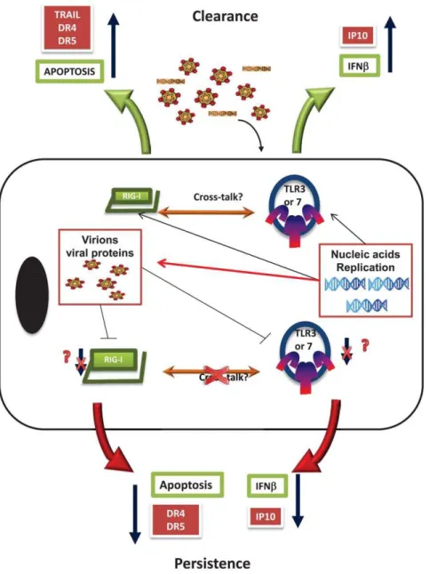
Figure 7. Schematics of the initial interaction of HCV in hepatocytes.Virus binds and gets in the cells were nucleic acid or replication
induces the host cells innate immunity through the induction of apoptosis (mediated by RIG-I) and IFNb(through TLR3 engagement). While
the replication of the virus leads to the development of viral proteins like NS34A that interact downstream of these effects (red arrow line) there is a possible earlier evasion strategy performed by virion proteins that lead to the down regulation of RIG-I and TLR3 preventing
apoptosis and the induction of IFNb. Apoptosis is particularly prevented by down regulating TRAIL receptors, DR4 and DR5, and IFNbby
preventing NF-kB pathways such as the one that induces IP-10. Together all of these factors help the cell survive and the virus to persist in the
host’s hepatocytes.
doi:10.1371/journal.pone.0021186.g007
References
1. Liang TJ, Rehermann B, Seeff LB, Hoofnagle JH (2000) Pathogenesis, natural history, treatment, and prevention of hepatitis C. Ann Intern Med 132: 296–305.
2. Shepard CW, Finelli L, Alter MJ (2005) Global epidemiology of hepatitis C virus infection. Lancet Infect Dis 5: 558–567.
3. Kanda T, Steele R, Ray R, Ray RB (2007) Hepatitis C virus infection induces the beta interferon signaling pathway in immortalized human hepatocytes. J Virol 81: 12375–12381.
4. Thomas A, Laxton C, Rodman J, Myangar N, Horscroft N, et al. (2007) Investigating Toll-like receptor agonists for potential to treat hepatitis C virus infection. Antimicrob Agents Chemother 51: 2969–2978.
5. Atencia R, Bustamante FJ, Valdivieso A, Arrieta A, Rinon M, et al. (2007) Differential expression of viral PAMP receptors mRNA in peripheral blood of patients with chronic hepatitis C infection. BMC Infect Dis 7: 136. 6. Villacres MC, Literat O, DeGiacomo M, Du W, Frederick T, et al. (2008)
Defective response to Toll-like receptor 3 and 4 ligands by activated monocytes in chronic hepatitis C virus infection. J Viral Hepat 15: 137–144.
7. Iwasaki A, Medzhitov R (2004) Toll-like receptor control of the adaptive immune responses. Nat Immunol 5: 987–995.
8. Malmgaard L (2004) Induction and regulation of IFNs during viral infections. J Interferon Cytokine Res 24: 439–454.
9. Servant MJ, Grandvaux N, Hiscott J (2002) Multiple signaling pathways leading to the activation of interferon regulatory factor 3. Biochem Pharmacol 64: 985–992.
10. Kawai T, Akira S (2006) Innate immune recognition of viral infection. Nat Immunol 7: 131–137.
11. Hiscott J, Lin R, Nakhaei P, Paz S (2006) MasterCARD: a priceless link to innate immunity. Trends Mol Med 12: 53–56.
12. Seth RB, Sun L, Ea CK, Chen ZJ (2005) Identification and characterization of MAVS, a mitochondrial antiviral signaling protein that activates NF-kappaB and IRF 3. Cell 122: 669–682.
13. Barton GM, Medzhitov R (2004) Toll signaling: RIPping off the TNF pathway. Nat Immunol 5: 472–474.
14. Natoli G, Austenaa LM (2008) A birthday gift for TRADD. Nat Immunol 9: 1015–1016.
15. Thompson AJ, Locarnini SA (2007) Toll-like receptors, RIG-I-like RNA helicases and the antiviral innate immune response. Immunol Cell Biol 85: 435–445.
16. Zhu H, Dong H, Eksioglu E, Hemming A, Cao M, et al. (2007) Hepatitis C virus triggers apoptosis of a newly developed hepatoma cell line through antiviral defense system. Gastroenterology 133: 1649–1659.
17. Lindenbach BD, Evans MJ, Syder AJ, Wolk B, Tellinghuisen TL, et al. (2005) Complete replication of hepatitis C virus in cell culture. Science 309: 623–626. 18. Wakita T, Pietschmann T, Kato T, Date T, Miyamoto M, et al. (2005) Production of infectious hepatitis C virus in tissue culture from a cloned viral genome. Nat Med 11: 791–796.
19. Zhu H, Nelson DR, Crawford JM, Liu C (2005) Defective Jak-Stat activation in hepatoma cells is associated with hepatitis C viral IFN-alpha resistance. J Interferon Cytokine Res 25: 528–539.
20. Zhu H, Butera M, Nelson D, Liu C (2005) Novel type I interferon IL-28A suppresses hepatitis C viral RNA replication. Virol J 2: 80.
21. Zhu H, Shang X, Terada N, Liu C (2004) STAT3 induces anti-hepatitis C viral activity in liver cells. Biochem Biophys Res Commun 324: 518–528. 22. Livak KJ, Schmittgen TD (2001) Analysis of relative gene expression data using
real-time quantitative PCR and the 2(-Delta Delta C(T)) Method. Methods 25: 402–408.
23. Shang XZ, Zhu H, Lin K, Tu Z, Chen J, et al. (2004) Stabilized beta-catenin promotes hepatocyte proliferation and inhibits TNFalpha-induced apoptosis. Lab Invest 84: 332–341.
24. Li K, Foy E, Ferreon JC, Nakamura M, Ferreon AC, et al. (2005) Immune evasion by hepatitis C virus NS3/4A protease-mediated cleavage of the Toll-like receptor 3 adaptor protein TRIF. Proc Natl Acad Sci U S A 102: 2992–2997. 25. Sumpter R, Jr., Loo YM, Foy E, Li K, Yoneyama M, et al. (2005) Regulating intracellular antiviral defense and permissiveness to hepatitis C virus RNA replication through a cellular RNA helicase, RIG-I. J Virol 79: 2689–2699. 26. Lin R, Heylbroeck C, Genin P, Pitha PM, Hiscott J (1999) Essential role of
interferon regulatory factor 3 in direct activation of RANTES chemokine transcription. Mol Cell Biol 19: 959–966.
27. Zorde-Khvalevsky E, Abramovitch R, Barash H, Spivak-Pohis I, Rivkin L, et al. (2009) Toll-like receptor 3 signaling attenuates liver regeneration. Hepatology 50: 198–206.
28. Yeruva S, Ramadori G, Raddatz D (2008) NF-kappaB-dependent synergistic regulation of CXCL10 gene expression by IL-1beta and IFN-gamma in human intestinal epithelial cell lines. Int J Colorectal Dis 23: 305–317.
29. Meylan E, Curran J, Hofmann K, Moradpour D, Binder M, et al. (2005) Cardif is an adaptor protein in the RIG-I antiviral pathway and is targeted by hepatitis C virus. Nature 437: 1167–1172.
30. Bowie AG, Unterholzner L (2008) Viral evasion and subversion of pattern-recognition receptor signalling. Nat Rev Immunol 8: 911–922.
31. Kaukinen P, Sillanpaa M, Kotenko S, Lin R, Hiscott J, et al. (2006) Hepatitis C virus NS2 and NS3/4A proteins are potent inhibitors of host cell cytokine/ chemokine gene expression. Virol J 3: 66.
32. Miettinen M, Sareneva T, Julkunen I, Matikainen S (2001) IFNs activate toll-like receptor gene expression in viral infections. Genes Immun 2: 349–355. 33. Yoneyama M, Kikuchi M, Natsukawa T, Shinobu N, Imaizumi T, et al. (2004)
The RNA helicase RIG-I has an essential function in double-stranded RNA-induced innate antiviral responses. Nat Immunol 5: 730–737.
34. Wang N, Liang Y, Devaraj S, Wang J, Lemon SM, et al. (2009) Toll-like receptor 3 mediates establishment of an antiviral state against hepatitis C virus in hepatoma cells. J Virol 83: 9824–9834.
35. Schroder M, Bowie AG (2005) TLR3 in antiviral immunity: key player or bystander? Trends Immunol 26: 462–468.
36. Nishimura M, Naito S (2005) Tissue-specific mRNA expression profiles of human toll-like receptors and related genes. Biol Pharm Bull 28: 886–892. 37. Kawai T, Akira S (2008) Toll-like receptor and RIG-I-like receptor signaling.
Ann N Y Acad Sci 1143: 1–20.
38. MacParland SA, Pham TN, Guy CS, Michalak TI (2009) Hepatitis C virus persisting after clinically apparent sustained virological response to antiviral therapy retains infectivity in vitro. Hepatology 49: 1431–1441.
39. Garcia M, Dogusan Z, Moore F, Sato S, Hartmann G, et al. (2009) Regulation and function of the cytosolic viral RNA sensor RIG-I in pancreatic beta cells. Biochim Biophys Acta 1793: 1768–1775.
40. Kang DC, Gopalkrishnan RV, Lin L, Randolph A, Valerie K, et al. (2004) Expression analysis and genomic characterization of human melanoma differentiation associated gene-5, mda-5: a novel type I interferon-responsive apoptosis-inducing gene. Oncogene 23: 1789–1800.
41. Kang DC, Gopalkrishnan RV, Wu Q, Jankowsky E, Pyle AM, et al. (2002) mda-5: An interferon-inducible putative RNA helicase with double-stranded RNA-dependent ATPase activity and melanoma growth-suppressive properties. Proc Natl Acad Sci U S A 99: 637–642.
42. Mengshol JA, Golden-Mason L, Rosen HR (2007) Mechanisms of Disease: HCV-induced liver injury. Nat Clin Pract Gastroenterol Hepatol 4: 622–634. 43. Bantel H, Schulze-Osthoff K (2003) Apoptosis in hepatitis C virus infection. Cell
Death Differ 10 Suppl 1: S48–58.
44. Lai MM (2002) Hepatitis C virus proteins: direct link to hepatic oxidative stress, steatosis, carcinogenesis and more. Gastroenterology 122: 568–571. 45. Afford SC, Adams DH (2005) Following the TRAIL from hepatitis C virus and
alcohol to fatty liver. Gut 54: 1518–1520.
46. Mundt B, Wirth T, Zender L, Waltemathe M, Trautwein C, et al. (2005) Tumour necrosis factor related apoptosis inducing ligand (TRAIL) induces hepatic steatosis in viral hepatitis and after alcohol intake. Gut 54: 1590–1596. 47. Mundt B, Kuhnel F, Zender L, Paul Y, Tillmann H, et al. (2003) Involvement of
TRAIL and its receptors in viral hepatitis. Faseb J 17: 94–96.
48. Li K, Chen Z, Kato N, Gale M, Jr., Lemon SM (2005) Distinct poly(I-C) and virus-activated signaling pathways leading to interferon-beta production in hepatocytes. J Biol Chem 280: 16739–16747.
49. Eksioglu EA, Bess JR, Zhu H, Xu Y, Dong HJ, et al. (2009) Hepatitis C virus modulates human monocyte-derived dendritic cells. J Viral Hepat.
50. Weber F (2007) Interaction of hepatitis C virus with the type I interferon system. World J Gastroenterol 13: 4818–4823.
51. Lee SH, Kim YK, Kim CS, Seol SK, Kim J, et al. (2005) E2 of hepatitis C virus inhibits apoptosis. J Immunol 175: 8226–8235.