BJRS
RADIATION SCIENCES
07-03 (2019) 01-17ISSN: 2319-0612 Accepted: 2019-05-27
Evaluation of cytotoxicity through MTS test of 2-ACBs
(2-dDCB AND tDCB) after exposure to hepatic cells –
Studies
in vitro
Barbezan
a, A. B., Carvalho
a, L. R., Vieira
a, D. P., Machado-Santelli
b, G. M.,
Martins
a, R., Villavicêncio
a, A. L. C. H.
aIPEN-CNEN / SP (Nuclear and Energy Research Institute, National Nuclear Energy Commission), Department
CTR, 03178-200, São Paulo, SP, Brazil
bUniversidade de São Paulo, Department of Cell Biology and Development of the Institute of Biomedical Sciences,
03178-200, São Paulo, SP, Brazil Author contact: [email protected]
ABSTRACT
Food irradiation has been approved in more than 60 countries for many applications in a wide variety of foods. 2-Alkylcyclobutanones (2-ACBs) are the only known radiolytic products formed when foods containing fatty acids are irradiated. Despite the importance of food irradiation, the toxicological potential of 2-ACBs in irradiat-ed food is still no fully understood. In this study we investigatirradiat-ed the cytotoxic effects of irradiatirradiat-ed palmitic and stearic fatty acids byproducts, 2-dDCB and 2-tDCB, in hepatic cell lines (HepG2, BRL3A and HTC). The cyto-toxic effects of 2-dDCB and 2-tDCB were evaluated at 100, 300 and 500 μM for 24 and 48 hours and the cell viability was measured using the MTS assay. While no toxicity was observed for 2-tDCB in all cells for all tested conditions, 2-dDCB was found to be toxic to BRL3A cells (at 100 µM after 48 hours) and HTC cells (at 24 hours in all tested concentrations). HepG2 cells on the other hand, were found to be resistant to 2-dDCB-induced tox-icity. Overall our data shows that the byproduct 2-tDCB is not toxic for hepatic cells while 2-dDCB can be used has a potential marker for food irradiation-induced toxicity.
1. INTRODUCTION
1.1 Food IrradiationFood irradiation is usually performed with X-rays from an electron accelerator, accelerated elec-tron beams, or with the gamma radiation generated by the radioactive isotopes Cobalt-60 and Ce-sium-137 (60Co or 137Cs) (Kim et al., 2004).
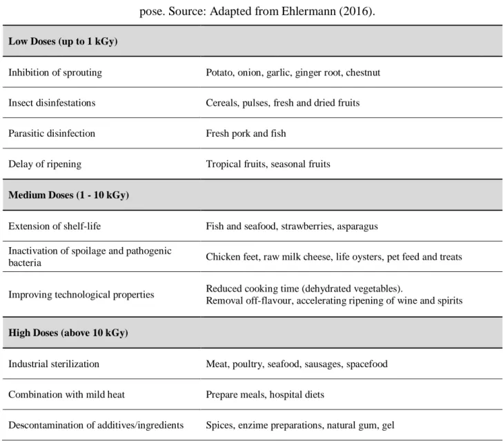
Depending on the absorbed dose of radiation, several effects can be achieved resulting in reduced storage losses, increased shelf life and improved food safety, reducing the biological load of pathogenic microorganisms possibly present in raw foods (Farkas et al., 2014). TAB. 1 presents the recommended doses of radiation used for food irradiation.
1.1 2-Alkylcyclobutanones (2-ACBs)
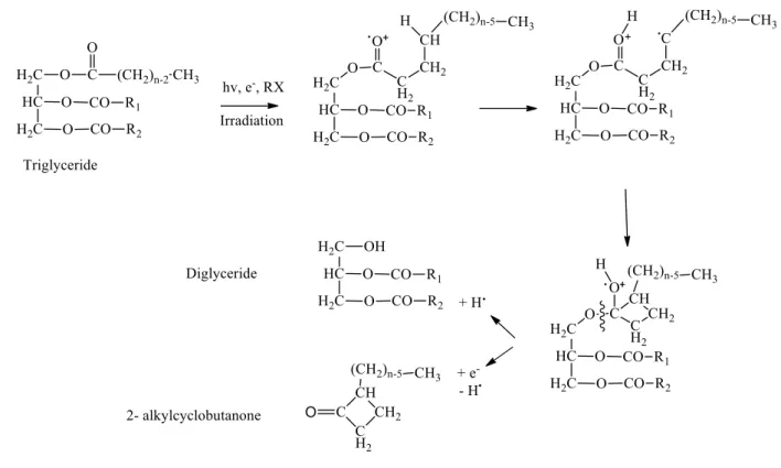
Alkylcyclobutanones (2-ACBs) are the only radiolytic products formed from irradiated foods containing triglycerides (fatty acids). 2-ACBs were first detected by Letellier and Nawar in 1972 in irradiated foods. At that time, it was found that when C6, C8, C10, C12, C14, C16, and C18-containing triglycerides were irradiated, the 2-alkyl group of alkylcyclobutanones was usually re-placed resulting in the formation of radiolytic products.
2-ACBs are cyclic compounds formed by the loss of an oxygen electron over the carbonyl of a fatty acid or triglyceride, followed by a rearrangement process to produce ionization resulting in the production of highly active free radicals. Palmitic acids, stearic, oleic, and linoleic are the primary fatty acids present in foods that, when subjected to irradiation process, are transformed into 2-dDCB (2-Dodecylcyclobutanone), 2-tDCB (2-Tetradecylcyclobutanone), 2 -tDeCB (2-tetradec-50-enylcyclobutanone), and 2-tDecBB2-tetradec-5'8'-di(2-tetradec-50-enylcyclobutanone), respectively (Song et al., 2014; Gadgil et al., 2002) Figure 1.
Table 1: Radiation doses applied in kGy in specific groups of foods according to the intended pur-pose. Source: Adapted from Ehlermann (2016).
Low Doses (up to 1 kGy)
Inhibition of sprouting Potato, onion, garlic, ginger root, chestnut Insect disinfestations Cereals, pulses, fresh and dried fruits
Parasitic disinfection Fresh pork and fish
Delay of ripening Tropical fruits, seasonal fruits
Medium Doses (1 - 10 kGy)
Extension of shelf-life Fish and seafood, strawberries, asparagus Inactivation of spoilage and pathogenic
bacteria Chicken feet, raw milk cheese, life oysters, pet feed and treats
Improving technological properties Reduced cooking time (dehydrated vegetables).
Removal off-flavour, accelerating ripening of wine and spirits
High Doses (above 10 kGy)
Industrial sterilization Meat, poultry, seafood, sausages, spacefood Combination with mild heat Prepare meals, hospital diets
Figure 1: Formation of 2-ACB from a triglyceride after radiation.
Source: Le Tellier and Nawar, 1972.
The 2-ACBs from irradiated food are usually present in trace amounts (in the range of parts per million). For example, the amount of 2-dDCB derived from palmitic acid, was estimated to be 12.9 μg per 100 g of chicken meat irradiated at 3 kGy (Yamakage et al., 2014).
The main concern regarding the consumption of irradiated foods is the promotion of cancer by 2-ACBs. For example, a study from Delincée and Pool-Zobel (1998) reported a genotoxic effect of ACBs in primary human and rodent colon cells. However, full studies of the possible effects of 2-ACBs on irradiated foods as well as their mechanism(s) of action on tumorigenesis are still needed to address public health concerns. In fact, an in-depth investigation on how the levels of 2-ACBs consumed by humans and animal models can impact the promotion of several types of cancers is extremely important before advising that irradiated foods may or may not promote cancer (Chin-thalapally, 2003).
A variety of short-term tests for toxicological studies have been used to evaluate the genotoxic and cytotoxic potential of food additives and chemicals formed by processing technologies (Tice et al., 2000).
The cytotoxicity tests aim to evaluate and screen the cellular viability using in vitro cell lines to observe cell growth, reproduction and morphological effects, after exposure to various compounds. Cytotoxic assays are the first-choice tests for toxicity evaluation since they are simple, rapid and highly sensitive (Li et al., 2015).
1.3 Cell Viability and 2-ACBs
Several authors have already addressed the cytotoxic effect of 2-ACBs in colon cells using sev-eral different techniques. The cellular viability was evaluated varying the type of compound, con-centrations and incubation times. TAB. 2 compiles all the in vitro cytotoxic studies using the 2-ACBs compounds performed so far. Moreover, it shows that human colon cells such as, HT-29 (de-rived from human colon carcinoma), 29 Clone A (differentiated tumor line de(de-rived from HT-29), LT-97 adenoma cells with preneoplastic characteristics) and mouse colon cells (not mentioned lineage), were mostly used in these studies. Taking into account that 2-ACBs can be deposited in adipose tissues, the use of colon cells can be explained because these cells contact directly with fat and is usually affected by its accumulation.
Table 2 – Authors and year in which the research was performed, compounds used, test of choice, cell line, evaluated concentration, incubation period and % of cell viability. The values referring to
the % of viability are approximate data, since they represent reading of graphic curves where the points were not expressed exactly.
30 min 60 min 90 min 120 min 24h 48h 2-DCB 0,25 mg/ml (1049 µM) 85% 0,50 mg/ml (2097 µM) 65% 0,75 mg/ml (3146 µM) 60% 1,00 mg/ml (4194 µM) 57% 1,25 mg/ml (5243 µM) 55% 0,25 mg/ml (1049 µM) 55% 0,50 mg/ml (2097 µM) 48% 0,75 mg/ml (3146 µM) 45% 1,00 mg/ml (4194 µM) 40% 1,25 mg/ml (5243 µM) 35% 2-tDCB MTT HT29 100 µmol 90% 80% 60% 200 µmol N/O 45% 17% 400 µmol N/O 5% 0% 2-tDeCB MTT HT29 100 µmol 90% 15% 10% 200 µmol 90% 10% 0% 400 µmol 85% 0% 0% MTT HT29 100 µmol <=100% 65% 25% 200 µmol <=100% 15% 0% 400 µmol 100% 5% 0% 2-dDCB MTT HT29 100 µmol <=100% 70% 20% 200 µmol <=100% 5% 0% 400 µmol 100% 0% 0% MTT HT29 100 µmol 95% 65% 40% 200 µmol 95% 15% 5% 400 µmol 95% 5% 0% 2-tDCB MTT 100 µmol <=100% 75% 50% 200 µmol <=100% 45% 15% 400 µmol <=100% 10% 5% 2-tDeCB MTT 100 µmol <=100% 75% 57% 200 µmol <=100% 30% 5% 400 µmol 90% 10% 5% MTT 100 µmol <=100% 55% 55% 200 µmol <=100% 30% 5% 400 µmol 85% 5% 0% 2-dDCB MTT 100 µmol <=100% 65% 30% 200 µmol 100% 25% 0% 400 µmol 95% 0% 0% MTT 100 µmol <=100% 55% 40% 200 µmol <=100% 25% 5% 400 µmol <=100% 23% 0% 2-tDCB WST HT29 100 µmol <=100% 100% <=100% 200 µmol <=100% 100% <=100% 400 µmol <=100% 25% 0% 2-tDeCB WST HT29 100 µmol 95% <=100% <=100% 200 µmol 90% 75% 15% 400 µmol 95% 20% 0% WST HT29 100 µmol <=100% <=100% 100% 200 µmol <=100% <=100% 35% 400 µmol <=100% 50% 5% 2-dDCB WST HT29 100 µmol 100% 100% <=100% 200 µmol <=100% 95% <=100% 400 µmol <=100% 25% 0% WST HT29 100 µmol <=100% <=100% <=100% 200 µmol <=100% 95% 95% 400 µmol <=100% 45% 0%
Author/ Year Compound Test Cell Line Concentration Period / % Viability Delincée at al / 1998 Tripan Blue Rat Cells Tripan Blue Human cells Delincée et al / 2002 2-tDeCB cis- pure 2-tDeCB trans- pure HT29 clone 19A HT29 clone 19A 2-tDeCB cis- pure HT29 clone 19A HT29 clone 19A 2-tDeCB trans- pure HT29 clone 19A 2-tDeCB cis- pure 2-tDeCB trans- pure
30 min 60 min 90 min 120 min 24h 48h 2-tDCB WST 100 µmol 98% <=100% <=100% 200 µmol 100% <=100% <=100% 400 µmol <=100% <=100% 80% 2-tDeCB WST 100 µmol <=100% 100% <=100% 200 µmol <=100% <=100% <=100% 400 µmol <=100% 85% 0% WST 100 µmol 95% <=100% <=100% 200 µmol <=100% <=100% <=100% 400 µmol <=100% <=100% 5% 2-dDCB WST 100 µmol 100% 90% <=100% 200 µmol <=100% <=100% <=100% 400 µmol <=100% <=100% 0% WST 100 µmol 80% <=100% <=100% 200 µmol 90% <=100% <=100% 400 µmol 98% <=100% 45% 2-TCB MTT HT29 100 µmol <=100% 80% 60% 200 µmol <=100% 40% 20% 400 µmol <=100% 10% 0% MTT 100 µmol <=100% 78% 50% 200 µmol <=100% 45% 18% 400 µmol <=100% 10% 3% 2-dDCB 150 µmol 94% 89% 92% 82% 300 µmol 94% 88% 89% 88% 600 µmol 90% 87% 87% 85% 1049 µmol 91% 88% 78% 85% 2097 µmol 88% 88% 79% 80% 2-dDCB LT 97 150 µmol 89% 74% 62% 19% 300 µmol 87% 72% 38% 10% 600 µmol 82% 69% 44% 24% 1049 µmol 80% 66% 34% 17% 2097 µmol 85% 64% 34% 8% 2-dDCB 150 µmol 77% 65% 34% 18% 300 µmol 67% 52% 27% 11% 600 µmol 70% 48% 19% 11% 1049 µmol 59% 45% 25% 9% 2097 µmol 67% 43% 14% 8% 2-dDCB 100 µmol <=100% 65% 35% 200 µmol <=100% 30% 0% 300 µmol <=100% 18% 0% 400 µmol <=100% 0% 0% Period / % Viability Delincée et al / 2002 HT29 clone 19A HT29 clone 19A 2-tDeCB cis- pure HT29 clone 19A HT29 clone 19A 2-tDeCB trans- pure HT29 clone 19A
Author/ Year Compound Test Cell Line Concentration
Hartwig at al / 2007 Tripan Blue HT29 clone 19A Delincée at al / 2002 HT29 clone 19A Knoll at al / 2006 Tripan Blue HT29 clone 19A Tripan Blue Tripan Blue Primary Human Colon
In this study, concerning cytotoxic studies of the 2-ACBs already performed, we verified that its effect on human colon cells (HT 29, HT 29 clone 19A, LT97 primary cells) and mouse colon cells
had been evaluated under doses as high as 2097 μM. This concentration is at a very high level, far above what is supposed to be consumed by humans in various periods, ranging from 30 minutes to 48 hours. The evaluation of the viability effect was performed through 3 cell viability assays (MTT, WST and Trypan Blue).
2. MATERIAL AND METHODS
2.1 Test Substance - Compounds 2-ACBs and Solutions
The chemical purity of 2-dDCB was 99.2% and 2-tDCB, 99.1% both were synthesized by Fluka Analytical and purchased from Sigma – Aldrich (EUA), CAS 35493-46-0 and CAS 35493-47-1.
2.2 Cell Lines
We used cells from hepatic lines of the BCRJ (Cell Bank of Rio de Janeiro). HepG2 (human he-patoma), BRL3A (normal hepatic cell) and HTC (rat hehe-patoma), available at ICB / USP (Institute of Biomedical Sciences of the University of São Paulo) in Cell and Tissue Biology and Development facilty, for use in micronucleus and cell viability tests.
• Defrosting cells
After removing the cryogenic tubes containing cells in liquid nitrogen they were thawed and their contents transferred to conical tubes containing culture medium (248 ml MilliQ water, 0.3 g sodium bicarbonate (NaHCO3) and 3.9 g of DMEM (Dulbecco's Modified Eagle Medium)
supple-mented with 10% fetal bovine serum (FBS-Cultilab, São Paulo, Brazil) and then taken for centrifu-gation for 5 minutes at 1500 RPM, after which the supernatant contents of the tube were discarded, and the pellet formed was resuspended in 1 ml of medium and then transferred to cell culture bot-tles containing 4 ml of fresh culture medium, after which the botbot-tles were conditioned in the oven under 5% CO2 at 37 ° C.
• Cell Peal and Maintenance
After reaching near confluence, the peal / subculture of these cells was done. The medium was removed from the bottles and then washed with 3 ml of PBS [deionized water, 0.2 g of potassium chloride (KCl), 0.2 g of potassium monobasic phosphate KH2PO4), 0.8 g of sodium chloride (NaCl)
and 1.15 g of anhydrous sodium phosphate (NaHPO4)], after the washing procedure, 1 ml of trypsin
(0.2% Trypsin in PBS, + EDTA (0.02%) solution) was added, and after complete cell detachment 1 ml of culture medium was added to inactivate the trypsin activity. New bottles were previously pre-pared with 5 ml of culture medium already supplemented and then the cell suspension was aliquot-ed, usually among 2 or 4 bottles. This procedure was performed with all 3 cell lines and only after the third passage that they were used in the experiments.
• Positive Control
A latex solution was prepared to be used as a positive control. The extract was made from 0.1 g / mL latex (0.5 g in 5 mL) incubated at 37 ° C overnight. After this procedure, the extract was filtered with a 0.22 μm membrane prior to use
2.3 Evaluation of Cytotoxicity
The evaluation of the cytotoxicity test was performed according to the protocol performed by Delincée et al., 2002, with some modifications such as the positive and negative controls used.
After resuspension of the cells in culture medium containing 10% FBS, they were then distribut-ed in 96 well plates at a final concentration of 6x10³ cells/well of HepG2, 4x10³ BRL3A and 2x10³ HTC, performed in quadruplicates. The final volume for distribution in the wells of the plates was 1.5 mL, respectively. After 24 hours of incubation, the media were withdrawn from the wells and then the culture medium was added to the wells with the diluted compounds at the concentrations of 100 μM, 300 μM and 500 μM. In the positive control latex (0,1 g/mL) was added, and in the nega-tive control culture medium was added.
Exactly 4 hours before the end of each compound incubation period (24 and 48 hours) plates were removed from the stove (without aspirating the well contents) and added to each well
CellTiter 96 Aqueous Non-radioactive Cell Proliferation Assay MTS (Promega Corporation G5421) previously prepared as reported by the manufacturer and then the first reading was per-formed on an EON Microplate Spectrophotometer microplate reader.
After reading the values of cell viability, data were evaluated through the GraphPad Prism pro-gram, analyzed and compared using ANOVA. Each experiment was repeated 3 times in non con-secutive days.
3. RESULTS
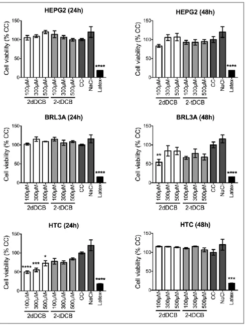
According to ISO 10993, in 5 studies published in 2009, if a compound causes the reduction of cell viability by more than 30%, it is considered to exert a cytotoxic effect. In this study, the cyto-toxic effects of the 2-dDCB and 2-tDCB at concentrations of 100, 300 and 500 μM were evaluated by MTS in BRL3A, HepG2 and HTC cell lines after 24 and 48 hours of exposure. As shown in Figure 2, 2-dDCB was found to be significantly toxic in BRL3A cells (at 100 µM after 48 hours) and HTC cells (at 24 hours in all tested concentrations). HepG2 cells on the other hand, were found to be resistant to 2-dDCB-induced toxicity. For 2-tDCB, no toxicity was observed in all tested con-ditions for all cell lines.
Figure 2 : Determination of the cytotoxic effects of 2-dDCB and 2-tDCB in different hepatic cell
lines. In the BRL3A line we can observe a cytotoxic effect only in the concentration of 100 μM in 2-dDCB for 48 hours period (B) (**): p <0.01. HepG2 cell line showed no cytotoxicity at any
concen-tration of 2-ACBs in any period evaluated (C; D). In the HTC cell line, the compound 2-dDCB showed cytotoxic effects in the 3 concentrations evaluated in 24 hours (E) (****): p <0.0001: (***): p <0.001: (*): p <0.05. However, in the 48-hour period no damage was observed in the
con-centrations of both compounds studied. Negative controls were Control Cells (CC), Sodium chloride (NaCl). Positive Control of cytotoxicity: Latex.
4. DISCUSSION AND CONCLUSION
In this study, a bibliographic survey was realized in order to study the cytotoxicity of 2-ACBs in different types of colon cells.
With the results and conclusions established, we decided to evaluate its effects in liver, consid-ering that it is an organ that may accumulate fat, processes toxic compounds and is also involved in the digestive process. Hence, we choose liver cells (HepG2, HTC and BRL3A) to perform this study, in 3 different concentrations (100, 300 and 500 μM, taking into consideration that a concen-tration of 500 μM is extremely high compared to what is consumed by human being), in 2 periods (24 and 48 hours). Evaluation of viability effect was performed by the cell viability assay using the MTS dye reduction method.
In the literature, the research conducted by Delinceé and Pool Zobel (1998) analyzed the cyto-toxicity of 2-DCB in colon cells of rats and humans obtained through biopsies and observed that cytotoxicity occurred with increasing concentrations. At 1.25 mg/ml (5.243 m/M or 5243 µM) there was a reduction in the percentage of viable cells. They realized that the greatest observed ef-fect occurred in human colon cells indicating to be more sensitive than rat cells. Viability was re-duced to less than 50% at higher concentrations of 2-DCB, clearly showing that inre-duced effects is dose dependent. Even with this important information, the test time was only 30 minutes, which is not the most adequate period. In 2002, Delinceé and colleagues continued to investigate the possible effects of 2-ACBs, now extending the period to 24 and 48 hours in the human colon tumor cell lines HT29 and HT29 clone 19A1 to evaluate the possible cytotoxic effect of compound 2-TCB. They found that, after prolonged action (1-2 days) and at higher concentrations, cytotoxic damages were observed; however, they pointed out that these damages only appeared at very high concentrations, when compared with human intake. Based on the data from the previous tests, Delincée and his team (2002a) decided to extend the scope of the tests, varying the compounds of the 2-ACBs family (2-tDeCB / 2-(tetradec'-enyl)-cyclobutanone, 2- dDCB in the same cell lines used in the previous
1 According to Delincée et al. (2002) Tumor cells from the human colon HT29 were obtained from a primary tumor of
an adenocarcinoma. An HT 29 cl 19A clone was obtained after treatment of HT 29 cells by sodium butyrate (Augeron and Laboisse, 1984). Undifferentiated HT 29 cells developed new properties, such as increased adhesion to the bottom of the vials and the possibility of forming "flat hearths". HT 29 cells, available in the differentiated or undifferentiated step, can now be tested for sensitivity to toxic compounds at various stages of their development.
test – HT29 and HT29 clone 19A), the type of test (MTT and WST), maintained the incubation time (24 and 48 hours) and the concentrations (100, 200 and 400 μM). No cytotoxic damage was ob-served within 30 minutes after incubation; however, at longer periods and concentrations (≥100μM), damages were observed.
Knoll et al. (2006) evaluated cytotoxicity of compound 2dDCB (150 to 2097 μM) in colon tu-mor human cells (HT29 clone 19A), adenoma cells (LT 97) and primary human colon cells. In that study, the primary cells were more sensitive to 2dDCB. There were reduction of viability in 120 minutes of in vitro culture and additionally as far as concentration of 2dDCB was increased, show-ing a dose dependent effect. In LT 97 lines, effects were observerd after 60 minutes in 600μM con-centration of 2dDCB
They concluded that, in both cases, at the maximum concentrations tested, the compound could be defined as "subtoxic" and therefore suitable for genotoxic assessments.
Hartwing et al. (2007), applying the 2-dDCB compound to human colon tumor cells (HT 29 clone 19A) at concentrations between 100 and 400 μM, also observed no change within the 30 mi-nute period, but in the same way as reported in other articles, cytotoxicity effects of the compound were seen as dose and time increased. The author reported that there were no significant differences observed in the viability of HT 29 and HT 29 clone 19A cells after incubation with 2dDCB using the MTT test (HT cell data 29 not shown in the paper).
After all the available literature, with the results and conclusions already established regarding the cytotoxic effects of 2-ACBs in colon cells, we can use it as a parameter and compare it to the study performed in the hepatic cells.
As per ISO 10993:5 standards (cytotoxic effect considered if cell viability drops to ~30%), it was observed that in the BRL3A cell line, only the 2-dDCB compound at concentration of 100 μM in 48 hours the period, has presented cytotoxic effect.However, at 300 and 500 μM none effect was observed. A possible explanation for this result may be the hormesis effect, which suggests that the same substance that has a certain action at high concentrations acts inversely at high dilutions (Cal-abrese, 2008). But we can not say that this is really the reason for our results. For this statement would be necessary other studies focusing on the possible hormesis effect.
The cell line derived from human hepatoma (HepG2) showed no inviability in any of the evalu-ated parameters, differently from what the authors Delinceé and Zobel (1998) and Hartwing et al. (2007) found. They reported in their study with human colon tumor cell line that, when concentra-tion of the compound was increased, damage increased as well, which suggested to be dose depend-ent. However in this experimental study, this dose dependence was not observed with human HepG2 line or with another type of lineage evaluated.
The HTC cell line showed cytotoxicity when exposed to compound 2-dDCB at the 3 concentra-tions studied after 24 hours. The same was not observed when cells were exposed to compound 2-tDCB at the same time. Cytotoxic damages were not noticed within 48 hours when the lineage was treated with the two compounds compounds at all concentrations and periods investigated.
Regarding compound 2-tDCB, although in some cases the cell viability did not present 100%, according to parameter ISO 10993:5, it was not cytotoxic in any of the evaluated situations.
As is known, the 2-ACBs ingested are part excreted through the feces and partly deposited in the adipose tissue. Until the accomplishment of this work had only been realized studies of the ac-tion of the 2-ACBs in colon cells, since this region is affected by the accumulaac-tion of fat.
However, there is another organ that is also usually affected by the accumulation of fat, the liv-er. In the last years it has presented pathological occurrences that would not be related to any genet-ic factor. So, this was the organ chosen to investigate whether 2-ACBs would have some cytotoxgenet-ic effect and, consequently, some promoter effect on liver cancer.
Colon cells produce mucus, which changes the interaction with substances, beyond to be epithe-lial. In addition, they do not possess activity capable of metabolizing compounds, which makes the responses different from liver cells. There is an in vitro study with canine cells that has shown that certain lipids (essential oils) are more cytotoxic in enterocytes than in hepatocytes (Monteiro-Riviere et al, 2015).
For this reason, the results presented here differ from the results found in the existing literature. This shows the novelty of this study, as there are no studies available in the literature regarding 2ACBs / liver cells.
After all these analyzes, observing each study, we can conclude that there is a cytotoxic effect of the 2-ACBs tested in colon cells. When concentrations and time were increased, their effects on sensitivity were also increasingly evident, showing to be totally dose - dependent, although the
greatest effect was observed at relatively high doses when compared to actual human consumption. We did not visualize similarity between the analyses observed with colon cells from bibliographic survey to the experimental study performed with liver cells.
Further additional studies should be performed regarding the cytotoxic potential of 2-ACBs, in-creasing the concentrations and the periods evaluated as well as the cell type to be studied, especial-ly when dealing with organs that have accumulation of fat and are involved in the digestive process, knowing that part of the 2-ACBs are excreted through the feces and partly remain deposited in adi-pose tissues, before categorically asserting that the alkylcyclobutanones provide or not a serious cytotoxic effect.
REFERENCES
[1] KIM, K. S.; S. E.O.; H. Y.; LEE, J. M.; PARK, E. R.; KIM, J. H.; HONG, C. H.; BYUN, M. W. Analysis of radiation-induced hydrocarbons and 2-alkylcyclobutanones from dried shrimps
(Penae-us aztec(Penae-us). J Food Prot. v. 67, p. 142–147, 2004.
[2] FARKAS, J.; EHLERMANN, D. A. E.; FARKAS, C. M. Food Technologies: Food Irradiation. Reference Module in Food Science – Enc. Food Safety. 3: 178-186, 2014.
[3] EHLERMANN, D. A. E. The early history of food irradiation. Radiat. Phy. Chem. 129: 10-12, 2016.
[4] LE TELLIER, P.; NAWAR, W. W. 2-Alkylcyclobutanones from radiolysis of triglycerides. Lipids, 7: 75-76, 1972.
[5] SONG, B. S. ; CHOI, S. J. ; JIN, Y. B. ; PARK, J. H. ; KIM, J. K. ; BYUN, E. B. ; KIM, H. ; MARCHIONI, E. “A critical review on toxicological safety of 2- alkylcyclobutanones”. Radiat. Phy. and Chem. v. 103, pp. 188-193, 2014.
[6] GADGIL, P.; HACHMEISTER, K.; SMITH, J. S.; KROPF, D. H. 2-Alkylcyclobutanones as Irradiation Dose Indicators in Irradiated Ground Beef Patties J. Agric. Food Chem, 50: 5746-5750, 2002.
[7] YAMAKAGE, K.; SUI, H.; OHTA, R.; TYOIZUMI, T.; KAWAKAMI, K.; MATSUMOTO, H.; TAKAHASHI, T.; SASAKI, K.; IKEZUMI, M.; NEGISHI, S.; IZUMI, K.; TODORIKI, S.; TAKASHI, K.; FURUTA, M. Genotoxic potencial and in vitro tumour-promoting potential of 2-dodecylcyclobutanone and 2-tetradecylcyclobutanone, two radiolytic products of fatty acids. Mutat Res. 770: 94-104, 2014.
[8] DELINCEÉ, H.; POOL-ZOBEL, B.L. Genotoxic properties of 2-dodecylcylobutanone, a com-pound formed on irradiation of food containing fat. Radiat. Phy. Chem. 52: 39-42, 1998.
[9] CHINTHALAPALLY, V. R. Do Irradiated Foods Cause or Promote Colon Cancer? (2003) Division for cancer prevention, American health Foundation-Cancer.
[10] TICE, R. R.; E. AGERELL; D. ANDERSON; B. BURLISON; A. HARTMANN; H. KOBA-YASHI; Y. MIYAMAE; E. ROJAS; J. C. RYU; Y. F. SASAKI. Single cell gel/comet assay: guide-lines for in vitro and in vivo genetic toxicology testing. Environ. Mol. Mutagen. 35: 206–221, 2000.
[11] LI, W.; ZHOU, J.; XU,Y. Study of the in vitro cytotoxicity testing of medical devices (Re-view). Biomed Rep. 3: 617-620, 2015.
[12] ISO 10993-5 International Standard. Biological evaluation of medical devices – Part 5: Tests for in vitro Cytotoxicity. Third edition, 2009.
[13] DELINCEÉ, H.; SOIKA, C.; HORVATOVICH, P.; HODAPP, C.; RECHKEMMER, G. Cytotoxicité et génotoxicité des 2-akkylcyclobutanones vis à vis des lignées de cellulas tu-morales humaines (HT 29 et HT 29 cl 19A). Bundesforschungsanstalt für Erährung
Karlsruhe. 57-72, 2002.
[14] DELINCEÉ, H.; SOIKA, C.; HORVATOVICH, P.; RECHKEMMER, G.; MAR-CHIONI, E. Genotoxicity of 2 alkylcyclobutanones markers for an irradiation treatment in fat-containing food – Part I: Cyto – and genotoxic potential of 2-Tetradecylcyclobutanone. Radiat. Phy. Chem. 63: 431-435, 2002a.
[15] KNOLL, N., WEISE, A., CLAUSSEN, U., SENDT, W., MARIAN, B., GLEI, M., POOL-ZOBEL, B. L. 2-dodecylcyclobutanone, a radiolytic product of palmitic acid, is genotoxic in prima-ry human colon cells and in cells from preneoplastic lesions. Mutat. Res. 594: 10-19, 2006.
[16] HARTWING, A., PELZER, A., BURNOUF, D., TITÉCA, H., DELINCÉE, H., BRIVIBA, K., SOIKA, C., HODAPP, C., RAUOL, F., MIESCH, M., WERNER, D., HORVATOVICH, P., MARCHIONI, E. Toxicological potential of 2-alkylcyclobutanones - specific radiolytic products in irradiated fat-containing food - in bacteria and human cell lines. Food Chem. Toxicol. 45: 2581-2591, 2007.
[17] CALABRESE, E.J., STANEK, E.J., NASCARELLA, M.A., HOFFMANN, G.R., "Hormesis Predicts Low-Dose Responses Better Than Threshold Models". Int J Toxicol. 27: 369-378, 2008. [18] MONTEIRO-REVIERI, N.A., ORTEGA, M.T., CHOI, K., KOSI, J., LIN, Z., JEFFERY, B., REVIEIRI, J.E., “Comparative in vitro Cytotoxicity of 20 Potential Food Ingredients in Canine Liver, Kidney, Bone Marrow-Dereived Mesenchymal Stem Cells, and Enterocyte-like Cells”. Appl In Vitro Toxicol. 1:4, 2015,