Frederico Miguel da Conceição Lourenço
Graduated in Biochemistry
Assignment of new roles for
malectin-like domains to understand their
divergent evolution
Dissertation to obtain Master degree in Biochemistry
Supervisor: Benedita Andrade Pinheiro, UCIBIO,
FCT/UNL
Jury:
Examiner: Doutor Jorge da Silva Dias Vowel: Doutora Benedita Andrade Pinheiro
III
Frederico Miguel da Conceição Lourenço
Graduated in Biochemistry
Assignment of new roles for malectin-like domains to
understand their divergent evolution
Dissertation to obtain Master degree in
Biochemistry
Supervisor: Benedita Andrade Pinheiro, UCIBIO, FCT/UNL
Jury:
Examiner: Doutor Jorge da Silva Dias Vowel: Doutora Benedita Andrade Pinheiro
I
“Assignment of new roles for malectin-like domains to understand their divergent evolution”
Copyright © em nome de Frederico Miguel da Conceição Lourenço, da Faculdade de Ciências e Tecnologia, Universidade Nova de Lisboa.
III
Acknowledgments:À minha orientadora, Doutora Benedita Pinheiro, por acreditar em mim e no meu trabalho.
Tive muita sorte em ter como orientadora, pelo apoio e pela aprendizagem.
À Professora Doutora Maria João Romão, não apenas como líder do grupo de investigação
XTAL da FCT-UNL, mas onde me recebeu e pela oportunidade de realizar o meu trabalho no Laboratório de Cristalografia de proteínas.
À Doutora Angelina Palma pelo ensinamento e orientação na técnica de microarrays de
glicanos.
À Doutora Ana Luísa pelo acompanhamento da difração de cristais in house.
À Viviana Correia que me apoiou e ajudou em toda a parte laboratorial, bem como sempre
disponível quando me surgia alguma dúvida.
À Professora Ten Feizi, ao Doutor Wengand Chai e à Doutora Lisete Silva do
Glycosciences Laboratory, Imperial College London pela colaboração na construção do robótico microarray de glicanos.
Ao Professor Manuel Coimbra, Doutora Cláudia Nunes e Carolina Pandeirada, da
Universidade de Aveiro, e à Professora Smestad Paulsen Berit da Universidade de Oslo pelo
fornecimento de amostras de polissacáridos para a construção dos microarrays.
A todos os colegas do grupo XTAL, pelo fantástico ambiente de trabalho, pela simpatia e
sempre disponíveis para ajudar em qualquer questão.
À Faculdade de Ciências e Tecnologia da Universidade Nova de Lisboa, uma faculdade
que me proporcionou toda a minha formação académica, e também a todos os docentes de licenciatura e mestrado em Bioquímica por todos os ensinamentos adquiridos.
Ao BAG Portugal pela oportunidade de recolher os dados de difração de cristais no European
Synchrotron Radiation Facility (ESRF), em Grenoble.
À Fundação para a Ciência e Tecnologia (FCT-MCTES) por ter financiado o trabalho
através dos projectos PTDC/BBB-BEP/0869/2014, PTDC/QUI-QUI/112537/2009 e RECI/BBB-BEP/0124/2012. A unidade de investigação UCIBIO (Unidade de Ciências e Biomoleculares Aplicadas) é financiada por fundos nacionais da FCT-MCTES (UID/Multi/04378/2013) e co-financiada pelo FEDER no âmbito do PT2020 (POCI-01-0145-FEDER-007728)
Um grande obrigado aos meus pais, por nunca terem duvidado de mim. Sem vocês não estava onde cheguei.
V
Abstract:Malectin is a highly-conserved animal lectin from the endoplasmic reticulum (ER), with a quality control function in the N-Glycosylation process. It has a β-sandwich core with long loops
connecting the β-sheets. Malectin binding-pocket is in the loops region. Several carbohydrate-binding modules (CBMs) discovered in other domains of life that shared sequence homology with the malectin, were classified and grouped as a novel CBM57 family by Carbohydrate-Active Enzymes (CAZy) database. The members of this family are expected to have a highly conserved
β-sandwich core, but high variance in the binding-pocket residues.
To investigate if the specificity of these modules is the same as the malectin, a bioinformatic analysis was performed with 315 members of the CBM57 family found in CAZy database. Several programs were used to predict the protein architecture and to analyse the conservation of amino acids sequences, especially in the binding-pocket. Based on this analysis, we predict animal CBM57 modules to have the same specificity as malectin. However, bacterial CBM57 modules in bacteria domain are predicted, after highlighting the modules associated with glycoside hydrolases from family 2, to have various specificities, and thus different biological functions. For verifying these assumptions, a total of 7 CBMs (family 57 and homologous) associated with glycoside hydrolases from family 2 and belonging to the human gut microbiome –Bacteroides ovatus and Bacteroides thetaiotaomicron- were chosen for characterization studies.
A re-cloning was initially performed for the recombinant DNAs, changing the His-tag position. Afterwards, expression tests were realized, in which 2 CBMs of different bacteria were expressed in soluble form. The production of the proteins was then performed at a larger scale, followed by affinity chromatography purification. By the analysis of the gels, the eluted samples had high purity and were suitable for characterization studies.
Glycan microarrays were performed for determining the binding-specificities of the 2 CBM modules. The CBM module from B.thetaiotaomicron revealed high specificity for pectin
polysaccharides, possible recognizing α 1-3 linked galacturonic acid and ramnose. For structural characterization by X-ray crystallography, several crystallization trials were performed. Crystals were obtained for the B.thetaiotaomicron CBM module, which diffracted to high resolution. The
structure is, yet, to be solved.
VII
Resumo:A malectina é uma lectina do retículo endoplasmático (ER), muito conservada em animais e com função de controlo de qualidade dos processos de N-glicosilação. A malectina tem uma
estrutura β-sandwich composta por folhas-β ligadas por longos loops que formam um pocket de
ligação. Vários módulos de ligação a hidratos de carbono (CBMs) descobertos noutros Domínios da Vida partilham homologia de sequência com a da malectina, sendo classificados e agrupados na base de dados Carbohydrate-Active Enzymes (CAZy) como uma nova família CBM57. Nesta família é expectável os módulos terem a estrutura β-sandwich conservada, mas com diferenças ao nível dos resíduos de ligação a açúcares.
Para investigar possíveis diferenças na especificidade destes módulos, uma análise bioinformática foi realizada para os 315 módulos de CBM57 encontrados na base de dados CAZy, usando vários programas para a previsão da arquitectura das proteínas e analise da conservação da sequência de aminoácidos. Com base nesta análise, é expectável a conservação de especificidade nos dominio CBM57 de animais. No entanto é previsto que domínios CBM57 de bactérias, principalmente quando associados a hidrolases glicosidicas da família 2, terem especificidades diferentes entre eles. Para averiguar tal hipótese, um total de 7 CBMs (membros da família 57 e homólogos) associadas a hidrolases glicosidicas da familia 2, encontradas em duas espécies de bactérias pertencendo ao microbioma humano, foram escolhidas para a caracterização bioquimica.
Uma re-clonagem inicial foi feita dos DNA recombinantes para a troca da posição da cauda de histidinas. Posteriormente, testes de expressão foram realizados, tendo 2 CBMs de duas bactérias foram expressos na forma solúvel. Feitos os crescimentos em maior escala destes 2 CBMs, foi feita a purificação por cromatografia de afinidade. Através da análise de géis, as concluiu-se que as amostras eluidas apresentavam uma grande pureza e suficiente para estudos de caracterização.
Para a determinação da especificidade de ligação dos 2 módulos CBM foram realizados microarrays de glicanos. O módulo CBM do B.thetaiotaomicron revelou alta especificidade para
pectinas, sendo os resíduos ácido galacturónico e ramnose ligados por uma ligação glicosíca α
1-3 o possivel epitopo reconhecido. Para a caracterização estrutural por cristalografia de raios-X, vários ensaios de cristalização foram realizados. Para o modulo CBM do B. thetaiotaomicron,
foram obtidos cristais que difrataram a uma alta resolução. No entanto, a sua estrutura está por resolver.
IX
Contest:Acknowledgments ... III Abstract ... V Resumo ... VII Contest: ... IX List of figures ... XIII List of tables: ... XV Abbreviations and Symbols: ... XVII
Chapter 1- General Introduction and objectives ... 1
1.1 Introduction to carbohydrates ... 3
1.2 Malectin ... 5
1.3 Carbohydrates-binding-modules (CBM): ... 7
1.3.1 Classification of CBMs: ... 8
1.4 Human gut microbiome: ... 9
1.4.1 The role of the Bacteroidetes in the human gut ... 9
1.5 Objectives: ... 10
Chapter 2- Phylogenetic studies ... 13
2.1 Introduction: ... 15
2.1.1 Distance methods: ... 17
2.1.1.1 Unweighted Pair Group Method Using Arithmetic Average (UPGMA): ... 17
2.1.1.2 Neighbor-Joining method ... 17
2.1.2 Discrete character methods: ... 18
2.1.3 Phylogenetic tree validation: ... 19
2.2 Materials and Methods ... 19
2.3 Results and Discussion ... 20
2.3.1 Analysis of 315 peptides sequences in different species: ... 20
2.3.2 Evolution of the sequences of the malectin-like modules: ... 21
2.3.2.1 Eukaryotic (excluding plants) malectin-like evolution: ... 22
2.3.2.2 Plants malectin-like modules evolution: ... 23
2.3.2.3 Bacteria malectin-like modules evolution: ... 24
2.3.3 Analysis of malectin-like modules from two organisms presents in microbiome human: Bacteroides Ovatus and Bacteroides Thetaiotaomicron: ... 26
2.4 Conclusion: ... 27
Chapter 3- Re-cloning, expression tests, production and purification of CBM modules ... 29
3.1 Introduction: ... 31
3.2 Materials and Methods: ... 31
3.2.1 Re-cloning of recombinant DNA into a new vector ... 32
3.2.1.1 Recombinant DNA production and isolation ... 32
3.2.1.1.1 Transformation: ... 32
3.2.1.1.2 Inoculation: ... 32
X
3.2.1.2 Re-cloning: ... 32
3.2.1.2.1 Primers design: ... 33
3.2.1.2.2 Amplification of protein encoding fragments: ... 33
3.2.1.2.3 Digestion of fragments and vectors: ... 34
3.2.1.2.4 DNA Ligation: ... 34
3.2.1.2.5 Colony PCR: ... 35
3.2.2 Expression tests: ... 35
3.2.2.1 Protocol for expression with IPTG-induction ... 36
3.2.2.2 Cell harvesting and lysis: ... 36
3.2.2.3 Analysis by polyacrylamide gel electrophoresis (SDS-PAGE): ... 36
3.2.3 Growth in large scale and purification: ... 36
3.2.3.1 Affinity chromatography: ... 37
3.2.3.2 Analysis by native polyacrylamide gel electrophoresis (Native-PAGE): ... 38
3.2.3.3 Size exclusion chromatography: ... 38
3.2.3.4 CBM modules Desalting and Concentration: ... 38
3.3 Results and Discussion: ... 39
3.3.1 Re-cloning of CBM modules: ... 39
3.3.2 Expression tests: ... 40
3.3.2.1 N-terminal His-tagged CBM modules expression: ... 40
3.3.2.2 C-terminal His-tagged CBM modules expression: ... 41
3.3.3 Large scale expression and purification: ... 42
3.3.3.1 Bacova03100_A module purification: ... 42
3.3.3.2 Bt0996_C module purification: ... 45
3.4 Conclusions: ... 46
Chapter 4- Specificity characterization of CBMs using glycan microarrays ... 47
4.1 Introduction: ... 49
4.1.1 Glycan sources: ... 49
4.1.2 Glycan immobilization types: ... 50
4.1.3 Glycan microarrays applications: ... 51
4.2 Glycan microarray assay method: ... 51
4.2.1 Description of array assay surface used in this thesis: ... 51
4.2.2 Choices of glycan libraries: ... 52
4.2.2.1 Manual assay construction: ... 52
4.2.2.2 Robotic assay construction: ... 53
4.2.3 Glycan microarrays binding assay: ... 55
4.2.3.1 Theory: ... 55
4.2.3.2 Experimental: ... 56
4.2.3.2.1 Manual array (slide of 2 pads): ... 56
4.2.3.2.2 Robotic array (slide of 16 pads): ... 56
4.2.4 Glycan microarrays analysis: ... 56
XI
4.3.1 Manual array results: ... 57
4.3.1.1 Proteins for quality control analysis: ... 58
4.3.1.2 Bt0996_C module analysis: ... 59
4.3.1.3 Comparison of the proteins signal spots ... 61
4.3.2 Robotic array analysis: ... 61
4.3.2.1 Proteins for quality control analysis: ... 61
4.3.2.2 CBM modules analysis: ... 63
4.3.2.3 Comparison of the proteins signal spots: ... 64
4.4 Conclusions ... 65
Chapter 5- Structural characterization of CBMs using X-ray crystallography ... 67
5.1 Introduction: ... 69
5.2 Materials and Methods: ... 70
5.2.1 Crystallization assay: ... 70
5.2.2 Crystals Harvesting: ... 71
5.2.3 Crystal x-ray diffraction: ... 72
5.3 Results and Discussion: ... 72
5.3.1 Protein crystallisation: ... 72
5.3.2 X-ray diffraction experiment: ... 73
5.4 Conclusions: ... 73
6. Global conclusions and future perspectives: ... 77
References: ... 79
XIII
List of figures:Figure 1.1- Schematic illustration of structure of 3 glucoses disaccharides. ... 4
Figure 1.2- Illustration of the intrinsic and extrinsic glycan binding proteins interactions. ... 4
Figure 1.3-Illustration of several carbohydrates in nature, by their symbol nomenclature ... 5
Figure 1.4- Tertiary structure of malectin with beta-sandwich fold ... 6
Figure 1.5- Illustration of the mechanism of nascent protein folding.. ... 7
Figure 1.6- Two types of interactions with a carbohydrate. ... 8
Figure 1.7- Schematic illustration of 3 types of CBMs with different binding to polysaccharides. . 8 Figure 1.8- Organization of genes that encodes proteins (and their orientation) for the degradation of RG II.. ... 9
Figure 2.1- An example of diagram tree... 15
Figure 2.2- Illustration the function of ClustalOmega (a multiple alignment program). ... 16
Figure 2.3- Neighbour-Joining tree phylogenetic construction process. ... 17
Figure 2.4- Illustration of Maximum Parsimony mechanism ... 18
Figure 2.5- Alignment of each malectin-like amino acid sequence in eukaryotes (excluding plants) compared to the human malectin. ... 22
Figure 2.6- Phylogenetic tree construction for all malectin-like in eukaryotes (excluding plants). ... 23
Figure 2.7- Prediction of proteins domains using the InterProScan program. ... 27
Figure 3.1- Schematic illustration of the re-cloning process.. ... 33
Figure 3.2- Schematic illustration of production and purification of our 2 CBM modules.. ... 37
Figure 3.3- An example of desalting chromatogram ... 38
Figure 3.4- Agarose gel (1.8%) electrophoresis intercalated with Safe Red of 7 PCR products from CBM modules of two Bacteroidetes species.. ... 39
Figure 3.5- Agarose gel (1.8%) electrophoresis intercalated with Safe Red of 5 fragments (A) and the pET28 vector digestion (B) ... 39
Figure 3.6- Agarose gel (1.8%) electrophoresis intercalated with Safe Red of the digestion recombinant (A) and the amplification of the fragments (B) ... 40
Figure 3.7- SDS-PAGE (10% acrylamide) analysis of the expression of the CBM modules.. ... 41
Figure 3.8- SDS-PAGE (10% acrylamide) analysis of the expression of the Bt0996_C module with C-terminal His-tag, induced at 37ºC. ... 41
Figure 3.9- Results from the purifications of Bacova03100_A ... 43
Figure 3.10- SDS-PAGE (10% acrylamide) analysis of IMAC purification without (A) and with DTT (B); Native-PAGE (12.5% acrylamide) of the IMAC purification without (C) and with DTT(D). ... 44
Figure 3.11- A and B- Results from purification of Bt0996_C, with N-terminal His-tag... 45
Figure 3.12- Result from purification of Bt0996_C-His, by size exclusion chromatography using E75 column. ... 46
Figure 4.1- Illustration of glycans synthesis... 50
Figure 4.2- A-Example of nitrocellulose matrix coated glass slides. ... 52
Figure 4.3- Illustration of glycans binding step by different proteins: antibodies, lectins and CBM modules. ... 55
Figure 4.4- Glycan microarray data analysis of proteins for que quality control of the microarray set. ... 58
Figure 4.5- Glycan microarray data analysis for our His-Bt0996_C module in study. ... 60
Figure 4.6- Heat-map analysis of the relative binding intensities calculated as the percentage of the fluorescence signal intensity given by the probe most strongly bound by each protein (normalized as 100%).. ... 61
Figure 4.7- Glycan microarray data analysis of proteins for que quality control of the microarray set (A) and of proteins for characterization studies (B) ... 62
Figure 4.8- Heat-map analysis of the relative binding intensities calculated as the percentage of the fluorescence signal intensity given by the probe most strongly bound by each protein (normalized as 100%).. ... 64
Figure 4.9- Schematic structure of RGI and RG II. RG I is associated with diverse pectins, such as arabinogalactan, pectin galactan and arabinan. ... 65
Figure 5.1- Schematic illustration of the steps to obtain the protein structure with the X-ray crystallography technique. ... 69
XIV
Figure 5.3- A-Schematic representation of hanging drop vapor diffusion technique; B- Phase diagram representing the concentration variation of protein and precipitant concentrations in crystallisation process. ... 71 Figure 5.4- Images of x-ray diffraction pattern of a salt (A) and a protein crystal (B). ... 72 Figure 5.5- The His-BT0996_C crystals obtained at 4ºC, with 0.2 M lithium sulphate and 20% (w/v) Polyethylene glycol 3350 at pH 2.97. ... 72 Figure 5.6- X-ray diffraction pattern from a His-Bt0996_C crystal. ... 73 Index Figure 1- Alignment of each malectin-like amino acid sequence in plants compared to the
human malectin……….98
Index Figure 2- Alignment of CBM57 modules associated with glycoside hydrolase family 2 compared to the human malectin. ... 102 Index Figure 3- Alignment of CBM57 modules associated with peptidase S8/S53 compared to the human malectin ... 103 Index Figure 4- Alignment of CBM57 modules associated with TolB-like compared to the human malectin ... 107 Index Figure 5- Alignment of CBM57 modules associated with PKD compared to the human malectin ... 110 Index Figure 6- Phylogenetic tree construction for one malectin-like module in each bacteria specie ... 112 Index Figure 7- The pET28 map, that carry an N-terminal His-tag and an C-terminal His-tag, the T7 promotor, the selection marker for kanamycin and the respective restriction sites. ... 113 Index Figure 8- Glycan microarray data analysis of proteins for que quality control of the
microarray set and of proteins for characterization studies.. ... 114 Index Figure 9- PEG Ion (A) and PEG Ion2 (B) commercial screens from Hampton Research used in crystallization trials. ... 116 Index Figure 10- Structure 1 (A) and Structure 2 (B) commercial screens from Molecular
XV
List of tables:Table 1.1- Illustration of 16 possible structures of hexoses by fisher projections. ... 3 Table 2.1- List of pros and cons from all the three methods described above. ... 19 Table 2.2- Principal domains and function associated with CBM57 family in Bacteria and distribution in life. ... 20 Table 2.3- List of clusters of malectin-like modules associated with a catalytic module. ... 24 Table 3.1- List of each recombinant protein information, including Locus tag, prevenient
organism, CBM family, theoretical isoelectric point, molecular weight and DNA length. ... 31 Table 3.2- List of concentrations of each component added in the PCR tubes to a final volume
of 50 μl. ... 34
Table 3.3- List of the performed PCR steps, describing the temperature, time and cycles. ... 34 Table 3.4- Description of digestion assay using restriction enzymes ... 34 Table 3.5- List of different conditions used in the expression tests of each recombinant protein in study. ... 35 Table 3.6- List of the best conditions for 2 CBM modules expression. ... 42 Table 4.1- Description of glycan libraries examples used in glycan microarrays assays. It is presented the number of glycans that compose the library, the research group, the source of the glycans and the immobilization method. ... 49 Table 4.2- List of all glycans probes used in the binding charts and in the matrix (heat-map), position and the predominant sequence/ monosaccharide composition. ... 53 Table 4.3- List of all glycans probes used in the binding charts and in the matrix (heat-map), position and the predominant sequence/ monosaccharide composition. ... 53 Index table 1- Carbohydrate binding modules (CBMs) for production to biochemical
XVII
Abbreviations and Symbols
:
% (w/v)- weight/volume percentage Å- Angstrom
B. ovatus- Bacteroides ovatus
B. thetaiotaomicron- Bacteroides thetaiotaomicron
CAZymes- Carbohydrate active enzymes CBM- Carbohydrate-binding module
E.coli- Escherichia coli
ERSF- European Synchrotron Radiation Facility GH2- Glycoside hydrolase from family 2
HEPES- 4-(2-hydroxyethyl)-1-piperazineethanesulfonic acid IMAC- Immobilized metal affinity chromatography
IPTG- Isopropyl β-D-1-thiogalactopyranoside KDa- Kilodalton
LB- Luria-Bertani
NMR- Nuclear Magnetic Resonance
MAD-Multiple Wavelength Anomalous Dispersion MIR- Multiple Isomorphous Replacement
MR- Molecular Replacement
O.D.600nm- Optical density at 600 nm PDB- Protein Data Bank
PKD- Polycystic Kidney disease RCA I- Ricinus Communis Agglutinin I
PEG - Polyethylene glycol PGA- Polygalacturonate acid RG I- Rhamnogalacturonan I RG II- Rhamnogalacturonan II Rpm– Rotations per minute
XIX
Chemical structures of monosaccharides present in glycan probes presented in this thesisThe symbol notation for monosaccharides is in accord with the proposed in the Glycosciences Laboratory, Imperial College(https://glycosciences.med.ic.ac.uk/docs/symbols.pdf).
1
1 and Objectives
3
1.1 Introduction to carbohydratesInitially, carbohydrates were usually associated with cell metabolism context and energy production. The evolution resulting from the sequencing of the human genome (and of other organisms) left lipids and carbohydrates excluded [Varki, et al, 2009], with their crucial biological
roles on physiological systems to be explained.
Carbohydrates are mono-, di-, oligo- and polysaccharides. Monosaccharides are the simplest carbohydrates. Giving an example the glucose, it is a hexose (6 carbon atoms). However, 4 of their 6 carbons are chiral, thus 16 chemical configurations are possible.
Table 1.1- Illustration of 16 possible structures of hexoses by fisher projections.
For example, only one difference on the C4 atom configuration results in 2 monosaccharides: glucose and galactose (figure 1.1) [Perez, 2014].
Figure 1.1- Stereochemistry of glucose and galactose. Red rectangle evidences the C4 of each
structure which is a mirror image.
Adding to the chirality of the monosaccharides, other modifications can occur. Continuing with the glucose structure, alteration of the 2-hydroxyl group with an acetylated amino group forms N-acetylglucosamine (GlcNAc), or the oxidation of C6 results in a carbohydrate acid, the glucuronic acid (GlcA) [Maureen, et al, 2003].
4
Disaccharides are other type of carbohydrates. They are composed of 2 monosaccharides joined by a glycosidic linkage, through a condensation reaction. However, from the same monosaccharides, several disaccharides structures are possible [Varki, et al, 2009]. Here we
show as examples 3 glucose disaccharides in the figure 1.2.
Figure 1.2 - Schematic illustration of structures of 3 glucoses disaccharides.
There are two variables in this context: 1) the glycosil linkage between two carbon atoms 2) the conformer of the anomeric carbon (C1 atom) in α or β.
The formation of different glycosil linkages has an incredibly importance in Biology. Malectin, for example, is able to recognize maltose and nigerose, although has a preference for nigerose due to the conformation of the disaccharide linkage in α -(1-3). On the other hand, malectin has a weaker interaction with cellobiose. Although maltose and cellobiose have the same carbons involved in the glycosylic linkage, the anomeric carbon has a different conformer, thus conferring the structure a different arrangement (further information about the malectin is in the section 1.2) [Schallus, et al, 2008].
Oligosaccharides have a number of monosaccharides usually less than 12 and can be covalently linked to macromolecules (glycoconjugate). As glycoconjugates, they have several biological roles such as in cell-cell interactions as well as for host-microbe interaction (figure 1.3), either through symbiotic relationships or pathogenicity [Varki, et al, 2009].
Figure 1.3- Illustration of the intrinsic and extrinsic glycan binding proteins interactions. Image
adopted from Essentials of Glycobiology book, chapter 6: Biological Roles of Glycans.
5
The biological roles of polysaccharides are in storage or structural. Glycogen and amylose are examples of storage polymers which are glucoses monosaccharides joined by alpha-linkages (figure 1.4; A and B).
Figure 1.4-Illustration of several carbohydrates in nature, by their symbol nomenclature. A and B-
Storage macromolecules of glucose in animals (glycogen) and plants (amylose). C- Cellulose, a cell wall component of plants. D-Galactomannan, a hemicellulose, other cell wall component of plants. E and F- Pectins, from the simple structure (galacturonan) to high complex structures (rhamnogalacturonan I).
Cellulose is another polysaccharide that exists in plant cell walls, composed of glucose monosaccharides joined together by β-linkages (figure 1.4; C) [Bledzki, et al, 1999]. Human
enzymes lack the specificity for β-linkage cleavage, thus humans are unable to degrade this polysaccharide. More than celluloses, the plants cell walls may have hemicelluloses and pectins (figure 1.4; D, E and F), whose macromolecules are more complex. Hemicelluloses besides glucose includes xylose, mannose, galactose, rhamnose and arabinose [Scheller, et al, 2010].
Pectins, in turn, have in their backbone galacturonic acid. However, pectins can have several types of monomers decorating their backbone, hence their incredible diversity. Pectin can be simple structure such as galacturonans, which are linear chains of galacturonic acid linked by α -(1-4) linkage, or complex polysaccharides such as rhamnogalacturonan I (RG I). The RG I besides the galacturonic acid in the backbone, it has also α-(1-2) rhamnose. In addition, it has several branches that contain arabinan and galactan. Furthermore, there is another extremely complex pectin, rhamnogalacturonan II. It comprises a homogalacturonan backbone with side chains comprising 12 different monosaccharides linked by 20 linkages type. Although more complex than RG I, its size is smaller [Gorshkova, et al, 2010].
1.2 Malectin
Malectin was first found and described in Xenopus laevis pancreas, but it was soon revealed
6
Malectin is a membrane-anchored reticulum endoplasmic protein involved in the N-glycosylation pathway (a process in which the nascent glycoproteins have the assisted folding). It has a β-sandwich fold (figure 1.5) capable of binding to nigerose, maltose and di-glucosylated oligosaccharide, with nigerose as preferred ligand [Schallus, et al, 2008].
Figure 1.5 - Tertiary structure of malectin with beta-sandwich fold, solved by X-ray crystallography
technique. Pinheiro, et al, unpublished.
The important residues that have been observed for making carbohydrate interactions are: Ser80, Glu102, Lys138, Asp201 and Asn202 through direct hydrogen bonds; Tyr82, Glu129, Val130 and Glln137 through water mediated hydrogens bonds; Tyr104 and Tyr131 through π-CH interactions [Pinheiro, et al, unpublished].
For a better understanding of this protein role, a brief introduction will be made for the N-glycosylation pathway. The nascent glycoproteins are unfolded and are transported into the ER lumen. There, an oligosaccharide with 3 terminal glucoses is transferred to the protein with Asn-X-Thr/Ser sequence consensus [Lehle, et al, 2006]. In short, 2 glucoses are successively
hydrolysed by 2 α-glucosidases, and 2 chaperones proteins bind to mono-glucosylated proteins,
initiating the assisted-folding. After the process is completed, the glycoproteins are released from
the chaperone proteins, the α-glucosidase hydrolyses the third glucose residue and the
7
Figure 1.6- Illustration of the mechanism of nascent protein assisted-folding. Glucosidases I and IIhydrolyses two glucoses residues of oligosaccharide, so this glycoprotein is recognized by chaperones proteins Calnexin and Calreticulin. Image adopted from Protein Glycosylation, Conserved from Yeast to Man: A Model Organism Help Elucidate Congenital human diseases review.
Malectin participates in this process before the hydrolyse of the second glucose. Here, the malectin is suspected to function as a quality control. In a study, where the cells were transfected with the DNA encoding hemagglutinin to enhance the stress of the ER, it showed that the expression of the malectin increased [Galli, et al, 2011]. Since there is a delay in the hydrolysis
of the second glucose, the malectin can protect these glycoproteins to prevent their degradation [Schallus, et al, 2010]. Furthermore, the same study has shown the Malectin doesn´t affect the
kinetic of the chaperone lectins but might identify the misfolded glycoproteins for their degradation, thus preventing the secretion of misfolded proteins to the Golgi and avoiding the formation of aberrant proteins [Galli, et al, 2011].
Interestingly, the malectin sequence shares similarity with some sequences of carbohydrates binding modules (CBMs) [Schallus, et al, 2008].
1.3 Carbohydrates-binding-modules (CBM):
Carbohydrate-Binding Modules (CBMs) are proteins with the ability to bind to glycans. CBMs were originally classified as cellulose-binding, but the term soon evolved with the discovery of new CBMs that had other specificities [Shoseyov, et al, 2006]. Although they are present in all
kingdoms of life, they predominate in prokaryotes.
CBMs are defined as non-catalytic modules that are appended to carbohydrate-active enzymes [Shoseyov, et al, 2006]. CBMs have a range of 30 to 200 amino acids, are in their
8
Figure 1.7- Two types of interactions with a carbohydrate. A-Hydrogen bonds between polar aminoacids chain to hydroxyl group with the cordination of the calcium. B-Hydrogen bonds between polar amino acids chain to hydroxyl group and aromatic amino acid chain reoriented for π/C-H interaction. Arrows represents hydrogen bonds. Yellow circle represents calcium cation. Image adopted from Lectin structure to functional glycomics: principle of the sugar code article.
The affinity of the CBMs for carbohydrates is relatively low, generally around 105-104 M-1 [Schallus, et al, 2010]. Studies have shown that the addition of a CBM in a protein increased the
catalytic activity while the removal of the CBM decreased the efficiency [Shoseyov, et al, 2006].
However, CBM modules are not always appended to catalytic modules, but can be individualized in one single protein. Occasionally, a CBM module can have multiple binding-sites [Boraston, et al, 2004].
1.3.1 Classification of CBMs:
Nowadays, there are 81 CBM families in CAZy database based on their sequence similarity,
with 7 possible folds: β-sandwich, β-trefoil, Cystein knot, Unique, OB fold, Hevein fold or mixture
of Uique and Hevein fold. The β-sandwich is so far, the major structure of CBMs. It comprises two
β-sheets, each with 3 to 6 antiparallel β-strands. In addition, the CBMs are divided into three different types accordingly to their binding typology (figure 1.8): 1) type A, glycan surface-binding; 2) type B, glycan chain binding; 3) type C, small sugar binding [Boraston, et al, 2004].
Figure 1.8- Schematic illustration of 3 types of CBMs with different binding to polysaccharides.
9
The type-A CBMs have the most distinct binding-interaction, with a planar binding site that binds to the surfaces of cellulose or chitin. The type-B CBM have groves that can accommodate various glycan units, while the type-C CBMs have short pockets for recognition of mono-, di- or tri- saccharides [Boraston, et al, 2004]. However, in type-C CBMs, the solved structure doesn´t
imply knowledge of the function, since members of the same CBM family may have different specificities [Taylor, et al, 2014].
In addition, the CBMs modules can have 4 different roles: 1) targeting effect; 2) proximity effect; 3) disruptive effect; 4) adhesion. In the targeting effect, the CBM target the enzyme to distinct zones within a larger glycan, either at the terminals or internal parts of polysaccharides chains. In the proximity effect, the CBMs directs the substrate to the enzyme to increase the efficiency of polysaccharide degradation. In the disruptive effect, the CBMs disrupt the surface of packed polysaccharides (fibres or granules), exposing the substrate more easily to the catalytic module. In the adhesion, the CBMs adhere onto the surface of the bacterial cell wall glycans while the enzyme has an activity on neighbour glycans [Shoseyov, et al, 2006].
1.4 Human gut microbiome:
Recently, several CBMs that share sequence homology with malectin were classified by CAZy as the novel CBM57 family. In addition, members of CBM6 and CBM35 families, both type-C CBMs (although family 6 CBMs can also be from type-B) also share sequence similarity with members of the CBM family 57. These CBMs exists in the genome of bacteria, some of them in our gut.
Human microbiome is composed of several bacteria microorganisms that live in our bodies, a superior number of 10 times more than our cells [Lynch, et al, 2016]. These microorganisms have
several impacts in the physiologic functions for our health and disease, since they protect us against pathogens and educate our immune system. The human gut has the most diverse population, with Bacteroidetes and Firmicutes as dominant phyla [Shreiner, et al, 2015]. Here, we
will focus on Bacteroidetes phylum.
1.4.1 The role of the Bacteroidetes in the human gut
Our organism can easily absorb carbohydrate monomers and cleave some disaccharides, but several polysaccharides, such as plant cell walls components, are resistant to our digestive enzymes (example of cellulose given in the section 1.1). Members of the Bacteroidetes phylum have the necessary tools for the degradation mechanism of these polysaccharides, thus complementing the eukaryotic genome [Thomas, et al, 2011].
B. thetaiotaomicron and B. ovatus are most known for adopting the Polysaccharide Utilization
Loci (PULs) system as a strategy for glycans degradation. B.thetaotaomicron has several PULs
that B.ovatus doesn´t have, but on the other hand, B.ovatus has unique PULs for hemicelluloses
degradation [Martens, et al, 2011]. Since these 2 bacteria are in distinct zones of the gut, this
characteristic may due to the adaptation of nutrient availability.
Here, we show one PUL organization of B.thetaiotaomicron in figure 1.9, which includes some
CBM modules used in our studies.
10
This PUL is specific for RG II degradation, which encodes a starch-utilization system-like (Sus-like), a polysaccharide lyase, carbohydrate esterases and several glycoside hydrolases [Ndeh, et al, 2017]. The Sus-like system is composed of at least one pair of outer membrane proteins
homologous to SusC and SusD, crucial for the transport and degradation of polysaccharides [Shipman, et al, 2000]. In periplasm, the polysaccharide lyase cleaves the backbone of the RG II,
proceeded by the action of carbohydrates esterases and glycoside hydrolases, which is coordinated [Ndeh, et al, 2017]. Carbohydrates esterases hydrolyse the esters group into acid
and alcohol groups [Nakamura, et al, 2017], while the glycoside hydrolases cleave the specific
glycosidic linkage [Naumoff, et al, 2011].
The CBM modules from this PUL, and used in our study, are appended to the Bt0996 protein
that has 2 distinct catalytic modules: 1) a glycoside hydrolase family 2 with β-D-glucuronidase
activity; 2) a novel glycoside hydrolase family 137 with β-L-arabinofuranosidase activity. The CBM
modules, as mentioned in the section 1.3.1, can have different roles and different specificities.
1.5 Objectives:
Decoding the specificities of the novel members of the CBM57 family and homologues (CBM6 and CBM35 families) is important for a better understanding of the evolutionary relationship between the malectin, a well conserved lectin in animal domain, with these modules identified in prokaryotes.
Moreover, these modules are present in two bacteria of the human gut, appended to glycoside hydrolases. Assigning their specificities is a crucial step to understand efficient degradation of complex polysaccharides such as Rhamnogalacturonan II. However, the identification of the binding specificity to these modules and the reveal of their roles is challenging.
Here we addressed the problem using first bioinformatic tools to observe the level of amino acid conservation, in particular of the putative binding residues and second to select the CBM modules, for combining glycan microarray with X-ray crystallography to study glycan-CBM interactions.
This Thesis has the aim to elucidate glycan specificities of identified CBMs in the genomes of two human gut bacteria: Bacteroide ovatus and Bacteroide thetaiotaomicron. The main goals of
this thesis are summarized in the following topics:
• To investigate the amino acid sequences using bioinformatic tools and compare with the human malectin sequence to predict carbohydrate-interacting residues and select some CBMs of the human gut bacteria for bio-characterization studies. This is detailed in Chapter 2.
• Re-cloning of the recombinant proteins and use of various expression tests to discover the best condition for a larger-scale production of CBMs, followed by a purification process. This is detailed in Chapter 3.
• To perform two types of glycan microarrays with the successfully produced CBM modules. First, to identify possible novel glycans and second, to determinate the epitope recognition of the CBM modules. This is detailed in Chapter 4
• To perform crystallization assays to explore the 3D structure and compare with the malectin structure. This is detailed in the Chapter 5
11
2017-May, Lourenço, F; Leote, C; Correia, VG; Brás, JL; Fontes, CMGA; Romão; MJ; Carvalho, AL; Palma, AS; Pinheiro, BA. Assignment of biological roles for CBM57 modules domains in human gut bacteria. 6th Meeting of Portuguese Synchrotron Radiation Users. LNEG, Lisbon.
13
2Chapter 2- Phylogenetic studies
15
2.1 Introduction:The accumulation of mutations in genes encoding proteins is an evolution mechanism. Usually the mutations are functionally and structurally silent, not having influence in the final protein. In some amino acids, the change may cause function loss, being the mutation called deleterious and eliminated by natural selection. On the other hand, the residue change may be neutral (similar residue) or the protein activity increased, and the mutation accumulated in the protein sequence, relating the evolution history [Soskine, et al, 2010].
Aligning molecular sequences (protein or DNA) of homologous proteins from all kingdoms of life helps to understand their evolution history, further helping the identification of conservation and variation sites [Yang, et al, 2012].
For several proteins (modules), the structure and, specially, the function role remains unknown. Phylogenetic studies help the prediction of structurally homologous proteins and the alignment of the molecular sequences, the identification of conserved and varied sites, help the prediction of the conservation (or not) of the protein function and specificity. Phylogenetic studies use bioinformatic tools (molecular sequence alignments and tree diagrams) to understand the gene evolution on various living organisms.
The tree diagram (figure 2.1) is composed of branches, and their sizes describe the molecular sequence divergences. The tips correspond to the molecular sequence of living organisms (taxa) while the nodes represent their respective ancestor and the root node the common ancestor [Yang, et al, 2012].
Figure 2.1- An example of diagram tree. A-E represent taxa, internal nodes represent the ancestor and
the root represents the common ancestor for all the taxa. Image adopted from Essential Bioinformatics book, chapter 10.
The tree diagram itself is the topology, the relationship of each taxon displayed in the phylogenetic tree (which taxon descended first and the node that belongs). It is possible in the final to have more than one topology, because various solutions are plausible to describe one evolution history [Yang, et al, 2012].
Considering that the protein evolution is divergent, if the ancestor descends to two taxa, then the tree diagram must bifurcate. However, if the tree diagram is multifurcating (the previously ancestor descends to more than two taxa), it is difficult to predict which of the two taxon descended first or if three taxa were instantly descended from the ancestor, instead of two taxa only. This process is known as radiation [Xiong, et al, 2006]. In addition, the generated tree can
be rooted or unrooted. In the case of a rooted phylogenetic tree, it is assumed that all taxa have a common ancestor (and a root node is showed up) [Yang, et al, 2012].
16
For phylogenetic tree construction, first the type of molecular sequences must be chosen (nucleotides or amino acids sequences) and then retrieved from the databases.
When related organisms are studied, the mutations in the molecular sequence are relatively low and for a better evolution history, DNA sequences should be used (the gene evolves faster than the protein)[Xiong, et al, 2006]. On the other hand, if the organisms in study are divergent,
the amino acids sequences should be used, otherwise it will be too complex to understand the evolution [Yang, et al, 2012].
The molecular sequences can be retrieved from diverse databases, for example, CAZy or SwissProt. The function prediction of each protein can be achieved by using the InterPro Scan program, which identifies the protein domains. This tool analyses each retrieved sequence individually and compares to sequences from the databases called signatures. After the analyse, the program presents the predicted domains [Jones, et al, 2014]. In case we want to clone a DNA with right nucleotides, other programs must be simultaneously used to see the agreement, or otherwise, the gene cloned could be codifying a truncated protein.
Obtained the sequences, alignments programs are used to analyse and compare them. When constructing a phylogenetic tree, it is important to use several state-of-the-art alignment programs such as ClustalOmega, T-Coffee or Muscle. Wrong alignments usually lead to a misinterpretation of an adaptive evolution process and, consequently, to an untruthful phylogenetic tree. The alignment programs align amino acid sequences by homology and add gaps in amino acids sequences when necessary, making sure that the amino acids sites aligned are identical (by chain propriety) as much as possible [Xiong, et al, 2006].
Giving ClustalOmega as example of a multiple alignment program function, the distance matrix is calculated using all taxa sequences and calculating a guide tree. The closest sequences pairs related in the guide tree are aligned. Each sequence pair aligned is considered as a consensus sequence and alignments of every consensus sequence are performed, generating a new consensus sequence. The sequences unpaired are aligned to the consensus sequence and the alignment performed by ClustalOmega is completed (figure 2.2) [Xiong, et al, 2006].
Figure 2.2- Illustration the function of ClustalOmega (a multiple alignment program).
17
There are two categories in the construction of phylogenetic tree: one based on distances, which calculates the distance matrix and one based on discrete characters (analyses each character site) [Yang, et al, 2012].
2.1.1 Distance methods:
Distance is defined as a count of substitutions present in two sequences aligned. Distance methods analyses the evolutionary distance between the sequences using the scores in the alignment to construct the distance matrix and derive the phylogenetic tree. The most common distance methods are UPGMA and Neighbor-Joining, that are detailed below [Xiong, et al, 2006].
2.1.1.1 Unweighted Pair Group Method Using Arithmetic Average (UPGMA):
UPGMA is the easiest distance method. As mentioned above, the distance matrix is calculated and the two sequences with the smallest value are joined together and a node between them is added. The pair sequence is treated as a consensus sequence and the distance matrix is recalculated. Again, another molecular sequence is clustered and the cycle is repeated until the last molecular sequence taxon is joined in the tree, creating a rooted tree [Xiong, et al, 2006].
UPGMA assumes that the molecular sequences evolve at constant rate, proportional to the accumulation of mutations (known as Molecular Clock technique). However, mutations usually
don’t occur at a constant rate, which can lead to a UPGMA phylogenetic tree creation that is not
entirely true. Despite this limitation, it is still a very fast method for constructing a phylogenetic tree [Xiong, et al, 2006].
2.1.1.2 Neighbor-Joining method
Neighbor-Joining method is another option for using a distance method for the construction of phylogenetic trees. Opposite to UPGMA, it uses an evolutionary rate correction formula [Xiong,
et al, 2006].
The Neighbour-Joining tree is based on the minimum evolution principle, where the branch length must be the lowest as possible. In the beginning, the tree has a star form with all branch lengths at the same value and the sum of their lengths is then calculated. Then, a pair of taxa is clustered together into a neighbour (node) with the sum of all branches length being recalculated. The pair of taxa with smaller sum value is joint in the node. This process is repeated until the final tree is generated (figure 2.3) [Xiong, et al, 2006].
Figure 2.3- Neighbour-Joining tree phylogenetic construction process. The final tree has lower sum of
18
The final constructed tree must have the lower sum of branch lengths. The branch lengths between nodes is representative of the amino acids differences between the sequences [Xiong,
et al, 2006].
The main limitation is that only a single topology is analysed. When trying to understand the evolution stages from (highly) divergent species, the final tree may not be completely correct. However, its advantage is the same as in UPGMA, since it has a fast computation time [Yang, et al, 2012].
2.1.2 Discrete character methods:
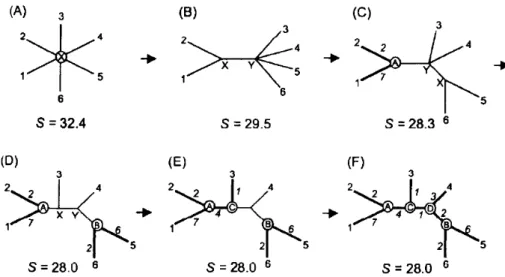
Discrete character methods analyse directly the character sites (nucleotide or amino acid site) in the alignment sequences and don´t involve distance matrix calculations. Maximum-Parsimony (MP) is one of the most known method based on discrete character [Yang, et al, 2012].
Maximum-Parsimony analysis molecular sequences sites with large divergence due to the information given about the evolution (mutations in the site). The non-informative sites are scrapped. The amount of changes at each informative site is summed and every possible phylogenetic tree is calculated [Yang, et al, 2012].
The discrete character method, opposite to the distance methods, gives the information about the molecular sequence from the common ancestor, which it is already extinct and impossible to retrieve [Yang, et al, 2012].
Every possible phylogenetic tree has the sequence of his own molecular common ancestor, with the most plausible solution being the phylogenetic tree that requires the minimum number of substitutions in the molecular sequence. This is done by analysing each informative site. For example, if the molecular sequence used is DNA and the taxa in a specific informative site have guanine, thymine or adenine, the nucleotide present in the ancestor common is the one requiring less substitutions. In a site, if six taxa are analysed and three have thymine, two have adenine and one has guanine, for the minimum substitutions the common ancestor sequence site must be thymine (figure 2.4) [Yang, et al, 2012].
Figure 2.4- Illustration of Maximum Parsimony mechanism, predicting the ancestor characters in two steps. The first step is to count all possible characters at each node. The second step is choosing the character in common ancestor that involve minimum number of substitutions. In this case, the common ancestor character must be T so the total number of substitutions is minimum. Choosing any other character would lead to an increase of substitutions. Image adopted from Essential Bioinformatics book, chapter 11.
19
The following table describes in general the pros and cons of each method referred above:
Table 2.1- List of pros and cons from all the three methods described above.
Name Based method Pros Cons
UPGMA Distance -Fast computation
-Easiest of all methods -Assume the evolution rate is constant (Molecular Clock) Neighbour Joining Distance -Fast computation
-Do not assume the evolution rate is constant
-Constructs only a possible tree
Maximum Parsimony Discrete character -More accurate phylogenetic trees compared to the methods based on distance
-Computation time demanding
-Cannot be applied to high divergence sequences
2.1.3 Phylogenetic tree validation:
After the phylogenetic tree construction, there are statistic criteria to evaluate the confidence of the made tree, such as bootstrap, jackknifing and others[Yang, et al, 2012].
Bootstrap has the principle of causing perturbations in the alignments, which can be done by the random replacement of sites. From here, there is a re-alignment and reconstruction of the phylogenetic tree (using the previously used method). This test is used 100 to 1000 times and the phylogenetic trees are clustered into a consensus tree. Bootstrap relies in the assumption that if the phylogenetic tree is strongly robust, the bootstrap consensus tree will be identical to the original tree and the evolution history is statistically correct. Moreover, the statically values added above each node in the phylogenetic tree represents the confidence of two taxa descended from the same ancestor. Values equal or superior of 70% are considered with confidence [Yang, et al,
2012].
Jackknifing is another validation method. This method relies in randomly scraping-out half of the sites and re-aligning and reconstructing the phylogenetic tree. However, using this approach has the counterpart that the sequence alignments are shorter and thus the original tree may no longer be replicated [Yang, et al, 2012].
Molecular Evolutionary Genetic Analysis (MEGA) is one of the programs for phylogenetic tree construction. It has the option of using DNA or protein alignments, constructing the phylogenetic tree using one of the three methods mentioned. The validation is then done by performing 100 to 1000 bootstrap tests [Kumar, et al, 2016]. If necessary, it is possible to edit the obtained alignment
for a better phylogenetic tree construction.
2.2 Materials and Methods
To evaluate the divergent evolution of CBM57, 315 protein sequences from bacteria and eukaryote were retrieved from the carbohydrate active enzymes (CAZy) database to date (06/09/2016). The domain architecture determination of each protein was performed using the InterProScan program [Jones, et al, 2014].
The alignments were done using the program Clustal Omega [Sievers, et al, 2011]. To
facilitate interpretation, the alignment of three groups of protein sequences were made: bacterial, plants and eukaryotes (non-plants). The alignments were then analysed using the tertiary structure of the human malectin domain. For checking the conserved regions, the software ESPript 3.0 [Robert, et al, 2014] was used. Since the binding site is present in the loops, these
20
Since the alignment of protein sequences gives a higher signal-to-noise in alignments and phylogenetic analysis, they were used to study the evolution of malectin in this widely divergent group of organisms (bacteria and eukaryotes). Also, we had the advantage of having the tertiary structure of two of the proteins to be used in the study, which served as guide lines for the alignment. For improvement, highly divergent sequences were removed from the alignment.
The phylogenetic trees were done using the software MEGA7.0 [Kumar, et al 2016]. The
Neighbor-Joining tree building method was used for phylogenetic tree construction. This method allows the analysis of several divergent protein sequences at the same time, using small computation times when compared to other tree construction methods, but still giving a satisfactory final tree.
The variation of amino acid residues in the sequence was estimated by the p-distance model, the simplest distance measure, which the value is a division of the number of amino acids variation by the total number of amino acids compared [Nei, et al, 2006]. Gaps presented in amino
acids sequences were treated using the pairwise-deletion option (the total deletion option loses some information about gene evolution) and the validation of phylogenetic tree was done by performing 1000 bootstrap replications.
2.3 Results and Discussion
2.3.1 Analysis of 315 peptides sequences in different species:
Understanding the evolution of malectin in the different species from all domains of life, may reveal important to predict novel specificities for CBM57 family members and the adaptive nature of this carbohydrate-protein interaction. These malectin-like modules are present in most of the kingdoms of life. They are found in Archaea (1), prokaryotes (176) and eukaryotes (140).
In plants, the malectin-like module is associated with kinases, being probably involved in signalling and regulatory processes. In other eukaryotes, the malectin-like module is an entire protein, probably involved in the N-glycosylation pathway. Interestingly, the malectin-like module is absent in fungi. One hypothesis is that during the evolution history the fungi kingdom, their members lost the gene coding for malectin.
In prokaryote kingdom, the malectin-like modules have a widespread distribution. These modules are part of proteins present both in Gram-positive and Gram-negative bacteria and in several ecologic niches (water, gut soil). Analysing the architecture of bacterial proteins with the CBM57 module, most have the sequence homology to the membrane transporter Tonb dependent receptor like-module or a catalytic module in their structure (table 1).
These modules (presented in table 2.2) may give a hint of possible specificities for the CBM57 module to which they are appended to.
Table 2.2- Principal domains and function associated with CBM57 family in Bacteria and distribution in life.
Domain Function Number of
proteins present
Number of organisms present
Type of organisms present Glycoside hydrolase (GH2) Hydrolyses the glycosylic bond between carbohydrates
49 38 Gram - Gram+
Pectin Lyases Degradation of
pectin 15 6 Gram -
Peptidase (S8/S52) Serine Protease; cleaves serine of N-
or C-terminal, depending the protein family
10 10 Gram+ Gram-
Quinoprotein alcohol dehydrogenase
Elimination of
21
Galactoseoxidase/kelch beta propeller
Oxidation of the hydroxyl group at
C6 position in galactose
12 10 Gram- Gram+
Polycystic kidney disease (PKD)
Unknown; predicted to interact with others proteins and
sugars
20 14 Gram-
TolB-like Associated for translocation of
group A and E colicins that penetrate and kill
cells
27 20 Gram- Gram+
PapDlike Periplasm
chaperone that mediates the attachment of
bacteria
9 8 Gram+ Gram-
Concanavalin A Activates
proliferation of cells 9 9 Gram -
Most of the modules attached to the CBM57 modules had a function prediction. However, some exceptions were observed. In 8 species of soil bacteria it was observed that the malectin-like modules are individualized, or the InterProScan program couldn´t simply predict the associated catalytic module.
While most of the proteins had only one malectin-like module, some proteins had two or more. They were found in 27 species, majority in soil or water, appended to several putative catalytic modules. More than one malectin-like module may be for increasing the affinity for a specific carbohydrate ligand or each malectin-like module may be for interacting with different epitopes from the same glycan molecule or from different glycan molecules. For this reason, a new set of alignments have been performed to see if there is conservation in the putative interacting residues present in the loop-region of these malectin-like modules.
2.3.2 Evolution of the sequences of the malectin-like modules:
Alignments of the malectin-like modules were performed using selected criteria groups: eukaryotes non-plants, plants and prokaryotes.
22
2.3.2.1 Eukaryotic (excluding plants) malectin-like evolution:Malectin-like modules from Eukaryotes are highly conserved, having minor differences in their amino acid sequences (figure 2.5).
Figure 2.5- Alignment of each malectin-like amino acid sequence in eukaryotes (excluding plants) compared to the human malectin, using ClustalOmega [Sievers, et al, 2011] and ESPript [Robert, et al, 2014] programs. The amino acids in red are conserved. The red symbols above the alignment represents the binding sites residues; Red triangles-by direct hydrogen bonds; Red squares-by hydrogen bonds mediated by water; red stars- by π/CH interactions. The black symbols mark the carbohydrate-interacting residues from malectin putative binding site.
The conservation among sequences of malectin-like modules is very high. The tertiary structure is conserved, except for 4 modules that have an extension of amino acids between β -sheets 11 and 12. The conservation of the amino acids involved in carbohydrate recognition is extremely high. Exceptions are for Caeorhabditis elegans and Cryptospodidium parvum species.
C. elegans from Animalia Kingdom has the Tyr35 replaced by histidine. This residue is polar
with positive charge and can as well to maintain the hydrogen bond with the carbohydrate. In addition, it is argued that Histidine also has an aromatic chain[Hudson, et al, 2015], suggesting
23
C.parvum from SAR Kingdom has 2 substituted residues: Tyr35 and Tyr84 replaced by Ser35and His84. Serine has an uncharged polar chain and, although weaker, capable of making hydrogen bonds with the carbohydrate. However, the substitution of Tyr84 by histidine may reflect
the loss of π-CH interactions, but a formation of a new hydrogen bond. This alignment result may
indicate the malectin-like for this specie has a different carbohydrate specificity, but due to the high conservation in the other interacting residues, it may still participate in the N-glycosylation pathway.
Some aromatic amino acids in the human malectin x-ray structure were observed to be exposed to the solvent and identified as putative binding sites 1 and 2. In the amino acid alignment, the putative binding site 1 includes 3 Phenylalanines at positions 20, 52 and 145. Except for the Phe145, the putative binding site is conserved and thus may be involved in carbohydrate interactions. On the other hand, the putative binding-site 2 with Phe152 and Phe153 shows a lower conservation level than the other putative binding-site.
To better understand the specification of malectin (excluding plants), a phylogenetic tree was constructed (figure 2.6), using the previous alignment.
Figure 2.6- Phylogenetic tree construction for all malectin-like in eukaryotes (excluding plants) using MEGA7 program. The method used was Neighbor-joining. Gaps presented in amino acids sequences were
treated using the pairwise-deletion option. The validation of phylogenetic tree was done by performing 1000 bootstrap replications.
The Caenorhabditis elegans and Cryptosporidium parvum are the most distant taxa, which
according to the alignments is due to the not conservation of the interacting residues. Moreover, the separation between Caenorhabditis elegans and Schistosoma japonicum point to the
possibility that these are ancestors and existed before the specification process that occurred for the remaining malectin modules that, like the human-malectin, are expected to recognize di-glucosylated oligosaccharides and participate in the N-glycosylation pathways.
2.3.2.2 Plants malectin-like modules evolution:
24
In comparison to the human malectin sequence, it has an extended N-terminal of a minimum of 20 to a maximum of 200 amino acids residues. However, few exceptions were found. Alignment of the N-terminal in malectin-like modules shows three conserved aromatic residues that may be part of a different interacting site. In addition to the fact that these malectin-like modules are associated with kinases, this suggests that the glycan recognized may be different from the recognized by the human malectin.
Despite the high variance, it was still possible to align their C-terminal part to the human malectin sequence. There are conserved structural elements with human malectin such as α
-helice 1 and β-sheets 5, 6, 7, 8 and 14. The interaction residues, on the other hand, aren´t
conserved with the human malectin, except for the Glu82 residue. Although the Lys91 in the human malectin has been changed to Arg91 in almost all malectin-like modules, it is a similar amino acid, so carbohydrate interaction maybe maintained.
However, there is some conservation of residues between the malectin-like module in plants. The Glu55, Tyr57 and Asn155 in human malectin are replaced by alanine, valine and glycine, respectably. Both have non-polar chains, thus the carbohydrate interaction in these residues are lost. On the other hand, Gln90 is replaced by a conserved lysine, with a larger and charged chain, capable of making the same type of interaction. This observation together with an extended N-terminal sequence may suggest that the binding-pocket changed of position in the tertiary structure to interact with other type of carbohydrates.
The conservation of the putative biding-sites is identical to the other eukaryotes, except the Phe20 in human malectin doesn´t align with several of these malectin-like modules.
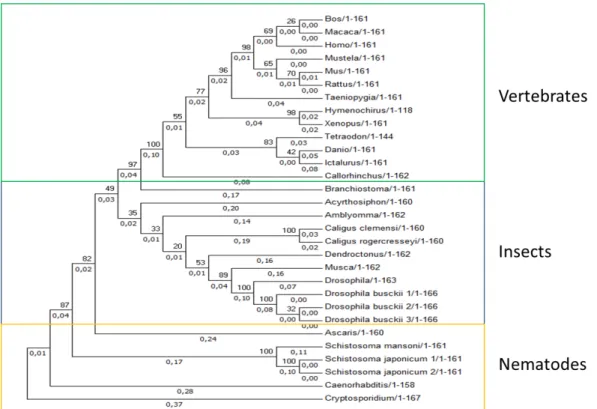
2.3.2.3 Bacteria malectin-like modules evolution:
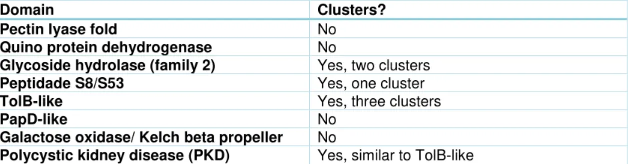
Malectin-like modules in this group are appended to a catalytic module. A phylogenetic tree (index figure 6) was made to visualize if these malectin-like modules evolved together with the catalytic module from one ancestors only, forming a cluster. Any clustering of related catalytic modules is summarized in table 2.3.
Table 2.3- List of clusters of malectin-like modules associated with a catalytic module.
Domain Clusters?
Pectin lyase fold No
Quino protein dehydrogenase No
Glycoside hydrolase (family 2) Yes, two clusters
Peptidade S8/S53 Yes, one cluster
TolB-like Yes, three clusters
PapD-like No
Galactose oxidase/ Kelch beta propeller No
Polycystic kidney disease (PKD) Yes, similar to TolB-like
Malectin-like modules associated with Glycoside Hydrolase family 2 (GH2), peptidase S8/S53, TolB-like and polycystic kidney disease (PKD) have clusters in the phylogenetic tree. The existence of clusters may indicate that the malectin-like modules have co-evolved with the catalytic module, gaining specific carbohydrate specificities. For this reason, 4 alignments of malectin-like modules with their respective associated catalytic module (GH2, Peptidase S8-S53, TolB-like, PKD) have been performed and are discussed below.
Alignment of family 2 of glycoside hydrolases associated Malectin-like modules:


![Figure 2.5- Alignment of each malectin-like amino acid sequence in eukaryotes (excluding plants) compared to the human malectin, using ClustalOmega [Sievers, et al, 2011] and ESPript [Robert, et al, 2014] programs](https://thumb-eu.123doks.com/thumbv2/123dok_br/16482986.732596/46.892.133.785.198.830/alignment-malectin-sequence-eukaryotes-excluding-compared-malectin-clustalomega.webp)