A protein with amino acid sequence
homology to bovine insulin is present in
the legume
Vigna unguiculata
(cowpea)
Laboratório de Química e Função de Proteínas e Peptídeos, Centro de Biociências e Biotecnologia,
Universidade Estadual do Norte Fluminense, Campos dos Goytacazes, RJ, Brasil
T.M. Venâncio, A.E.A. Oliveira, L.B. Silva, O.L.T. Machado, K.V.S. Fernandes and J. Xavier-Filho
Abstract
Since the discovery of bovine insulin in plants, much effort has been devoted to the characterization of these proteins and elucidation of their functions. We report here the isolation of a protein with similar molecular mass and same amino acid sequence to bovine insulin from developing fruits of cowpea (Vigna unguiculata) genotype Epace 10. Insulin was measured by ELISA using an anti-human insulin antibody and was detected both in empty pods and seed coats but not in the embryo. The highest concentrations (about 0.5 ng/µg of protein) of the protein were detected in seed coats at 16 and 18 days after pollination, and the values were 1.6 to 4.0 times higher than those found for isolated pods tested on any day. N-terminal amino acid sequencing of insulin was performed on the protein purified by C4-HPLC. The
significance of the presence of insulin in these plant tissues is not fully understood but we speculate that it may be involved in the transport of carbohydrate to the fruit.
Correspondence
J. Xavier-Filho
Laboratório de Química e Função de Proteínas e Peptídeos Centro de Biociências e Biotecnologia, UENF
28013-600 Campos dos Goytacazes, RJ Brasil
Fax: +55-22-2726-1520 E-mail: xavier@uenf.br
Research supported by CAPES, CNPq, PRONEX, FINEP, and Universidade Estadual do Norte Fluminense (FENORTE).
Received October 14, 2002 Accepted May 21, 2003
Key words
•Insulin •Insulin-like •Cowpea •Vigna unguiculata •Developing fruits •Sequence homology •Evolution
Introduction
The peptide hormone insulin was discov-ered in 1921 in connection with the treat-ment of diabetes mellitus and was isolated from the pancreas of dogs (1). Later, it was shown by others to be a small protein of about 6 kDa (2). Insulin was one of the first proteins to be crystallized (3) and was the first to be sequenced in 1955 (4). Right after the discovery of insulin, members of the Canadian group involved in the work re-ported the presence of similar hypoglycemic activity in plant materials such as the green tops of onions, lettuce and bean leaves,
bar-ley and beet roots, and others (5,6). Collip (5) coined the word glucokinin to refer to the plant-isolated material because he did not accept that a protein of plant origin should be named insulin (from the Latin insula) since plants do not possess the pancreatic islets (of Langerhans) from which insulin was iso-lated.
fruits of the bitter gourd (Momordica cha-rantia) that exerted positive effects on dia-betic patients and showed properties similar to those of insulin. Following these reports, Collier et al. (11) reported on the isolation from spinach, rye and Lemna gibba of pro-teins with molecular masses, chromato-graphic properties, immunological identity and biological activities identical to those of vertebrate insulins. No structural information was given for these molecules at the time.
Recently, in our laboratory we discov-ered by chance that a protein isolated from the seed coats of the legume Canavalia ensi-formis (jack bean) has the same molecular mass and amino acid sequence as that of bovine insulin. We further showed that the purified protein reacts with anti-vertebrate insulin antibodies and lowers blood glucose levels in diabetic animals. We also found associated with this protein a 15-amino acid sequence fragment, which showed a high sequence homology with a receptor protein kinase from humans (12). We have also shown that insulin-like antigens are present in the leaves of several green plants, fungi and a cyanobacterium, suggesting a wider distribution of this protein than is commonly thought (13).
It is well known that insulin is part of signaling pathways involved in the internal-ization of glucose in several types of verte-brate cells (14,15). Insulin also has effects on protein synthesis and gene transcription (16). In addition, insulin has been detected in tissues of members of several other phyla, suggesting that these pathways have been evolutionarily conserved (17-19). In plants, apart from many reports on the lowering of blood sugar levels in diabetic animals by extracts of plant parts (20,21), no place for insulin as a member of signaling pathways has been suggested by workers interested in the study of glucose mobilization or trans-port (22-24). Nevertheless, it has been shown that addition of bovineinsulin increases ger-mination of some seeds (25-27), accelerates
synthesis of ribosomal proteins in germinat-ing maize embryos (28,29), and increases the activity of glyoxysomal enzymes which catalyze the conversion of fat to carbohy-drate in some fat-storing seeds (25).
In the present paper we report on the isolation and characterization of insulin from the fruit of cowpea (Vigna unguiculata) as well as changes in its concentration during fruit development.
Material and Methods
Plant material
Cowpea (Vigna unguiculata (L.) Walp.) seeds of the Epace 10 genotype were ob-tained from the Departamento de Fitotecnia, Universidade Federal do Ceará, Fortaleza, CE, Brazil. Seeds were sown in pots contain-ing locally collected soil at the Centro de Ciências e Tecnologias Agropecuárias, Uni-versidade Estadual do Norte Fluminense, Campos dos Goytacazes, RJ. After 10 days, seedlings were transferred to soil in a green-house. Flowers were tagged at opening and fruits were collected at 4, 6, 8, 10, 12, 14, 16, 18 and 20 days after pollination (DAP). At 10 DAP, fruits were divided into empty pods and seeds (cotyledons plus seed coats); from 12 to 20 DAP fruits were divided into empty pods, cotyledons and seed coats. All parts were weighed before and after freeze-dry-ing. After drying, materials were finely pow-dered and kept at -20ºC for analysis.
Insulin extraction
water and 40 ml 95% ethanol were added and the preparation was again shaken for another 20 min when the pH was adjusted to 1.7 with sulfuric acid. The suspension was then filtered and the filtrate taken to pH 3.0 with ammonium hydroxide. To this new sus-pension 150 ml of 95% ethanol and 200 ml of diethyl ether were added. After standing for 12 h at 4ºC the suspension was centri-fuged at 3,000 g for 10 min and the sediment was washed with acetone and diethyl ether before dissolving in 25% ethanol, pH 8.5. To this solution 100 µl of 1 M zinc chloride was added and the preparation was left to stand for 18 h at 25ºC to precipitate insulin and other insulin-immunoreactive proteins. The precipitate was collected by centrifugation.
Antibodies
A highly purified antibody (Cat. No. GGG7303/971577) against human insulin raised in guinea pigs was purchased from Peninsula Laboratories, San Carlos, CA, USA. A peroxidase-conjugated guinea pig anti-IgG antibody (A5545) raised in goats was from Sigma, St. Louis, MO, USA.
Protein determination
Total protein in the extracts was meas-ured by the method of Bradford (30) using ovalbumin as standard.
ELISA
Insulin was measured by ELISA using a guinea pig anti-human insulin antibody (Pen-insula Laboratories) applied to separated parts of the V. unguiculata fruit (see above) according to the following procedure. The samples were freeze-dried and extracted with 0.1 M carbonate/bicarbonate buffer, pH 9.6 (15 mg/ml). After centrifugation for clarifi-cation, the extracts were diluted with car-bonate/bicarbonate containing 50 mM EDTA at 4ºC. The wells of a 96-well Maxisorp type
plate (Nunc, Roskilde, Denmark) were treated overnight with 100 µl of a solution containing 15 µg of total protein from each sample diluted in carbonate/bicarbonate buf-fer, pH 9.6. We then treated the wells with a solution of 2% gelatin in sodium phosphate-buffered saline (PBS) containing 0.05% Tween 20. Next we treated the wells with 50 µl anti-insulin IgG (1:5000) for 1 h at 37ºC. The wells were then submitted to frequent washes with PBS 0.05% Tween 20 solution. As a second antibody we employed an anti-IgG + peroxidase (1:3000). A standard curve for bovine insulin was always run alongside the samples. Negative controls were per-formed employing the IgG-peroxidase com-plex without addition of the anti-insulin IgG and a blank well with no added antibody. The reaction was developed with ortho-phen-ylenediamine and stopped with 3 N H2SO4.
The plates were read at 492 nm in a spectro-photometer.
Polyacrylamide gel electrophoresis and Western blotting
Samples (50 mg) were extracted overnight in buffer (0.1 M Tris, 10% sucrose, 0.1% SDS, and 0.005% bromophenol blue) and after clari-fication they were heated to 100ºC for 5 min. SDS-PAGE was done under nonreducing con-ditions. After 15% acrylamide gel electropho-resis (31), protein bands were visualized by Coomassie brilliant blue and silver staining. Western blotting (32) was done on nitrocellu-lose membranes utilizing a semi-dry device for electrotransfer of proteins and a buffer made up of 25 mM Tris, 192 mM glycine and 20% methanol. Transfer was performed at 1 mA/cm2 for 4 h. After transfer and blocking
Reverse-phase HPLC
Proteins were separated and purified by RP-HPLC using a 50 x 5 mm C4 Vydac column with a 0-80% acetonitrile in 0.1% TFA gradient. The fraction obtained by zinc chloride precipitation (50 µg of protein) was
dissolved in 500 µl of 0.1% TFA containing 0.1 M EDTA and applied to the column. The column was operated at 0.7 ml/min and pro-teins were detected by absorbance at 280 nm. Bovine insulin (ca. 300 µg of protein in 500 µl) was used as a standard to compare retention times.
N-terminal sequencing
N-terminal amino acid sequencing was performed on a Shimadzu PPSQ-10 Auto-mated Protein Sequencer (Kyoto, Japan) by Edman degradation (33). Sequences were determined for the RP-HPLC peak (C4 col-umn) with the same retention time to that of bovine insulin. About 200 pmol of V. ungui-culata insulin was submitted to N-terminal sequencing. Proteins were not reduced/alky-lated in order to prevent loss of cysteine residues, and thus we observed two phenylthiohydantoin (PTH)-amino acids per cycle. PTH-amino acids were detected at 269 nm after separation on a reverse-phase C18 column (4.6 mm x 2.5 mm) under isocratic conditions according to the manu-facturer instructions (Shimadzu PPSQ-10 Protein Sequencer). The sequences obtained were compared to sequences reported in amino acid data banks and submitted to au-tomatic alignment, which was performed using the NCBI-BLAST search system (34).
Results
Presence of insulin antigen in developing cowpea fruits
An immunoreactive insulin, as demon-strated by ELISA utilizing an anti-human insulin antibody, was shown to be present in developing whole fruits of cowpea from 4 to 20 DAP. The levels of insulin per fruit in-creased up to 16 DAP and then fell at 20 DAP when fruits were dry (Table 1). When the results were plotted as ng insulin per µg protein (Table 2) no difference in the insulin
Table 2. Insulin levels per protein and dry weight of plant part in fruits of cowpea (Vigna unguiculata) as a function of time after pollination.
Days after pollination Insulin
(ng insulin/µg protein) (ng insulin/mg dry weight)
4 (whole fruit) 0.48 ± 0.04 7.27 ± 0.656
6 (whole fruit) 0.47 ± 0.015 8.95 ± 0.278
8 (whole fruit) 0.46 ± 0.017 10.05 ± 0.346
10 (empty pod) 0.47 ± 0.023 11.4 ± 0.548
12 (empty pod) 0.46 ± 0.012 7.8 ± 0.173
14 (empty pod) 0.48 ± 0.015 8.65 ± 0.28
16 (empty pod) 0.44 ± 0.07 5.1 ± 0.751***
16 (seed coat) 0.51 ± 0.053 18.15 ± 1.89**
18 (empty pod) 0.35 ± 0.015* 4.1 ± 0.18***
18 (seed coat) 0.56 ± 0.04 20.2 ± 1.5**
20 (seed coat) 0.36 ± 0.01* 5.4 ± 0.15***
Insulin was measured by ELISA using an anti-human insulin antibody. In samples 4, 6 and 8 days after pollination (DAP), insulin was measured in the whole fruit. In sample 10 DAP, fruits were divided into seeds (plus seed coats) and empty pods. In samples 12 DAP to 20 DAP, fruits were divided into seed coats, cotyledons and empty pods. Seeds (cotyledons plus axes) of 10, 12, 14, 16, 18 and 20 DAP fruits did not contain any insulin as measured by ELISA. The same number of asterisks indicates that the
data do not differ statistically (P < 0.05, Student t-test; N = 7).
Table 1. Total insulin in fruits of cowpea (Vigna
unguiculata) as a function of time after pollination.
Days after Insulin
pollination (µg insulin per fruit)
4 0.456 ± 0.039
6 1.90 ± 0.09
8 3.27 ± 0.11
10 4.635 ± 0.221
12 7.1 ± 0.173
14 9.67 ± 0.306*
16 12 ± 1.428*
18 10.84 ± 0.691*
20 1.28 ± 0.026
Data are reported as mean ± SD for 7 whole fruits. Insulin was measured by ELISA with anti-human insulin antibodies. The asterisks indicate that these three measurements do not differ
concentrations was observed in fruits up to 16 DAP. At 18 DAP, insulin concentration decreased in empty pods and increased in seed coats. At 20 DAP the antigen was de-monstrable in seed coats but not in empty pods. This could be due to the inability to solubilize insulin from the dried pods by the buffer utilized (Table 2). No insulin was detected in seeds from 10 to 14 DAP fruits or in the cotyledons of any developmental stages of the cowpea fruit (data not shown). When we plotted specific activity of insulin (ng) per tissue dry weight (mg) (Table 2) we observed that the values found for seed coats from 16 and 18 DAP fruits were 1.6 to 4.0 times higher than the values found for iso-lated pods at the same stages (Table 2).
Identification of the immunoreactive insulin
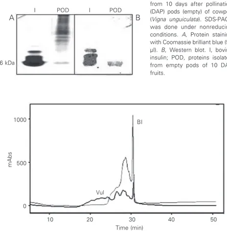
When the extracts (10 DAP empty pods) containing the insulin antigen were submit-ted to SDS-PAGE we detecsubmit-ted a protein band in the same position as a bovine insulin band with a molecular mass of approximately 6 kDa (Figure 1A). The insulin standard has more than one band probably because of the property of insulin to form aggregations. When a similar gel was treated with the anti-insulin antibody employed by us, the band at the same position as insulin (ca. 6 kDa) stained positive, indicating the presence of insulin-like antigens in the plant materials (Figure 1B). The same insulin-positive bands were seen in gels originating from extracts obtained from materials of different DAP (data not shown). These results indicate that the insulin antigen present in cowpea fruits has the same molecular weight as vertebrate insulins.
The insulin-like protein isolated as de-scribed in Methods (9) was also submitted to RP-HPLC, using commercial bovine insulin as standard. The insulin-like material iso-lated from cowpea fruits and commercial bovine insulin presented similar chromato-graphic behavior (Figure 2).
The definitive proof of the identity of the molecule came when we submitted it to N-terminal amino acid sequencing. The protein present in the major peak (absorbance at 280 nm, 31-min retention time) obtained by RP-HPLC (Figure 2) was used for automatic Edman sequencing. The resulting sequence was shown to be identical to the sequence of bovine insulin (Figure 3). Since the insulin had not been reduced and alkylated by se-quencing, the molecule had intact disulfides and thus no PTH-cysteine was obtained. Note that all the X’s in Figure 3 correspond to cysteine in native insulin. Thus, we can con-clude that the sequence of the V. unguiculata
protein is identical to bovine insulin.
Figure 2. Reverse-phase HPLC separation of bovine insulin (BI, 300 µg) and of insulin
isolated from empty pods of 10 days after pollination fruits of Vigna unguiculata (VuI, 50
µg). The column was operated at 0.7 ml/min and proteins were detected by absorbance at 280 nm and a gradient of acetonitrile and 0.1% TFA was applied to the column as described in Methods. The light line indicates the elution profile of insulin and the dark line, the profile of the cowpea extract.
I POD I POD
A B
6 kDa
Figure 1. SDS-PAGE and West-ern blot of proteins extracted from 10 days after pollination (DAP) pods (empty) of cowpea (Vigna unguiculata). SDS-PAGE was done under nonreducing
conditions. A, Protein staining
with Coomassie brilliant blue (50
µl). B, Western blot. I, bovine
insulin; POD, proteins isolated from empty pods of 10 DAP fruits.
mAbs
1000 BI
Vul 500
0
10 20 30 40 50
Discussion
It is well known that during fruit develop-ment in legumes such as cowpea and others, photosynthate enters the embryo (sink) through the pod and the seed coat (35). It is also thought that sucrose is mostly hydro-lyzed to glucose and fructose by invertase in order to enter the cotyledons (35). The isola-tion of a protein from the developing fruits of cowpea (V. unguiculata) with positive immunoreactivity to anti-human insulin an-tibody and amino acid sequence identity with bovine insulin would suggest that insulin might be involved in the transport of sugars (e.g., glucose) to the embryo, a role similar to the one exerted by this hormone in verte-brates (14) and inverteverte-brates alike (36). Strong data suggest that insulin enhances the metabolism of germinating maize seeds (27) and also of some fat-storing seeds (25). Re-cently, our group demonstrated that insulin stimulates the germination of Canavalia en-siformis (Oliveira AEA, Ribeiro ES, Da Cunha M, Gomes VM, Fernandes KVS and Xavier-Filho J, unpublished results) and Pha-seolus vulgaris seeds (Santos VO, Silva LB, Oliveira AEA, Da Cunha M, Fernandes KVS
and Xavier-Filho J, unpublished results). Further identification of the insulin anti-gen with bovine insulin was obtained by RP-HPLC of the insulin-like protein isolated by the method of Khanna et al. (9). Comparison of its retention time with that for bovine insulin showed an identical chromatographic profile, suggesting once again that the mole-cule we isolated was the same as bovine insulin (Figure 2).
The levels of insulin-like protein in fruits first increased and then decreased with the development of the fruits (Table 1). The phase of decreased concentration of insulin corresponds to the ripening phase, when fruits lose water and the seeds become dry and metabolically less active (37). The presence of insulin in higher amounts when the fruit is metabolically active is an indication of a possible role for insulin in fruit. Insulin in plants may be involved in signaling events, as suggested by us and by others (12,13,38). It is interesting to note that the sequence of the protein found in developing cowpea fruits and reported here is identical to the sequence of the insulin we have isolated from seed coat tissues of the jack bean, another legume plant (12). The presence of insulin-like antigens in plant tissues was re-ported previously by Collier et al. (11) and more recently by us (13).
The findings reported in the present study of yet another plant which contains insulin strengthen the view that the peptide hor-mone insulin is evolutionarily conserved throughout living organisms, including mi-crobes (18), insects (36) and plants (11,12, 27), probably participating in similar signal-ing pathways.
α chain of bovine insulin
protein isolated from V. unguiculata
ß chain of bovine insulin
protein isolated from V. unguiculata
Figure 3. Alignment of the sequence found by automatic amino acid sequencing of the
fraction isolated by RP-HPLC of cowpea (Vigna unguiculata) insulin with the sequence of
bovine insulin (gi229095). Sequences were compared using the NCBI-BLAST search system. Cysteine residues were not detected since proteins were not reduced/alkylated; an X appearing in the sequence indicates that the amino acid at that position is probably a cysteine residue.
References
1. Banting FG & Best CH (1922). The internal secretion of the
pan-creas. Journal of Laboratory and Clinical Medicine, 7: 465-480.
2. Harfenist EJ & Craig LC (1952). The molecular weight of insulin.
Journal of the American Chemical Society, 74: 3087-3089.
3. Abel JJ (1926). Crystalline insulin. Proceedings of the National
Academy of Sciences, USA, 12: 132-136.
4. Sanger F (1959). Chemistry of insulin. Science, 129: 1340-1344.
5. Collip JB (1923). Glucokinin. A new hormone present in plant tissue.
Preliminary paper. Journal of Biological Chemistry, 56: 513-543.
7. Gray AM & Flatt PR (1997). Nature’s own pharmacy: The diabetes
perspective. Proceedings of the Nutrition Society, 56: 507-517.
8. Khanna P, Nag TN, Jain SC & Mohan S (1974). Extraction of insulin
from a plant source. 3rd International Congress on Plant Tissue and
Cell Cultures, July 21-26, Leicester, UK.
9. Khanna P, Nag TN, Chandrajaia S & Mohan SV (1976). Process for
isolation of insulin from plant source. United States Patent. Patent
number 3,945,988.
10. Khanna P, Jain SC, Panagariya A & Dixit VP (1981). Hypoglycemic
activity of polypeptide-P from a plant source. Journal of Natural
Products, 44: 648-655.
11. Collier E, Watkinson A, Cleland CF & Roth J (1987). Partial purifica-tion and characterizapurifica-tion of an insulin-like material from spinach and
Lemna gibba G3. Journal of Biological Chemistry, 262: 6238-6247. 12. Oliveira AEA, Machado OLT, Gomes VM, Xavier-Neto J, Pereira AC,
Vieira JGH, Fernandes KVS & Xavier-Filho J (1999). Jack bean seed coat contains a protein with complete sequence homology to
bo-vine insulin. Protein and Peptide Letters, 6: 15-21.
13. Silva LB, Santos SSS, Azevedo CR et al. (2002). The leaves of green plants as well as a cyanobacterium, red alga, and fungi contain
insulin-like antigens. Brazilian Journal of Medical and Biological
Research, 35: 297-303.
14. Baumann CA, Ribon V, Kanzaki M, Thurmond DC, Mora S, Shigematsu S, Bickel PE, Pessin JE & Saltiel AR (2000). CAP de-fines a second signalling pathway required for insulin-stimulated
glucose transport. Nature, 407: 202-207.
15. Brüning JC, Gautmam D, Burks DJ, Gillette J, Schubert M, Orban PC, Klein R, Krone W, Müller-Wieland D & Kahn CR (2000). Role of brain insulin receptor in control of body weight and reproduction.
Science, 289: 2122-2125.
16. Alper J (2000). New insights into type 2 diabetes. Science, 289:
37-39.
17. LeRoith D, Delahunty G, Wilson GL, Roberts Jr CT, Shemer J, Hart C, Lesniak MA, Shiloach J & Roth J (1986). Evolutionary aspects of
the endocrine and nervous systems. Recent Progress in Hormone
Research, 42: 549-587.
18. LeRoith D, Shiloach J, Heffron R, Rubinovitz C, Tanenbaum R & Roth J (1985). Insulin-related material in microbes: similarities and
differences from mammalian insulins. Canadian Journal of
Bio-chemistry and Cell Biology, 63: 839-849.
19. Chan SJ & Steiner DJ (2000). Insulin through the ages: phylogeny of
a growth promoting and metabolic regulatory hormone. American
Zoology, 40: 213-222.
20. Ernst E (1997). Plants with hypoglycemic activity in humans.
Phy-tomedicine, 4: 73-78.
21. Platel K & Srinivasan K (1997). Plant foods in the management of diabetes mellitus: vegetables as potential hypoglycaemic agents.
Nahrung, 41: 68-74.
22. Ward JM, Kühn C, Tegeder M & Frommer WB (1998). Sucrose
transport in higher plants. International Review of Cytology, 178:
41-71.
23. Lemoine R (2000). Sucrose transporters in plants: update on
func-tion and structure. Biochimica et Biophysica Acta, 1465: 246-262.
24. Williams LE, Lemoine R & Sauer N (2000). Sugar transporters in
higher plants - a diversity of roles and complex regulation. Trends in
Plant Sciences, 5: 283-290.
25. Goodman DBP & Davis WL (1992). Insulin accelerates the post
germinative development of several fat storing seeds. Biochemical
and Biophysical Research Communications, 190: 440-446. 26. Wobus U & Weber H (1999). Sugars as signal molecules in plant
seed development. Biological Chemistry, 380: 937-944.
27. Oliveira AEA, Azevedo CR, Venâncio TM et al. (2001). Insulin in
plants. Plant Biology-2001, Providence, RI, USA, July 21-25, 1999,
29 (Abstract).
28. Sánchez de Jiménez E, Beltrán-Penã E & Ortíz-López A (1999). Insulin-stimulated ribosomal protein synthesis in maize embryonic
axis during germination. Physiologia Plantarum, 105: 148-154.
29. Dinkova TD, Aguilar R & Sánchez de Jiménez E (2000). Expression of maize eukaryotic initiation factor (eIF) iso4E is regulated at the
translational level. Biochemical Journal, 351: 825-831.
30. Bradford MM (1976). A rapid and sensitive method for the quantita-tion of microgram quantities of protein utilizing the principle of
protein-dye binding. Analytical Biochemistry, 72: 248-254.
31. Laemmli KA (1970). Cleavage of structural protein during assembly
of the head of bacteriophage T4. Nature, 227: 680-685.
32. Towbin H, Staehelin NT & Gordon J (1979). Electrophoretic transfer of proteins from polyacrylamide gels to nitrocellulose sheets;
pro-cedures and some applications. Proceedings of the National
Acade-my of Sciences, USA, 176:4350-4354.
33. Edman P (1950). Method for determination of amino acid sequence
in peptides. Acta Chemica Scandinavica, 4: 283-293.
34. Altschul SF, Gish W, Miller W, Myers EW & Lipman DJ (1990).
Basic local alignment search tool. Journal of Molecular Biology,
215: 403-410.
35. Offler CE & Patrick JW (1993). Pathway of photosynthate transfer in
the developing seed of Vicia faba L - A structural assessment of the
role of transfer cells in unloading from the seed coat. Journal of
Experimental Botany, 44: 711-724.
36. Sower SA, Suzuki K & Reed KL (2000). Perspective: Research activity of enteropancreatic and brain/central nervous system
hor-mones across invertebrates and vertebrates. American Zoology,
40: 165-178.
37. Bewley JD & Black M (1994). Seeds: Physiology of Development
and Germination. 2nd edn. Plenum Press, New York.
38. Ryan CA, Pearce G, Scheer J & Moura DS (2002). Polypeptide