RELAÇÃO ENTRE SELEÇÃO IN VITRO E EM PLÂNTULA PARA RESISTÊNCIA À Bipolaris sorokiniana EM TRIGO
ROSA MARIA DE LUJÁN OVIEDO DE CRISTALDO', FERNANDO 'RAJÁ FELIX DE CARVALHO', MAN MOHAN KOHLF, ROSA LIA RAREIEM', LUIZ CARLOS FEDERIZZF, CARLOS PIEROBOM6
RESUMO — Doze genótipos de trigo (Triticum aestivum L.) foram avaliados in vivo e in vitro para resistência à helmintosporiose, moléstia induzida pelo fungo Bipolaris sorokiniana, com o objetivo de verificar a existência de uma correlação entre a resposta apresentada por calos e por plântulas . A partir de dois isolados foram obtidos filtrados tóxicos para serem adicionados ao meio de cultura onde se desenvolveram os calos de trigo; também foram produzidas suspensões de esporos para serem pulverizados a plfintulas. A reação dos calos fOi quantificada através do crescimento daqueles expostos aos filtrados em comparação aos que não receberam este tratamento. A resposta das plântulas foi medida pela porcentagem de lesões necróticas presentes nas folhas. Foi verificado que um menor crescimento de calo correspondeu a uma maior porcentagem de lesões em plântulas.
Palavras-chave: Bipolaris sorokiniana, Triticum aestivum, filtrados tóxicos, cultura de tecidos.
RELATIONSHIP BETWEEN IN VITRO AND SEEDLING SELECTION FOR RESISTANCE TO SPOT BLOTCH
(Bipolaris sorokiniana) IN WHEAT
ABSTRACT — Twelve wheat (Triticum aestivum L.) genotypes were assessed, in vitro and in vivo, for resistance to spot blotch induced by Bipolaris sorokiniana, with the objective of verify correlation between calli and seedling responses. Toxic filtrates were obtained from two fungai isolates and added to the culture mediam where the wheat callus developed; spore suspensions were also produced and applied to wheat seedlings. The callus reaction was quantified by the difference between growth of those exposed to the filtrates and the untreated cheks. The response of the seedlings was measured by the percentage of necrotic lesions present on the leaves. A smaller callus growth corresponded to a higher percentage of lesions on the seedling leaves.
Key words: Bipolaris sorokiniana, Triticum aestivum, toxic filtrates, tissue culture. INTRODUCTION
The spot blotch of wheat induced by Bipolaris sorokiniana diminishes seed germination, increases premature seedling mortality, dries the aerial photosyn-thetic paris, prevenis the fomiation of cariopsis, develops lesions in the forro of stains on the leaves and glumes, and rot in the roots and stalk base resulting in yield reduction (011USTENSEN, 1925). The fungus is found in
the plant tissues, and in the soil (MERONUK and PEPPER,
1968). The pathogen multiplies mainly in the residues of the host species and on the wild and cultivated grasses, carrying inoculum from one year to another (Diem.„ 1982; REIS, 1982). Reaction to spot blotch fungus depends on the pathogen specialization (Mana, 1981,b), moisture during incubation (Luz 1982), and temperature
(Luz and BERGSTROM, 1986). B. sorokiniana produces
toxins which are essential for spot blotch development in rye, wheat and oat seedlings (LuDwiNG, 1957). These toxins act as an inhibitor of the electron transfer process in the oxidative phosphorylation in the mitochondria, affecting cellular respiration (Tnumucm and \\turre , 1967).
The use of tissue culture allows easier analysis of the host-pathogen interaction that may help breeders to select disease resistam cultivais. A great advantage of this technique is the control of the variable effects of the environment which is reduccd in field studies caused by the variation between locations and yearly climatic fiuctuations. Furthermore, in vitro cultures allow the use of a great number of genotypes in a relatively small space. An in vitro selection combining tissue culture
1. Eng. Agr., Ph.D. — Ministerio de Agricultura, Assuncion, Paraguay.
2. Eng. Agr., Ph.D. — Departamento de Fitotecnia, Faculdade de Agronomia Eliseu Maciel, Universidade Federal de Pelotas (UPPel). Caixa Postal 354, 96001-970 Capa° do Leso — RS/BRASIL Autor para correspondência.
3. Eng. Agr., Ph.D. — Centro Internacional de Mejoramiento de Mais y Trigo (CIMMYT), Montevideo, Uruguay.
4. Biál., M.Sc. — Curso de Pós-Graduaçâo em Genética e Biologia Molecular, Instituto de Biologia, Universidade Federal do Rio Grande do Sul (UFRGS), Porto Alegre, RS/BRASIL.'
5. Eng. Agr., Ph.D. — Departamento de Plantas de Lavoura, Faculdade de Agronomia da UFRGS. Caixa Postal 776, 91501-970 Porto Alegre — RS/BRASIL.
- 6. Eng. Agr., Ph.D. — Departamento de Fitossanidade, Faculdade de Agronomia Eliseu Maciel, UFPeI, 96001-970 Canino do Leilo — RS/BRASIL. Recebido para publicaçâo em 10/06/1997
ROSA MARIA DE LUJAN OVIEDO DE CRISTALDO, FERNANDO IRMA FELIX DE CARVALHO, MAN MOHAN KOHLI, ROSA LIA BARBIERI, LUIZ CARLOS FEDERIZZI, CARLOS PIEROBOM
with the use of toxins or pathogen toxin filtrates may be successful if the disease resistance is expressed at a cellular levei (Dnen, 1986). Thus, the resistance mechanisms should not depend on organized structures or organized tissues, such as flowers or cuticle (MEnEorni, 1984). The manifestation of resistance or susceptibility should be easily identifiable among the individuais exposed to selection. Furthermore, the existence of a correlation between resistance to a pathogen and resistance to its toxin is a fundamental requirement for the use of phytotoxins in in vitro cultures (BEHNKE, 1980).
The use of B. sorokiniana toxic tilintes applied to young wheat and oat calli has been shown as an efficient approach to identify resistant genotypes, by avaliation of callus growth (CRISTALDO, 1993; BARBIERI, 1995; HANDEL, 1996). This study was developed to test the correlation between the in vitro and in vivo responses of
12 wheat cultivars to B. sorokiniana.
MATERIAL AND METHODS
Twelve wheat genotypes, selected for their known resistance or susceptibility to the spot blotch pathogen, were assessed in field trials during severa] years. The fúngal isolates were obtained from wheat grains from the cultivars IAC 5 -Maringá and Trigo BR 35 infected with B. sorokiniana and showing the most characteristic symptom of the disease: a black point.
ks varo selection
The toxin filtrates were obtained from the colonies of the purified isolates using the methodology describcd by AiAm (1989) (Figure 1).
colonies of fungus in solid PDA medium (7 days)
9
Fries modified liquid medium (21 days) filtering
4.
concentration to 10% of volume (45 °C)9
addition of 70% ethanol4.
overnight at 4 °C 1 ffitering9
concentration to 10% of initial volume (40 °C) FIGURE 1 — Scheme for obtaining B. sativum toxin
filtrates from the isolates IAC 5 -Maringá and Trigo BR 35
Immature wheat embryos were used as controls. After being removed, they were placed on Petri dishes containing the callus induction medium MS (MURASHIGE and Sxooc, 1962) with 2.0 mg/1 of 2.4-D (dichlorophenolacetic acid), 3% saccharose and 0.8% carrageen. The calli were kept in this medium for four weeks ala temperature of 25 °C± 1 °C. Atter this period, they were cut in pieces of approximately 1.0 mm and transferred to the MS maintenance and callus growth medium with 0.5 mg/1 of 2.4-D, 3% saccharose and 0.8% carrageen, containing toxin filtrates of Lhe fungus in the cotios of 1:8 or 1:16. The plates were placed under the light and temperature conditions mentioned above. The calli were measured twice: when placed in the growth and maintenance mediam and atter four weeks. The difference between these two measurements was considered as the measurement of callus growth.
Seedling selection
The fungai isolates Trigo BR 35 and IAC 5-Maringá were placed in Petri dishes containing PDA medium covered with a damp filter paper. The plates were kept at 24 °C ± 2 °C in the dark, for three weeks.
The conidial suspension was prepared by adding 1 ml of sterilized distilled water to the plates containing the spores and agitating until the spores were detached from the paper surface. This concentrated conidial suspension was diluted to obtain the concentration used in inoculation studies. Dilutions were carried out in an Erlenmeyer flask with 400 ml of sterilized distilled water. The suspension was added until reaching a concentration 1 x 10⁵ conidia per ml of solution. A drop of the emulsifying agent TWeen 20 for each 200 ml of suspension was added. The standardization of the inoculum concentration was done by counting the conidia in a Neubauer chamber (MATsumuna, 1991).
This experiment was conducted in a growth chamber. The 12 wheat genotypes were sown, atter disinfestation, in plastic vases containing sterilized vermiculite. Germinated seeds were thinned to four seedlings per vase and were watered periodically with the nutritive solution (4.2 g of MgSO 47H 20; 1.4 g of 1C2HPO4; 5.8 g of KNOJ and Ca(NO 3)2.4H20, diluted in 10 liters of water) and kept in the growth cabinet at a temperature of 20± 1° C, with a 12 hour photoperiod, until reaching the stage of three expanded leaves. At this stage, the vases were put in a damp chamber (metal cabinet completely closed with plastic) and inoculated with the spore suspension from Trigo BR 35 and IAC 5-Maringá isolates, while the controls were treated with sterilized distilled water (Lngong, 1989; MtasuiviunA, 1991). A continuous 10 pounds pressurejet spray obtained with a compressor motor was used to ensure uniform inoculation. The plants were sprayed until their leaves were completely covered with the suspension. Atter
RELAÇÃO ENTRE SELEÇÃO IN VITRO E EM PLÂNTULA PARA RESISTÊNCIA À Bipolaris sorokiniana EM TRIGO inoculation, half of the plants were kept in (hese
chambers for 18 hours and the other half for 36 hours under relative humidity near to saturation point (± 100%) ata temperature of 24 ± 2° C. The incubated plants were then transferred to the growth chamber at the same temperature and ± 70% relative humidity, for six days, when the infection indexes were scored (Mnasumusn, 1991).
Assessment of the disease intensity was carried out by considering the percentage of necrotic lesion on the second leaf of each seedling (JAmes, 1971).
Experimental Design
The experiment with 1,11e callus and the toxin filtrates was carried out in an 3 x 12 x 3 factorial design; three toxins (Trigo BR 35 toxin, IAC 5-Maringá toxin and control), 12 genotypes and three replications. Each replication was made up of a plate with 10 calli. The analysis was done considering the mean of each replication.
The test with seedlings and fungal spore suspension was in a 2 x 2 x 12 x 3 factorial design: two isolates (Trigo BR 35 and IAC 5-Maringá), two incubation periods (18 and 36 hours of nearly 100% relative humidity) and 12 genotypes in three replications. Each replication was made up of three vases with four seedlings. The evaluations (% of infected leaf arca) were made only on the second leaf of each piara. The mean of each replication was used in the analysis without transformation of the data.
RESULTS AND DISCUSSION
The results were obtained from the analysis of two experiments; one using B. sorokiniana toxin filtrates in tissue culture, and the other carried out in the growth chamber with seedlings and spore suspension from the same isolates.
The first provided data on the genetic variability among the wheat genotypes using fungal toxin filtrates, while the second allowed an seedling assessment of trait resistance or susceptibility to the spot blotch. Correlations between the responses at in vitro callus and seedling reaction were established.
Genotype response to the toxin action
The genotypes were distinct in their callus production potential and had different response in the presence of toxins from different origins (such as IAC 5-Maringá and Trigo BR 35). The genotypes sensitive to the presence of toxins showed different reactions to the Trigo BR 35 and IAC 5-Maringá toxins, while the resistant genotypes had a similar response to both toxins.
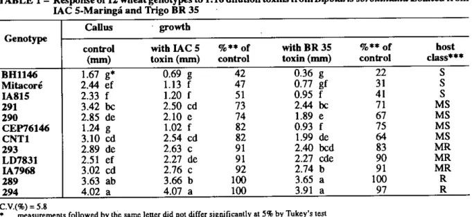
The growth measurement of the calli from 12 genotypes treated with the 1AC 5-Maringá and Trigo BR 35 toxins at 1:16 dilution were significantly different among each other and in relation to the control treatment. The toxins delayed the callus growth, where the genotypes BH 1146, Mitacoré and IA 815 were the most affected, producing callus growth 50% inferior in size, when compared with the callus of the same genotypes without toxin. CEP 76146, CNT 1, line 290 and line 291 developed callus growth varying between 60 and 80%, while IA 7968, LD 7813 and Tine 293 had callus growth greater than 90% compared to the treaunent without toxins. On the olhei- hand, the callus development of line 289 and line 294 were similar to those observed on the callus without toxins (Table 1). The genotypes were classitied in four distinct groups. Genotypes 289 and 294 were considered resistant; IA 7968, LD 7831 and 293 as moderately resistant; genotypes CEP 76146, CNT 1, 290 and 291 as moderately susceptible and BH 1146, IA 815 and Mitacoré as susceptible (Table 1).
TABLE 1 - Response of 12 wheat genotypes to 1:16 dilution toxins from Bipolaris sorokiniana isolated from IAC 5-Maringá and Trigo BR 35
Callus • growth Genotype
control with IAC 5 %** of with BR 35 %** of host
(mm) toxin (mm) control toxin (mm) control class***
BH1146 1.67 g* 0.69 g 42 0.36 g 22
s
Mitacoré 2.44 ef 1.13 f 47 0.77 gf 31 IA815 2.33 f 1.20 f 51 0.95 f 41 291 3.42 bc 2.50 cd 73 2.44 bc 71 MS 290 2.85 de 2.10 e 74 1.89 e 67 MS CEP76146 1.24 g 1.02 f 82 0.93 f 75 MS CNT1 3.10 cd 2.54 cd 82 1.99 de 64 MS 293 2.89 de 2.63 c 91 2.40 bcd 83 MR LD7831 2.51 ef 2.27 de 91 2.27 cde 90 MR IA7968 3.02 cd 2.76 c 92 2.74 b 91 MR 289 3.63 ab 3.66 b 100 3.65 a 100 R 294 4.02 a 4.07 a 100 3.91 a 97 R* measurements followed by the same letter did not differ significantly at 5% by Tukey's test •• growth percentage in relation to the treatment without toxin
*•* phenotypic classification of the host according to scale established for percentage callus growth relative to the control without toxin: R (resistant) > 95%, MR (moderately resistant) = 80% to 94%, MS (moderately susceptible) = 60% to 79%, and S (susceptible) <60%
nron • rinevn gn • (7I, II• ..11 ... 7 ... 1A7_1C1 1007 149
ROSA MARIA DE LUJÁN OVIEDO DE CRISTALDO, FERNANDO IRAM FELIX DE CARVALHO, MAN MOHAN KOHL', ROSA LIA BARBIERI, LUIZ CARLOS FEDERIZZI, CARLOS PIEROBOM
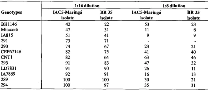
No interaction among lhe tested genotypes and the toxins was observed. Trigo BR 35 toxin, however, caused a more intense reaction than the IAC 5-Maringá toxin showing differences in aggressiveness among the isolates. The difference in aggressiveness of these isolates was clearly visible among moderately susceptible and susceptible genotypes, especially in CNT 1 and BH 1146, than among the resistant or moderately resistant genotypes. Furthermore, the resistant and moderately resistant genotypes did not have differentiated response to the toxins of the same isolates. Callus growth treated with the 1:8 dilution was lower than that observed in the 1:16 dilution. All genotypes, those with a resistance, susceptible or intermediate reaction, drastically reduced the growth of their callus obtained in the 1:16 dilution. The only exception was CNT 1 where callus size remained unchanged in both dilutions. Besides smaller calb, the genotypes developed calli with a lot of necrosis. The toxin dilution of 1:8 did not allow genotype classification among host reaction (resistance and/or susceptibility) groups. Furthermore, genotypes IA 7968 and LD 7813, considered moderately resistant
at 1:16 dilution had callus growth dose to genotypes considered susceptible, such as IA 815 and Mitacoré, especially in the presence of the Trigo BR 35 toxin. Genotypes 289, 293, 290, CEP 76146 and BH 1146, with reactions ranging from resistant to moderately susceptible with 1:16 dilution behaved the same at 1:8 dilution. However, CNT 1 had similar reaction for both 1:8 and 1:16 dilutions and along with genotype 294 were considered the most resistant genotypes in the experiment (Table 2).
Significant differences were detected in the means of the two dilutions. There were also distinct effects among the toxin within each dilution, with Trigo BR 35 toxin baving a more drastic effect than
IAC 5-Maringá toxin (Table 2). As in the case of 1:16 dilution, no interaction was observed among the toxins and the genotypes of 1:8 dilution levei. However, the toxins showed their differences in aggressiveness, with the toxin from Trigo BR 35 isolate being more aggressive than that extracted from the IAC 5-Maringá isolate. Lack of genotype x isolate interaction is indicative of absence fo fungal specialization among isolates studied.
TABLE 2 — Percentage of callus growth exposed to two dilutions (1;8 and 1:16) of two toxin filtrates (IAC 5 Maringá and Trigo BR 35) relative to the growth of toxin free control
Genotypes 1:16 dilution 1:8 dilution IACS-Maringá isolate BR 35 isolate IACS-Maringá isolate BR 35 isolate BH1146 42 22 53 23 Mitacoré 47 31 11 6 IA815 51 41 9 9 291 73 71 290 74 67 23 21 CEP67146 82 75 41 40 CNT] 82 64 63 46 293 91 83 47 32 LD7831 91 90 26 11 IA7869 92 91 16 13 289 100 100 30 21 294 100 97 35 31
Evaluation of seedling response
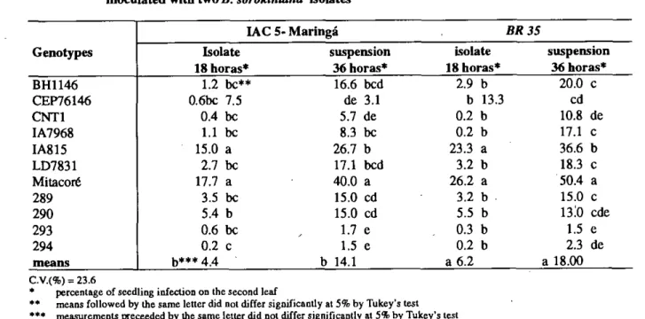
The seedling reaction caused by inoculation with the conidial suspension of the IAC 5-Maringá and Trigo BR 35 isolates demonstrated significant differences among genotypes at both leveis of incubation (18 and 36 hours) (Table 3).
Seedlings incubated for 18 hours, under high relative humidity conditions, allowed classification of genotypes in two categories: resistant, such as 294 or susceptible, such as IA 815 and Mitacoré (Table
1Sn
3). On the other hand, 36 hours of incubation period allowed identification of more than two classes. The isolates IAC 5-Maringá and Trigo BR 35 allowed the ranking of the tested genotypes in the categories: resistant (294, 293 and CNT 1); moderately resistant (289, 290 IA 7968 and CEP 76146); moderately susceptible (BH 1146 and LD 7831), and susceptible (IA 815 and Mitacoré). The differences in reaction were more intense in the presence of the Trigo BR 35 isolate.
RELAÇÃO ENTRE SELEÇÃO IN VITRO E EM PLÃNTULA PARA RESISTÊNCIA A Bipolaris sorokiniana EM TRIGO
TABLE 3 - Percentage of seedling infection on 12 wheat genotypes after 18 and 36 hours incubation period,
inoculated with two
B. sorokinianaisolates
Genotypes
IAC 5- Maringá
BR 35Isolate
18 horas*
suspension
36 horas*
isolate
18 horas*
suspension
36 horas*
BH1146
1.2 bc**
16.6 bcd
2.9 b
20.0 c
CEP76146
0.6bc 7.5
de 3.1
b 13.3
cd
CNT1
0.4 bc
5.7 de
0.2 b
10.8 de
IA7968
1.1 bc
8.3 bc
0.2 b
17.1 c
IM15
15.0 a
26.7 b
23.3 a
36.6 b
LD7831
2.7 bc
17.1 bcd
3.2 b
18.3 c
Mitacoré
17.7 a
40.0 a
26.2 a
50.4 a
289
3.5 bc
15.0 cd
3.2 b
15.0 c
290
5.4 b
15.0 cd
5.5 b
13.0 cde
293
0.6 bc
1.7 e
0.3 b
1.5 e
294
0.2 c
1.5 e
0.2 b
2.3 de
means
b*** 4.4
b 14.1
a 6.2
a 18.00
C.V.(%) .=
23.6percentage of scedling infection on the second leaf
•• means followed by the same letter did not differ significantly at 5% by Tukey's test *** measurcments preceeded by the same letter did not differ significantly at 5% by Tukey's test
Relationship between
in vitroand seedling
selection
A correlation analysis to check the existente of a
possible relationship among the genotypes selected by
the
in vitrotoxins and those classified by isolate conidial
suspension was carried out (Table 4).
Significant and negative correlation coefficients
(0.47 to -0.68) were observed between the toxin dilution
and the incubation period under high relative humidity.
Thus, a bigger callus growth was related to a lower
seedling infection. The highest correlation (-0.68) was
observed for the 1:8 dilution and 36 hours of incubation
period. On the other hand, the correlation coefficient
detectecl for the 1:16 dilution and 36 hours of incubation
(-0.51) was similar to the coefficient of the 1:8 dilution
and 18 hours of incubation (-0.52) (Table 4).
TABLE 4 - Correlation matriz between callus in 1:8 and 1:16 dilutions of
B. sorokiniana toxins and thepercentage of seedling infection assessed after 18 hours and 36 hours of incubation period
1:16 dilution
1:8 dilution
18 hours
36 hours
1:16 dilution
1:8 dilution
18 hours
36 hours
1.00
0.87*
-0.47*
-0.51*
100
-0.52*
-0.68*
1.00
0.85
1.00
P(0.05)
The experiments carried out
in vitroand
in vivoshowed a small but significant and negative correlation,
based on callus growth in toxin filtrate and seedling
infection measured in percentage of lesions. Other
parameters, such as number and size of lesions, and
chlorosis were not considered. The latter is an important
factor to determine the degree of susceptibility (MEIrrA,
1981,a). Thus, genotypes wit few and small lesions,
without chlorosis, would be consideredresimant, while
the genotypes wit the same number of lesion but with
pronounced chlorosis would belong to a distinct class.
However, the significance of the correlations indicated
that there were correspondente between the
in vitroresponse of the callus growth and the seedling reaction
to infection.
CONCLUSIONS
In vitro
selection of wheat callus for resistance to
spot blotch, using fungai toxin filtrates, was shown to
be a relatively simple and highly feasible technique.
Considering the elimination of the pathogen in the
ROSA MARIA DE LUJAN OVIEDO DE CRISTALDO, FERNANDO IRMA FÊLIX DE CARVALHO, MAN MOHAN KOHLI, ROSA LIA BARBIERI, LUIZ CARLOS FEDEFUZZL CARLOS PIEROBOM
filtrate under controlled environmental conditions, the assessment criteria of Lhe callus exposed to a 1:16 dilution of the toxin filtrates allowed an accurate screening of the genotypes according to their seedling reaction to the pathogen.
A smaller callus growth corresponded to a higher percentage of lesions on lhe seedling leaves, determining a negative correlation among
in
vitro andin vivo
selection. The existence of this correlation allows, therefore, the use of phytotoxins ofB. sorokiniana
as possible selection procedere in wheat callus tissue to determinate its resistance to the disease.REFERENCES
ALAM, B.K. Genetic regulation of host-selective toxin production; characterization of necrosis minus mutant of Pyrenophora tritici-repentis (DIED.) Drech. which has lost the ability to produce host-selective toxins in wheat. Columbia: University of Missouri, 1989. 128 p. Thesis (Ph.D), University of Missouri, 1989.
BARBIERI, R.L. Genética da resistência ao
Helminthosporium salivam em trigo: uso de filtrados tóxicos em cultura de tecidos. Porto Alegre:UFRGS,
1995.47 p. Dissertação (Mestrado) —Genética e Biologia Molecular, Departamento de Genética, Instituto de Biociências, UFRGS, 1995.
BEHNKE, M. General resistance to late blight o fSolanum
tuberosum plants regencrated from callus resistant to culture filtrates of Pyrenophora infestans. Theoretical and A pplied Genetics, Berlin, v. 56, p. 151-152, 1980.
CHRISTENSEN, 1.1. Physiologic specialization and mutation in Helminthosporium salivam. Phytopathology, St. Paul, v. 15, p. 785-795, 1925.
CRISTALDO, R.M.L.O. Uso de filtrados tóxicos para avaliar a resistência ao fungo Helminthosporium sativum em trigos hexaplóldes in vitro. Porto Alegre: UFRGS, 1993. 129 p. Tese (Doutorado) — Fitotecnia, Departamento de Plantas de Lavoura, Faculdade de Agronomia, UFRGS, 1993.
DAUB, M. Tissue culture and the selection of resistance to pathogens. Annual Review of Phytopathology,
Stanford, v. 23, p. 159-181, 1986.
DIEHL, J. Podridão comum. In: TRIGO no Brasil. Campinas: Fundação Cargill, v.2, cap. 12, p. 501-508,1982. HANDEL, C.L. Avaliação in vitro da resistência à
helminthosporiose em avela através do uso de filtrados tóxicos do fungo e do inseticida Methomyl. Porto Alegre: UFRGS, 1996. 69 p. Dissertação (Mestrado) — Fitotecnia, Departamento de Plantas de Lavoura, Faculdade de Agronomia, UFRGS, 1996.
JAMES, W.C. An illustrated series of assessment keys for plant diseases their preparation and usage. Canadian Plant
Disease Survey, Ottawa, v. 51, n. 2, p. 20-73, 1971.
LINDEN, A.R. Alterações na patogenicidade, morfologia e atividade isoesterásica de Hebnintosporium salivam
decorrentes de passagens sucessivas por duas cultivares de trigo. Porto Alegre: UFRGS, Faculdade de Genética, UFRGS, 1989. 116 p. Dissertação (Bacharelado) — Genética, Departamento de Genética, Instituto de Biociências, UFRGS, 1989.
LUDWING, R. A. Toxin production by Helminthosporium salivam P.K. & B. and its significance in disease development. Canadian inumai of Botaay, Otawa, v. 35, p. 291-303, 1957.
LUZ, W.C. Influência do período de umidificação pés-inoculação na reação de cultivares de trigo a mancha foliar (Cochliobolus salivas). Fitopatologia Brasileira,
Brasília, v. 7, p. 111-115, 1982.
LUZ, W.C.; BERGSTROM, G.C. Temperature-sensitive development of spot blotch in spring wheat cultivars differing in resistance. Fitopatologia Brasileira,
Brasília, v. 11, p. 197-204, 1986.
MATSUMURA, A.T. Variabilidade intraespecífica quanto a patogenicidade, características de cultura e padrão isoesterásico em populações naturais de Bipolaris sorokiniana (Helminthosporium sativum). Porto Alegre: UFRGS, 1991. 262 p. Tese (Doutorado) — Genética, Departamento de Genética, Instituto de Biociências, UFRGS, 1991.
MENTA, Y. R. Produção de conídios, período de esporulação e extensão da lesão por Helminthosporium salivam nas folhas bandeiras de trigo. Pesquisa Agropecuária Brasileira, Brasflia, v. 16, n. 1, p. 77-99, 1981,a. MENTA, Y. R. Identification of races of Helminthosporium
sativum of wheat in Brazil. Pesquisa Agropecuária Brasileira, Brasília, v. 16, n. 3, p. 331-336, 1981,6. MEREDITH, C.P. Selecting better crops from cultured cells. IN: STADLER GENETICS SYMPOSIUM 16., 1986, Columbia. Gene manipulation ia plant improvement. New York: Plenum Press, 1984. p. 149:172. 1984. MERONUCK, R.A.; PEPPER, E.H. Clamidospore formation
in condia of Helminthosporium salivam Phytopathology,
St. Paul, v. 58, p. 866. 1968.
MURASHIGE, T.; SKOOG, F. A revised medium for rapid growth biossays with tobacco tissue cultures. Physiologia Plantarum, Copenhagen, v. 15, p. 473-497, 1962. REIS, E. M. Levantamento de plantas cultivadas, nativas e
invasoras hospedeiras de fungos causadores de podridões radiculares em cereais de inverno e em outras culturas.
Summa Phytopathologica, Jaguariúna, v. 8, p. 134-140, 1982.
TANIGUCHI, E.; WHITE, G.A. Site of action of the phytotoxin helminthosporal. Biochemistry and Biophysical Research Communications, New York, v. 28, p. 879-885, 1967,