online | memorias.ioc.fiocruz.br
TNF/TNFR1 signaling up-regulates CCR5 expression by CD8
+T
lymphocytes and promotes heart tissue damage during
Trypanosoma cruzi infection: beneficial effects of TNF-a blockade
Karina Kroll-Palhares,Jaline Coutinho Silvério, Andrea Alice da Silva1, Vladimir Michailowsky2, Ana Paula Marino,Neide Maria Silva3, Cristiano Marcelo Espinola Carvalho,
Luzia Maria de Oliveira Pinto, Ricardo Tostes Gazzinelli2, Joseli Lannes-Vieira/+
Laboratório de Auto-imunidade e Imuno-regulação (LAIIR), Instituto Oswaldo Cruz-Fiocruz, Av. Brasil 4365, 21045-900 Rio de Janeiro, Brasil 1Laboratório de Imunologia, Departamento de Patologia, Universidade Federal Fluminense, Niterói, RJ, Brasil 2Laboratório de
Imu-nopatologia, Instituto Rene Rachou-Fiocruz, Belo Horizonte, MG, Brasil 3Departamento de Morfologia Instituto de Ciências Biomédicas,
Universidade de Uberlândia, Uberlândia, MG, Brasil
In Chagas disease, understanding how the immune response controls parasite growth but also leads to heart
damage may provide insight into the design of new therapeutic strategies. Tumor necrosis factor-alpha (TNF-α) is
important for resistance to acute Trypanosoma cruzi infection; however, in patients suffering from chronic T. cruzi
infection, plasma TNF-α levels correlate with cardiomyopathy. Recent data suggest that CD8-enriched chagasic
myocarditis formation involves CCR1/CCR5-mediated cell migration. Herein, the contribution of TNF-α, especially
signaling through the receptor TNFR1/p55, to the pathophysiology of T. cruzi infection was evaluated with a focus
on the development of myocarditis and heart dysfunction.Colombian strain-infected C57BL/6 mice had increased
frequencies of TNFR1/p55+ and TNF-α+ splenocytes. Although TNFR1-/- mice exhibited reduced myocarditis in the
absence of parasite burden, they succumbed to acute infection. Similar to C57BL/6 mice, Benznidazole-treated
TNFR1-/- mice survived acute infection. In TNFR1-/- mice, reduced CD8-enriched myocarditis was associated with
defective activation of CD44+CD62Llow/- and CCR5+ CD8+ lymphocytes.Also, anti-TNF-α treatment reduced the
fre-quency of CD8+CCR5+ circulating cells and myocarditis, though parasite load was unaltered in infected C3H/HeJ
mice. TNFR1-/- and anti-TNF-α-treated infected mice showed regular expression of connexin-43 and reduced
fibronectin deposition, respectively. Furthermore, anti-TNF-α treatment resulted in lower levels of CK-MB, a
cardiomyocyte lesion marker. Our results suggest that TNF/TNFR1 signaling promotes CD8-enriched myocarditis formation and heart tissue damage, implicating the TNF/TNFR1 signaling pathway as a potential therapeutic target
for control of T. cruzi-elicited cardiomyopathy.
Key words: heart disease - inflammation - Trypanosoma cruzi - CCR5 - TNFR1 - TNF-α
Chagas disease, caused by the hemoflagellate proto-zoan Trypanosoma cruzi, afflicts 15-16 million people in South America with 75-90 million people exposed to infection (Coura 2007). Approximately 30% of pa-tients develop a chronic disease typically characterized by myocarditis associated with prominent fibrotic scar-ring and organ dysfunction (Teixeira et al. 2006, Coura 2007). Although heart inflammatory cells contribute to control parasite growth, they are also involved in per-petuating heart disease (Freitas et al. 2005). CD8+ T cells
predominate in myocardium from cardiac patients (Hi-guchi et al. 1997) and T. cruzi-infected mice(dos Santos et al. 2001). In experimental T. cruzi infection, the prev-alence of CD8+ T cells in heart tissue has been linked
Financial support: CNPq (501561/2004-8, 301504/2005-9, 471518/2006-9), Faperj (E-26/151999/2000)
+Corresponding author: [email protected] Received: 17 March 2008
Accepted: 9 June 2008
with early production of the CC-chemokines CCL3/
MIP-1α, CCL4/MIP-1β, and CCL5/RANTES as well as
up-regulation of CCR5 on circulating leukocytes (Mari-no et al. 2004). Treatment with Met-RANTES, a partial CCR1/CCR5 antagonist, decreases cell infiltration in
T. cruzi-infected murine hearts and has a beneficial
effect on survival (Marino et al. 2004). In addition, infect-ed CCR5-/- mice have deficient recruitment of T cells and
macrophages to the heart(Hardison et al. 2006). These findings implicate CCR5 in the active development of
T. cruzi-elicited myocarditis.
Cytokines produced in the heart tissue of T. cruzi -infected individuals during the initial immune response can also influence the regulation of subsequent immune
reactions (Brener & Gazzinelli 1997). TNF-α is detect -able in inflamed hearts of chronic cardiomyophatic cha-gasic patients (D’Avila Reis et al. 1993), produced by T cells derived from endomyocardial biopsies (Abel et al.
2001). This suggests that TNF-α might be involved in the
maintenance of chronic myocarditis.
TNF-α signaling via TNFR1 (p55/60), but not TNFR2
TNF-a in Trypanosoma cruzi-induced cardiopathy • Karina Kroll-Palhares et al.
376
production, and cell mobilization to sites of infection (Aliberti et al. 2001). Importantly, TNF-α plasma lev -els directly correlate with the degree of heart dysfunc-tion in chronic chagasic patients (Ferreira et al. 2003, Perez-Fuentes et al. 2003). In this context, the present study was undertaken to better understand the
partici-pation of TNF-α, especially signaling via TNFR1/p55,
in the pathophysiology of T. cruzi infection, focusing on the mechanisms involved in cell activation, myocarditis development, and heart dysfunction.
MATERIALS AND METHODS
Animals- Five- to seven-week-old female C57BL/6
(H-2d) mice, B6.129-Tnfraf1a (p55/60)-deficient mice,
and C3H/HeJ (H-2k) mice obtained from the animal
fa-cilities of the Oswaldo Cruz Foundation (Fiocruz, Rio de Janeiro) were maintained under standard conditions and manipulated according to the institutional guide-lines for animal ethics and biosafety of Fiocruz (protocol #161/03). All B6.129-Tnfraf1a (p55)-deficient mice were genetically characterized using the primers and proto-cols described at The Jackson Laboratories home page (http://www.jax.org/).
Experimental infection - Mice were infected
intra-peritoneally with 5,000, 1,000, or 100 blood trypomas-tigotes (bt) of the low virulence Colombian strain of
T. cruzi isolated and maintained as previously described
(dos Santos et al. 2001). Parasitemia was estimated using 5 µl of blood obtained from the tail vein, and employed as a parameter to establish acute and chronic phases (dos Santos et al. 2001). In some experiments, the animals were treated with a subcurative dose of Benznidazole (100 mg/kg/day) from days 10 to 17 after infection (Mi-chailowsky et al. 2001).
TNF blocking protocol - After 14 days of infection,
C3H/HeJ (H-2k) mice were subcutaneously treated with
injection grade saline (BioManguinhos-Fiocruz, Brazil)
containing 10 µg of anti-human TNF-α blocking antibody
(Remicade®, Infliximab), a gift from Schering-Plough of
Brazil, at 48 h intervals over 14 days. Infliximab was
previously shown to block in vivo TNF-α biological
activities in murine models (Redlich et al. 2002).
Antibodies and reagents - A polyclonal antibody
rec-ognizing T. cruzi antigens was produced in our labora-tory (LAIIR). A polyclonal antibody recognizing mouse fibronectin was obtained from Gibco (USA). A poly-clonal antibody recognizing connexin-43 was purchased from Sigma (USA). Purified anti-F4/80 antigen (clone F4/80) antibody was purchased from Caltag Laborato-ries (USA). Supernatants used for immunohistochemis-try staining were produced in-house using anti-mouse CD8a (53-6.7) and anti-mouse CD4 (GK1.5) hybridomas. Biotin- and FITC-conjugated anti-mouse CD8a (53-6.7), PE- and biotin-conjugated anti-mouse CD4 (GK1.5), biotin-conjugated anti-CD62L (clone MEL-14), FITC-conjugated CD44 (IM7.8.1), PE-FITC-conjugated
anti-mouse TNF-α (MP6-XT22), PE-conjugated anti-anti-mouse
CCR5 (C34-3448), biotin-conjugated anti-ICAM-1 (CD54, clone 3E2), isotype control, and Cy-Chrome-streptavidin
were purchased from BD PharMingen (USA). Biotinylated anti-rat immunoglobulin was purchased from Dako (Denmark). Biotinylated anti-rabbit immunoglobulin and peroxidase-streptavidin complex were purchased from Amersham (England). Appropriate controls were prepared by replacing primary antibodies with purified rat immunoglobulin or normal rabbit serum.
Histopathology - Groups of 5-7 infected mice and 3-4
age-matched controls were killed under anesthesia at vari-ous time points post-infection. Hearts were removed and fixed in 4% buffered formalin. Sectionswere analyzed by light microscopy after paraffin-embedding andstandard hematoxylin and eosin (H&E) staining. Tissue parasit-ism was scored by counting the total amastigote nests in each section (50-80 microscopic fields, 250x magnifica-tion). For the inflammatory infiltrate score, the number of inflammatory foci present in 50 microscopic fields was counted in each section (dos Santos et al. 2001).
Immunohistochemistry (IHS) - The animals were
killed under anesthesia at various time points post-infection. The hearts were removed, embedded in tis-sue freezing medium (Tistis-sue Tek, Miles Laboratories, USA), and stored in liquid nitrogen. Serial 5 µm-thick serial cryostat sections were fixed in cold acetone and subjected to indirect immunoperoxidase staining or im-munofluorescence, as previously described (dos Santos et al. 2001). Sections of spleen were used as positive controls for lymphocyte staining. Cells stained for CD4, CD8, and F4/80 were counted using light microscopy, with positive cells identified by a partial or complete rim of dark color outlining the cell membrane. Three sections were counted for each animal and the data are present-ed as average and standard deviation of each group of animals. The expression of connexin 43 and fibronectin in heart tissue sections was also evaluated using a digital area evaluation. Images were digitized using a color view
XS digital video camera adapted to a Zeiss microscope.
The images were analyzed using the AnaliSIS 3.2 Soft Image; two sections per heart and 25 fields per section were analyzed. The results are expressed as percentage of positive area per 25 mm2 of area in the heart or as the
distance (µm) between gap junctions.
Flow cytometric analysis - The animals were killed
ex-clude dead cells from the analysis. At least 12,000 cells were acquired inside this gate. Fluorescence gates were cut in accordance to labeling controls, respecting curve inflexions. Cytometric analyses were performed using the program WinMDI Version 2.8.
Statistical analysis - Data are expressed as arithmetic
means ± standard error. Student's t test was used to ana-lyze the statistical significance of the observed differ-ences. The Kaplan-Meier method was used to compare the survival times of the studied groups. All statistical tests were performed with SPSS-8.0 (Michigan Univer-sity, USA). Differences were considered statistically sig-nificant when p < 0.05.
RESULTS
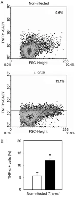
T. cruzi infection increases expression of TNFR1 and TNF-α- When C57BL/6 mice were infected with 100 bt of the low virulence Colombian strain, surface TNFR1
and intracellular TNF-α were up-regulated in spleno
-cytes at 28 days post-infection (dpi) (Figs 1a, 2b). TNF-α levels in serum and TNF-α mRNA in the heart tissue
were increased during acute infection (28 dpi) and re-mained elevated throughout the chronic stage (120 dpi), as previously described (Starobinas et al. 1991, dos San-tos et al. 2001).
TNFR1 and TNF-α participate but are not critical
for control of T. cruzi growth - TNFR1-/- mice infected
with 5,000 or 100 bt exhibited increased parasitemia (e.g., when animals received 100 bt, 4.7 ± 2.4 x1 04 bt/ ml
in C57BL/6 versus 19.2 ± 12.5 x 10⁴ bt/ ml in TNFR1-/- at
28 dpi) and died from 25 to 50 dpi or from 28 to 100 dpi, respectively. C57BL/6 mice survived the acute infection independent of the number of parasites injected (Table I). Although cardiac parasitism was decreased at 15 dpi in TNFR1-/- mice infected with 5,000 bt, when TNFR1-/-
mice received 100 bt a trend to increased parasitism was observed at 28 dpi. However, no parasite burden was observed in T. cruzi-infected TNFR1-/- mice when
com-pared with C57BL/6 mice (Table I). Interestingly, in T.
cruzi-infected C3H/HeJ mice treated with anti-TNF-α,
parasitemia (3.4 ± 3.2 x 10⁵ bt/ ml in non-treated versus
5.8 ± 3.8 x 10⁵ bt/ ml in anti-TNF-α-treated miceat 32 dpi)
and the numbers of heart parasite nests were similar to saline-injected controls (Table I). These data suggest that
TNF-α and TNFR1 participate in but are not essential to
the control of parasite growth. In fact, 100% of TNFR1
-/-animals infected with 5,000 bt and treated with a subcura-tive dose of Benznidazole (100mg/ kg/ day) from day 10 to 17 dpi survived the acute infection and had no parasite nests in the cardiac tissue at 100 dpi (Table I).
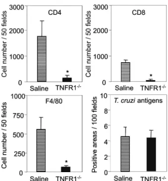
TNFR1 signaling is required for T. cruzi-elicited
CD8-enriched myocarditis formation - The importance
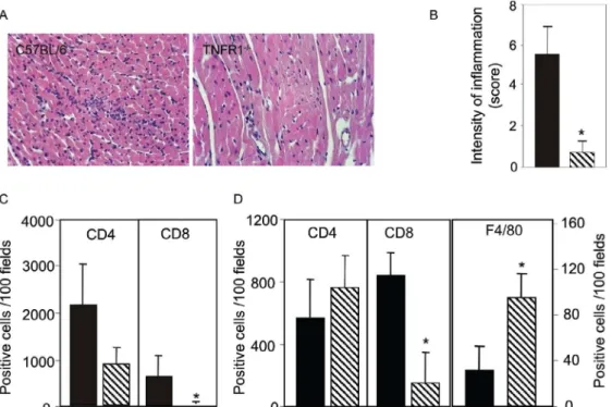
of TNF/TNFR1/p55 signaling in the development of myocarditis during acute T. cruzi infection was apparent in TNFR1-/- mice. In comparison with C57BL/6 mice,
when TNFR1-/- mice were infected with 5,000 bt,
myo-carditis formation was hampered at 15 dpi (Figs 2a, 2b), in association with a decrease in the numbers of CD4+
Fig. 1: increased expression of membrane TNFR1 and intracellular
TNF-α in splenocytes of T. cruzi infected C57BL/6 mice. Splenocytes were isolated at 28 days post infection and analyzed using flow-cytometry (gate R1). Representative flow-flow-cytometry plots show (A) increased TNFR1 expression in T. cruzi-infected mice. Intracellular
TNF-α expression was significantly enhanced in T. cruzi-infected mice (B). Vertical lines represent the standard deviations of the means of the results obtained from three to five mice. Asterisk indicates p < 0.05, T. cruzi-infected mice (black bars) in relation to non-infected controls (white bar).
and, mainly, CD8+ lymphocytes (Fig. 2c). Furthermore,
when TNFR1-/- mice infected with 5,000 bt were treated
TNF-a in Trypanosoma cruzi-induced cardiopathy • Karina Kroll-Palhares et al.
378
with 100 bt, hearts from C57BL/6 mice displayed a pro-gressively increased percentage of infiltrating CD8+ T
cells at 28 dpi, while few CD8+ cells infiltrated the hearts
from infected TNFR1-/- mice (Fig. 2d). In addition,
T. cruzi-infected TNFR1-/- mice had a higher
percent-age of F4/80+ cells (macrophages) among their heart
infiltrates than C57BL/6 mice (Fig. 2d). The decreased myocarditis was not associated with
re-compartmen-talization of inflammatory cells to other muscle tissues as low numbers of CD4+, CD8+, and F4/80+ cells were
found in the skeletal muscle of T. cruzi-infected TNFR1-/-
mice compared to C57BL/6 mice (supplementary data). Interestingly, in infected TNFR1-/- mice, diminished
myocarditis was associated with a decreased frequency of heart blood vessels expressing ICAM-1 (66.7 ± 4.5% in C57BL/6 versus 9.1 ± 6.8 % in TNFR1-/- at 28 dpi). Fig. 2: reduction of heart inflammation mainly due to decreased accumulation of CD8+ T cells in T. cruzi-infected TNFR1-/- mice. C57BL/6 and TNFR1-/- mice were infected with 5,000 (A, B, C) or 100 (D) blood trypomastigotes of the Colombian strain of T. cruzi. Heart sections of T. cruzi -infected C57BL/6 and TNFR1-/- mice sacrificed at 15 days post infection (dpi) (A) were stained with conventional hematoxylin and eosin stain
for analysis of inflammatory infiltrate. Original magnifications 200X.Inflammatory scores (B) were determined in blind by two independent
observers. T. cruzi-infected animals were sacrificed at 15 (C) or 28 (D) dpi, sections of frozen heart tissues were immunohistochemically stained for CD4+, CD8+ and F4/80+ cells and the numbers of positive cells were counted. Vertical lines represent the standard deviations of the means of the results obtained from five mice. Asterisks indicate p < 0.05, TNFR1-/- (striped bars) in relation to C57BL/6 (black bars) T. cruzi-infected mice.
TABLE I
Heart parasitism and survival of T. cruzi-infected C57BL/6 and TNFR1-/- mice and T. cruzi-infected C3H/HeJ mice treated with anti-TNF-α
Experimental groupa
(experimental day) Inoculum Heart Parasitism Survival (%)
C57BL/6 (15 dpi) 5,000 74.08 ± 5.64b 100
TNFR1-/- (15 dpi) 5,000 41.2 ± 12.2c 100
C57BL/6 (28 dpi) 100 23.15 ± 9.84 100
TNFR1-/- (28 dpi) 100 48.98 ± 20.18 90-100d
C57BL/6 (45 dpi) 100 40.53 ± 20.77 100
TNFR1-/- (45 dpi) 100 31.8 ± 16.27 45-5 c, d
C57BL/6 (100 dpi) 5,000 4.3 ± 0.9 100
C57BL/6 + Bz (100 dpi) 5,000 ND 100
TNFR1-/- (100 dpi) 5,000 Nt 0
TNFR1-/- + Bz (100 dpi) 5,000 ND 100c
C3H/HeJ (32 dpi) 100 14 ± 7.7 100
C3H/HeJ + anti-TNF-α (32 dpi) 100 11.5 ± 2.6 100
Signaling via TNFR1 is required for complete CD8+
T-cell activation - To understand the molecular
mecha-nisms hampering the formation of CD8-enriched myo-carditis in T. cruzi-infected TNFR1-/- mice, we evaluated
the numbers and activation phenotypes of mononuclear cells in the spleen and blood. T. cruzi infection of C57BL/6 mice resulted in a remarkable splenomegaly associated with enhanced cellularity; these effects were hindered in infected TNFR1-/- mice (Table II). Although
the frequencies of total B and T cells were similar in both C57BL/6 and TNFR1-/- infected mice during the
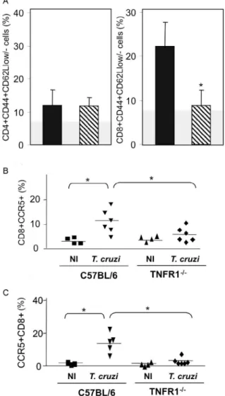
acute phase of infection (Table II), a significant accu-mulation of CD8+CD44+CD62Llow/- activated/memory
lymphocytes was observed in the spleens of TNFR1
-/-mice (4.56% ± 0.04% in C57BL/6 versus 11.67% ± 5.55% in TNFR1-/-), with a corresponding decrease of this cell
population in peripheral blood (Fig. 4a). This alteration was restricted to CD8+ T cells, as similar frequencies of
CD4+CD44+CD62-Llow/- activated/memory T cells were
seen in C57BL/6 and p55-deficient mice in both spleen (22.67% ± 2.04% in C57BL/6 versus 22.95% ± 10.69% in TNFR1-/-) and blood (Fig. 3a).
Since CCR5 is required to recruit T cells into car-diac tissue during T. cruzi infection (Marino et al. 2004, Hardison et al. 2006) and TNFR1-/- mice had defective
cardiac infiltration and splenic retention of CD8+ cells,
we studied the frequency of CD8+CCR5+ cells
avail-able for tissular recruitment in infected TNFR1-/- mice.
During T. cruzi infection, an increased frequency of CD8+CCR5+ cells was detected in the blood (Fig. 3b)
and spleen (Fig. 3c) of C57BL/6 mice, however these alterations were hampered in T. cruzi-infected TNFR1 -/- mice. Altogether, these findings show the requirement
for TNFR1 signaling for the acquisition of the full acti-vation phenotype associated with migration potential in CD8+ lymphocytes during T. cruzi infection.
Anti-TNF-α abrogates the up-regulation of CCR5
expression by CD8+ cells and myocarditis development
during T. cruzi infection - To test the participation of
TNF-α in controlling the expression of CCR5, particu -larly in CD4+ and CD8+ blood lymphocytes, anti-TNF-α
blocking antibody (Infliximab) was administered during acute infection of C3H/HeJ mice, a model in which the participation of CCR5+ cells in T. cruzi-induced
myo-carditis formation was described (Marino et al. 2004). During acute infection by T. cruzi, a substantial increase
Fig. 3: reduced frequency of CD8+CD44+CD62-LLow activated/memo-ry cells and CD8+CCR5+ cells in T. cruzi-infected TNFR1-/- mice. The frequencies (A) of CD4+CD44+CD62-LLow, CD8+CD44+CD62-LLow and CD8+CCR5+ T cells in peripheral blood (B) and spleen (C) of acutely (28 days post infection) T. cruzi-infected C57BL/6 and TNFR1-/- mice were analyzed using flow-cytometry. A: vertical lines represent the standard deviations of the means of the results obtained from five mice. The gray area represents the mean plus 2-fold standard deviation of non-infected mice in the analyzed phenotype. Asterisks indicate p < 0.05, TNFR1-/- (striped bars) in relation to C57BL/6 (black bars)
T. cruzi-infected mice. B, C: horizontal lines represent the means of the results obtained from four to six mice. Asterisks indicate p < 0.05,
T. cruzi-infected mice in relation to non-infected controls (NI).
TABLE II
Relative weight, cellularity and frequency of B and T cells in the spleen of T. cruzi-infected C57BL/6 and TNFR1-/- mice
Relative spleen
Experimental groupa weight (mg/g) Spleen cellularity (x 108) B cells (%) T cells (%) C57BL/6 normal 2.82 ± 0.65 35.5 ± 12.5 14.0 ± 2 62.7 ± 3.2 C57BL/6 28 dpi 16.5 ± 7.2b 144 ± 24.7b 16.6 ± 0.5 55.3 ± 8.2
TNFR1-/- normal 3.86 ± 1.61 49.5 ± 14.5 12.2 ± 1.7 78.5 ± 3.1 TNFR1-/- 28 dpi 5.2 ± 3.7b, c 35.4 ± 7.0 13.2 ± 2.4 73.3 ± 7.2
TNF-a in Trypanosoma cruzi-induced cardiopathy • Karina Kroll-Palhares et al.
380
in the numbers of circulating leukocytes was observed (4,000 ± 1,340 x 103/mm3 in non-infected versus 17,760
± 2,478 x 103/mm3 in T. cruzi-infected mice). Treatment
with anti-TNF-α did not alter the leukocytosis (13,100
± 2,673 x103/mm3 in saline-injected versus 15,650 ±
2,800 x 103/mm3 in anti-TNF-α-treated), showing that
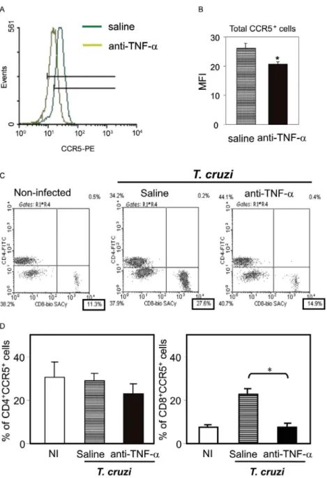
this treatment did not exacerbate the aberrant leukocyte activation. Interestingly, the up-regulation of CCR5, measured as mean fluorescence intensity, by circulating leukocytes in T. cruzi-infected mice was prevented when
the animals were treated with anti-TNF-α every 48 h
from 14 to 32 dpi (Figs 4a, 4b). The abrogation of CCR5 Fig. 4: modulation of TNF-α results in decreased expression of CCR5 by CD8+ cells in T. cruzi-infected mice. C3H/He mice were infected with 100 blood trypomastigotes of the Colombian strain of T. cruzi. After 14 days post of infection (dpi) the animals were treated with 10 µg of
anti-human TNF-α blocking antibody (black bars) or vehicle (striped bars), every 48 h during 14 days. The animals were anesthetized, blood was collected at 32 dpi. Representative flow-cytometry plot shows decreased CCR5 expression in PBMC of anti-TNF-α-treated infected mice
(A). The mean fluorescence intensity (MFI) confirms this finding (B). Vertical lines represent the standard deviations of the means of the
re-sults obtained from five mice. Asterisks indicate p < 0.05, anti-TNF-α-treated (black bars) in relation to vehicle-injected controls (striped bars).
Representative flow-cytometry plots (C) of PBMC lymphocytes (gate R1) showing CD4+ and CD8+ cells among CCR5+ lymphocytes (gate R1 and R4) show increased frequency of the CD8+CCR5+ subset in T. cruzi-infected animals in comparison with non-infected controls and
abro-gation of CCR5 up-regulation in anti-TNF-α-treated T. cruzi-infected. Frequencies (D) of CD4+CCR5+ and CD8+CCR5+ subsets in peripheral
blood of non-infected (white bars) and vehicle-injected (striped bars) and anti-TNF-α-treated (black bars) T. cruzi-infected mice. Vertical lines represent the standard deviations of the means of the results obtained from five mice. Asterisks indicate p < 0.05, vehicle-injected T. cruzi
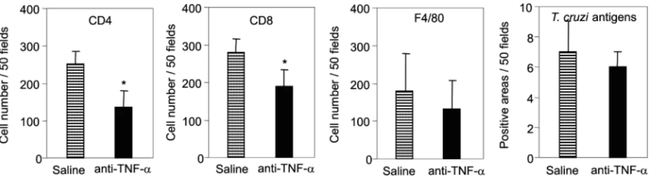
Fig. 5: reduction of myocarditis formation in anti-TNF-α-treated T. cruzi-infected mice in absence of parasite burden. C3H/He mice were in-fected with 100 blood trypomastigotes of the Colombian strain of T. cruzi and treated with anti-TNF-α or vehicle as described in Fig. 4. The animals were killed at 32 days of infection, the hearts were removed, embedded in freezing medium and tissue sections were submitted to im-munohistochemistry staining. The numbers of CD4+, CD8+, F4/80+ cells and T. cruzi antigen+ areas were counted. Significant reduction in the numbers of CD4+ and CD8+ cells was observed in the inflammatory lesions of anti-TNF-α-treated infected mice. Similar numbers of F4/80+ cells and T. cruzi antigen+ areas were detected in heart sections of vehicle-injected or anti-TNF-α-treated infected mice. Vertical lines represent
the standard deviations of the means of the results obtained from five mice. Asterisks indicate p< 0.05, anti-TNF-α-treated infected mice (black
bars) in relation to vehicle-injected T. cruzi-infected mice (striped bars).
up-regulation was mainly observed in CD8+ T cells (Fig.
4c) and the frequency of CD8+CCR5+ T cells in
anti-TNF-α-treated infected mice resembled that found in
non-infected controls (Fig. 4d).
We next tested the effect of TNF-α blocking in
myocarditis formation. A significant reduction in the numbers of CD4+ and CD8+ T cells was observed in the
cardiac tissue of animals that received anti-TNF-α. In
addition, the numbers of macrophages and areas positive for parasite antigens were similar in both groups (Fig. 5).
These results indicate that TNF-α is essential for CCR5
up-regulation involved in recruitment of lymphocytes to the heart during T. cruzi infection, and reinforce the hy-pothesis that massive heart inflammation is unrelated to the control of parasite growth.
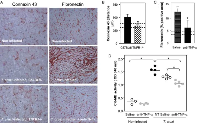
TNF/ TNFR1 signaling is involved in cardiac
tis-sue damage during T. cruzi infection - TNF-α is poten
-tially harmful to heart tissue in chronic Chagas disease (Ferreira et al. 2003, Perez-Fuentes et al. 2003). Thus, the functional integrity of myocardium was checked in TNFR1-/- and anti-TNF-α-treated infected mice through
IHS using markers that are altered during T. cruzi infec-tion, the gap junction marker connexin 43 (Campos-de-Carvalho et al. 1992) and fibronectin deposition (Ma-rino et al. 2004). In non-infected controls, the expression of connexin 43 was regular and organized. However in
T. cruzi-infected C57BL/6 mice, the expression of
con-nexin 43 was reduced, disorganized, and more scattered, as characterized by increased distance between connexin spots. In contrast, in T. cruzi-infected TNFR1-/- mice, the
expression of connexin 43 resembled that of non-infected controls (Fig. 6a). Moreover, the distance between gap junctions in T. cruzi-infected TNFR1/p55-deficient mice was similar to non-infected mice, contrasting with T. cru-zi-infected C57BL/6 mice (Fig. 6b). When compared to non-infected controls, a remarkable increase in deposi-tion of the extracellular matrix component fibronectin was detected in T. cruzi-infected C3H/HeJ mice.
How-ever, in anti-TNF-α-treated infected mice, the expres -sion of fibronectin was significantly reduced and similar to non-infected controls (Fig. 6a, b). More importantly, inspection of CK-MB levels, one of the markers of myo-cardial injury (de Souza et al. 2000), revealed that
anti-TNF-α-treated mice had lower CK-MB levels compared
to non-treated or saline-injected T. cruzi-infected C3H/ HeJ mice (Fig. 6d). Altogether, these results demonstrate that the TNF/TNFR1 signaling pathway is involved in heart tissue damage characterized by alterations in fi-bronectin deposition, electric conductivity, and cardio-myocyte injury during T. cruzi infection.
DISCUSSION
A collection of data implicates TNF-α in the progres -sion of chagasic cardiomyopathy, yet the contribution of this cytokine to the pathophysiology of T. cruzi infection has not been elucidated. In the present study, we provide
evidence that TNF-α, signaling through TNFR1/p55, is
an inducer of cell activation, myocarditis development, and heart tissue damage during experimental T. cruzi in-fection. We observed an increase in TNFR1-bearing and
TNF-α-expressing cells during infection of C57BL/6
mice with the low virulence Colombian strain of T. cruzi.
TNF-α production was previously shown to be enhanced
in T. cruzi-infected mice in association with
susceptibil-ity (Starobinas et al. 1991), and in chronically infected patients in association with heart dysfunction (Ferreira et al. 2003, Perez-Fuentes et al. 2003). T. cruzi-derived molecules, including
glycosylphosphaditidylinositol-anchored mucins, have been shown to induce TNF-α production (Ropert et al. 2002). TNF-α synthesis can
also be controlled by different intrinsic stimuli such as hormones, NO, cytokines, and other inflammatory me-diators (Aggarwal et al. 2002). However, since several of these molecules are up-regulated in T. cruzi infec-tion (Brener & Gazzinelli, 1997, Silva et al. 2003), the
molecular circuit leading to TNF-α overproduction in
TNF-a in Trypanosoma cruzi-induced cardiopathy • Karina Kroll-Palhares et al.
382
Previous studies using TNFR1-FcIgG3 transgenic and TNFR1-/- mice showed that TNF-α is required for the con
-trol of parasite burden during T. cruzi infection (Santos-Lima et al. 1997, Aliberti et al. 2001). TNFR1-/- mice
treat-ed from 10-17 dpi with a subcurative dose of Benznidazole survived the acute infection and exhibited reduced heart inflammation during the chronic phase, contrasting with
IFN-γ-/- and IL12-/- mice, which succumbed shortly after
treatment cessation with intense parasitemia, parasitism, and inflammatory processes (Michailowsky et al. 2001).
Therefore, though important, TNF-α is not crucial for the
control of parasite dissemination.
Our results show that TNFR1 is crucially involved in the development of T. cruzi splenomegaly. Splenomegaly induced by T. cruzi infection has been linked to aber-rant B and T cell activation (Minoprio 2001), especially CD8+ T-cell dysregulation (dos Santos et al. 2001,
Tzele-pis et al. 2007). Additionally, immunological abnormali-ties were linked to T. cruzi persistence and toxic lesions (DosReis et al. 2005). Thus, our data suggest that TNF/
TNFR1 signaling might play a detrimental role during
T. cruzi infection by contributing to the development of
immunological abnormalities, mainly involving CD8+ T
cell activation, and heart injury.
We have previously proposed that CD8-enriched
T. cruzi-elicited myocarditis formation is dependent on
the differential activation of the CD8+ T-cell population
after infection (dos Santos et al. 2001). In support of this model, the paucity of CD8+ T cells in cardiac tissues of
TNFR1-/- infected mice correlated with a decreased
fre-quency of activated/memory CD44+CD62LlowCD8+ cells
in the blood and retention of this cell subset in the spleen. These data indicate that TNF/TNFR1 signaling is not es-sential for the full differentiation of this T-cell subset, as indicated by the expression of the activation/ memory markers CD44 and CD62L, but somehow controls CD8+
T-cell compartmentalization.
CCR5 up-regulation, particularly by CD8+ T cells,
oc-curs during T. cruzi infection (Marino et al. 2004). How-ever, when TNF signaling was disrupted in TNFR1-/- mice
Fig. 6: beneficial effects of abrogation of TNF-α signaling via TNFR1 and TNF-α modulation by anti-TNF-α treatment in heart of T. cruzi -infected mice. A: C57BL/6, TNFR1-/- and C3H/HeJ mice were infected with 100 blood trypomastigotes of the Colombian strain of T. cruzi.
C3HeJ mice were treated with anti-TNF-α or vehicle as described in Fig. 4. The animals were killed at 28 or 32 days of infection, the hearts were
removed, embedded in freezing medium and tissue sections were submitted to immunohistochemistry staining for detection of connexin 43 or fibronectin deposition. Representative sections show that when compared with non-infected controls decreased expression of connexin 43 was observed in T. cruzi-infected C57BL/6 mice. This connexin loss was abrogated in TNFR1-/- . Original magnifications: 200X. B: quantification of the distance between connexin plaques shows that in TNFR1-/- mice (black bars) the distances resemble non-infected controls (dashed lines show the distance intervals found in non-infected controls). Asterisks indicate p< 0.05, TNFR1-/- (striped bars) in relation to C57BL/6 (black bars)
T. cruzi-infected mice. A: representative sections show that when compared with non-infected controls enhanced deposition of fibronectin was observed in T. cruzi-infected C3H/HeJ mice. This over deposition of fibronectin was blocked in anti-TNF-α-treated T. cruzi-infected. Original
magnifications: x100. C: quantification of interstitial fibronectin deposition (percentage of occupied area) shows that anti-TNF-α treatment
blocks fibronectin deposition in comparison with vehicle injection in T. cruzi-infected mice, resembling the amounts detected in non-infected mice (dashed lines). D: cardiomyocyte lesion was evaluated by creatine kinase cardiac isoenzyme MB (CK-MB) activity in serum samples from
or by anti-TNF-α treatment of T. cruzi-infected mice, a decrease in the frequency of CD8+CCR5+ T lymphocytes
was observed in the peripheral blood and spleen, and was associated with reduced heart inflammation. Thus, our results indicate that TNF/TNFR1 signaling is a pre-requisite for CCR5 expression by CD8+ T cells,
promot-ing CD8-enriched myocarditis formation. Nonetheless, future studies are required to elucidate the correlation
between TNF-α and CCR5 modulation, including the
involvement of intracellular signaling and transcriptional factors such as AP-1 and NF-kB, both of which are parts of the TNFR1 signaling pathway (Baud & Karin 2001).
T cells, mostly CD8+, present in the myocardium of T.
cruzi-infected mice express CCR5 (Marino et al. 2004)
in an environment enriched in TNF-α and CC-chemok -ines (dos Santos et al. 2001, Marino et al. 2004). CCR5 up-regulation by CD8+ T cells allows their attraction to
sites where the cognate chemokines are produced (Cas-tellino et al. 2006). Partial blockade of CCR5 in T. cruzi -infected mice decreased myocarditis without hampering the control of parasite growth (Marino et al. 2004). In the present study, we demonstrated that interfering with
the biological effects of TNF-α using Infliximab, an an
-tibody that blocks soluble and membrane-bound TNF-α
(Wong et al. 2008), led to CCR5 down-modulation and ameliorated heart inflammation without interfering with parasite control. On the other hand, infection of CCR5-/-
mice results in higher parasite burden, mortality, and impaired macrophage and T cell influx into the cardiac tissue (Hardison et al. 2006). Therefore, we believe that the total absence of either TNFR1 or CCR5 signaling impairs the entrance of parasite-controlling cells in the
heart, while the partial reduction of TNF-α and CCR5
signaling promotes beneficial effects for cardiomyopa-thy. Furthermore, the decreased recruitment of cells to cardiac tissue in TNFR1-/- infected mice was associated
with diminished ICAM-1 expression by heart endothe-lial cells. Thus, it seems that the TNF/TNFR1 signaling pathway plays a crucial role in inflammatory cell ex-travasation to cardiac tissue by controlling the expres-sion of CCR5 and ICAM-1, molecules involved in the establishment of T. cruzi-elicited myocarditis (Marino et al. 2004, Michailowsky et al. 2004).
The reduced myocarditis observed in TNFR1-/- and
anti-TNF-α-treated infected mice compared to the appro -priate controls was associated with diminished tissue dam-age, indicated by preserved myocardial cell connectivity and decreased fibronectin deposition, resembling non-infected controls. The expression of connexin 43, a com-ponent of cardiac gap junctions leading to myocardium electric connectivity (Delmar et al. 2004), is profoundly altered in cardiomyocytes infected in vitro with T. cruzi
(Campos-de-Carvalho et al. 1992). However, it remains to be clarified whether this is a direct effect of the parasite. Herein, we observed that abrogation of TNF/TNFR1 sig-naling preserves connexin 43 patterns in the cardiac tissue
of T. cruzi-infected mice. TNF-α has important roles in
the depression of heart function by down-regulating con-nexin 43 (Fernandez-Cobo et al. 1999). In this context, our data suggest that during T. cruzi infection TNF-α might
play a role in reducing connexin 43 expression.
Enhanced deposition of extracellular matrix compo-nents in the inflamed cardiac tissue has been described
in T. cruzi infection (Andrade et al. 1991, Marino et al.
2004). We observed that heart fibronectin deposition
was diminished in anti-TNF-α-treated infected mice,
in parallel with a reduction in inflammation. Interest-ingly, in Verapamil-treated T. cruzi-infected mice, in
situ modulation of TNF-α and IL-1β correlates with re -ductions in heart fibrosis and inflammation (Huang et al. 1999). These findings reinforce the hypothesis that
TNF-α blockade might exert a beneficial effect during
T. cruzi infection by modulating heart fibrosis or
fibro-genic inflammation.
Lastly, we found that during T. cruzi infection,
anti-TNF-α treatment partially hampered the development
of cardiomyocyte lesions, characterized by the release of CK-MB (de Souza et al. 2000), reinforcing the role of TNF-α as a contributor to cardiac injury. Recently,
TNF-α blockade has been shown to decrease necrotic
areas in the spleen during acute T. cruzi infection (An-drade et al. 2008). Conversely, treatment with Etanercept
(soluble human TNFR2/p75 that binds TNF-α and lym
-photoxin α) aggravated chronic chagasic cardiomyopa -thy in hamsters, leading to the claim that the absence of
TNF-α signaling may be deleterious to the failing heart
in Chagas disease cardiomyopathy (Bilate et al. 2007). This apparent paradox emphasizes the need for an under-standing of the modes of action and the limiting factors
of emerging novel therapeutic tools that target TNF-α (Wong et al. 2008). Furthermore, TNF-α is involved in
cytoprotective signals that prevent and/or delay the de-velopment of cardiomyocyte apoptosis (Kurrelmeyer et al. 2000). However, it is clear that prolonged exposures
to high levels of TNF-α have deleterious effects on cardiac function (Sarzi-Puttini et al. 2005). In fact, chronic cha-gasic patients with left ventricular dysfunction had 2-fold
higher TNF-α levels than patients without ventricular
dysfunction (Ferreira et al. 2003). In addition, cumulative
and complementary effects of TNF-α and NO levels cor -relate with adverse prognosis in chagasic patients (Perez-Fuentes et al. 2003), suggesting that susceptibility to se-vere chagasic cardiomyopathy is a multi-factorial process
in which TNF-α plays a protagonist role.
Thus, the biological roles played by TNF-α (and other inflammatory cytokines such as lymphotoxin α)
in T. cruzi-elicited cardiomyopathy deserve to be
fur-ther explored for the development of rational fur-therapeutic strategies. In this context, our results suggest that the detrimental effects of TNF-α in T. cruzi infection might
be determined by the degree and duration of TNF-α
production. Therefore, modulation (but not complete
abrogation) of TNF-α production or its biological action
might have a beneficial effect by keeping parasite dis-semination under control, while impairing the develop-ment of harmful myocarditis and cardiomyocyte lesions during T. cruzi infection.
REFERENCES
TNF-a in Trypanosoma cruzi-induced cardiopathy • Karina Kroll-Palhares et al.
384
Aggarwal BB, Shishodia S, Ashikawa K, Bharti AC 2002. The role of TNF and its family members in inflammation and cancer: les-sons from gene deletion. Curr Drug Targets Inflamm Allergy1: 327-341.
Aliberti JC, Souto JT, Marino AP, Lannes-Vieira J, Teixeira MM, Farber J, Gazzinelli RT, Silva JS 2001. Modulation of chemokine production and inflammatory responses in interferon-gamma- and tumor necrosis factor-R1-deficient mice during Trypanoso-ma cruzi infection. Am J Pathol 158: 1433-1440.
Andrade SG, Freitas LA, Peyrol S, Pimentel AR, Sadigursky M 1991. Experimental chemotherapy of Trypanosoma cruzi infection: persistence of parasite antigens and positive serology in parasito-logically cured mice. Bull World Health Organ 69: 191-197.
Andrade SG, Magalhaes LA, Pessina DH 2008. Importance of TNF-α
in the course of acute infection with Trypanosoma cruzi: in-fluence of its inhibition by pentoxiflyline treatment. Mem Inst Oswaldo Cruz 103: 21-26.
Baud V, Karin M 2001. Signal transduction by tumor necrosis factor and its relatives. Trends Cell Biol 11: 372-377.
Bilate AM, Salemi VM, Ramires FJ, de Brito T, Russo M, Fonseca SG, Faé KC, Martins DG, Silva AM, Mady C, Kalil J, Cunha-Neto E 2007. TNF blockade aggravates experimental chronic Chagas disease cardiomyopathy. Microbes Infect 9: 1104-1113.
Brener Z, Gazzinelli RT. 1997. Immunological control of Trypanoso-ma cruzi infection and pathogenesis of Chagas’ disease. Int Arch Allergy Immunol 114: 103-110.
Campos-de-Carvalho AC, Tanowitz H, Wittner M, Dermietzel R, Roy C, Hertzberg EL, Spray DC 1992. Gap junction distribution is altered between cardiac myocytes infected with Trypanosoma cruzi. Circ Res 70: 733-742.
Castellino F, Huang AY, Altan-Bonnet G, Stoll S, Scheinecker C, Germain RN 2006. Chemokines enhance immunity by guiding naïve CD8+ T cells to sites of CD4+ T cell-dendritic cell interac-tion. Nature 440: 890-895.
Coura, JR 2007. Chagas disease: what is known and what is needed - A background article. Mem Inst Oswaldo Cruz 102: 113-122.
D’avila Reis D, Jones EM, Tostes Jr S, Lopes ER, Gazzinelli G, Colley DG, Mc Curley TL 1993. Characterization of inflammatory infil-trates in chronic chagasic myocardial lesions: presence of tumor necrosis factor-a+ cells and dominance of granzyme A+, CD8+ lymphocytes. Am J Trop Med Hyg 48: 637-644.
Delmar M, Coombs W, Sorgen P, Duffy HS, Taffet SM 2004. Struc-tural bases for the chemical regulation of Connexin43 channels.
Cardiovasc Res 62: 268-275.
de Souza AP, Olivieri BP,de Castro SL, Araújo-Jorge TC 2000. Enzy-matic markers of heart lesion in mice infected with Trypanosoma cruzi and submitted to Benznidazole chemotherapy. Parasitol Res 86: 800-808.
dos Santos PVA, Roffê E, Santiago HC, Torres RA, Marino APMP, Pai-va CN, SilPai-va AA, Gazzinelli RT, Lannes-Vieira J 2004. PrePai-valence of CD8+α T cells in Trypanosoma cruzi-elicited myocarditis is as-sociated with acquisition of CD62LLowLFA-1HighVLA-4High
activation phenotype and expression of IFN-γ-inducible adhesion
and chemoattractant molecules. Microbes Infect 3: 971-984.
DosReis GA, Freire-de-Lima CG, Nunes MP, Lopes MF 2005. The importance of aberrant T-cell responses in Chagas disease.
Trends Parasitol 21: 237-243.
Fernandez-Cobo M, Gingalewski C, Drujan D, De Maio A 1999. Downregulation of connexin 43 gene expression in rat heart dur-ing inflammation. The role of tumor necrosis factor. Cytokine 11: 216-224.
Ferreira RC, Ianni BM, Abel LC, Buck P, Mady C, Kalil J, Cunha-Neto E 2003. Increased plasma levels of tumor necrosis factor-alpha in asymptomatic/”indeterminate” and Chagas disease car-diomyopathy patients. Mem Inst Oswaldo Cruz 98: 407-411.
Freitas HF, Chizzola PR, Paes AT, Lima AC, Mansur AJ 2005. Risk stratification in a Brazilian hospital-based cohort of 1220 out-patients with heart failure: role of Chagas’ heart disease. Int J Cardiol 102: 239-247.
Hardison JL, Wrightsman RA, Carpenter PM, Kuziel WA, Lane TE, Manning JE 2006. The CC chemokine receptor 5 is important in control of parasite replication and acute cardiac inflammation following infection with Trypanosoma cruzi. Infect Immun 74: 135-143.
Higuchi, ML, Reis MM, Aiello VD, Benvenuti LA, Gutierrez PS, Bellotti G, Pileggi F 1997. Association of an increase in CD8+ T cells with the presence of Trypanosoma cruzi antigens in chronic, human chagasic myocarditis. Am J Trop Med Hyg 56: 485-489.
Huang H, Chan J, Wittner M, Jelicks LA, Morris SA, Factor SM, Weiss LM, Braunstein VL, Bacchi CJ, Yarlett N, Chandra M, Shirani J, Tanowitz HB 1999. Expression of cardiac cytokines and inducible form of nitric oxide synthase (NOS2) in Trypano-soma cruzi-infected mice. J Mol Cell Cardiol 31: 75-88.
Kurrelmeyer KM, Michael LH, Baumgarten G, Taffet GE, Peschon JJ, Sivasubramanian N, Entman ML, Mann DL 2000. Endog-enous tumor necrosis factor protects the adult cardiac myocyte against ischemic-induced apoptosis in a murine model of acute myocardial infarction. Proc Natl Acad Sci USA 97: 5456-5461.
Marino AP, da Silva A, dos Santos P, Pinto LM, Gazzinelli RT, Tei-xeira MM, Lannes-Vieira J 2004. Regulated on activation, nor-mal T cell expressed and secreted (RANTES) antagonist (Met-RANTES) controls the early phase of Trypanosoma cruzi-elicited myocarditis. Circulation 110: 1443-1449.
Michailowsky V, Celes MR, Marino AP, Silva AA, Vieira LQ, Rossi MA, Gazzinelli RT, Lannes-Vieira J, Silva JS 2004. Intercellular adhesion molecule 1 deficiency leads to impaired recruitment of T lymphocytes and enhanced host susceptibility to infection with
Trypanosoma cruzi. J Immunol 173: 463-470.
Michailowsky V, Silva NM, Rocha CD, Vieira LQ, Lannes-Vieira J, Gazzinelli RT 2001. Pivotal role of interleukin-12 and interferon-gamma axis in controlling tissue parasitism and inflammation in the heart and central nervous system during Trypanosoma cruzi
infection. Am J Pathol 159: 1723-1733.
Minoprio P 2001. Parasite polyclonal activators: new targets for vac-cination approaches? Int J Parasitol 31: 588-591.
Perez-Fuentes R, Guegan JF, Barnabe C, Lopez-Colombo A, Salgado-Rosas H, Torres-Rasgado E, Briones B, Romero-Diaz M, Ramos-Jimenez J, Sanchez-Guillen M del C 2003. Severity of chronic Chagas disease is associated with cytokine/antioxidant imbalance in chronically infected individuals. Int J Parasitol 33: 293-299.
Redlich K, Hayer S, Maier A, Dunstan CR, Tohidast-Akrad M, Lang S, Türk B, Pietschmann P, Woloszczuk W, Haralambous S, Kol-lias G, Steiner G, Smolen JS, Schett G 2002. Tumor necrosis fac-tor alpha-mediated joint destruction is inhibited by targeting os-teoclasts with osteoprotegerin. Arthritis Rheum 46: 785-792.
Ropert C, Ferreira LRP, Campos MAS, Procópio DO, Travassos LR, Ferguson MAJ, Reis LFL, Teixeira MM, Almeida IC, Gazzinelli RT 2002. Macrophage signaling by glycosylphosphatidylinositol-anchored mucin-like glycoproteins derived from Trypanosoma cruzi trypomastigotes. Microbes Infect4: 1015-1025.
Sarzi-Puttini P, Atzeni F, Doria A, Iaccarino L, Turiel M 2005. Tumor necrosis factor-alpha, biologic agents and cardiovascular risk.
Lupus 14: 780-784.
Silva JS, Machado FS, Martins GA 2003. The role of nitric oxide in the pathogenesis of Chagas disease. Front Biosci 8: s314-325.
Starobinas N, Russo M, Minoprio P, Hontebeyrie-Joskowicz M 1991. Is TNF alpha involved in early susceptibility of Trypanosoma cruzi-infected C3H/He mice? Res Immunol 142: 117-122.
Teixeira ARL, Nascimento RJ Sturm NR 2006. Evolution and pathology in Chagas disease: a review. Mem Inst Oswaldo Cruz 101: 463-491.
Tzelepis F, Persechini PM, Rodrigues MM 2007. Modulation of CD4+ T cell-dependent specific cytotoxic CD8+ T cells differentiation and proliferation by the timing of increase in the pathogen load.
PLoS ONE 2: e393.
Wong M, Ziring D, Korin Y, Desai S, Kim S, Lin J, Gjertson D, Braun
Fig. S1: reduction of skeletal muscle inflammation in
T. cruzi-infected TNFR1-/- mice. C57BL/6 and TNFR1-/-
mice were infected with 100 blood trypomastigotes of the Colombian strain of T. cruzi. The animals were killed at 45 dpi, the skeletal muscle were removed, embedded in freezing medium and tissue sections were submitted to immunohistochemistry staining. The numbers of CD4+,
CD8+, F4/80+ cells and T. cruzi antigen+ areas were counted.
Vertical lines represent the standard deviations of the means of the results obtained from five mice. Asterisks indicate p < 0.05, TNFR1-/- (striped bars) in relation to