Assessment of genetic integrity, splenic phagocytosis and cell death potential
of (
Z
)-4-((1,5-dimethyl-3-oxo-2-phenyl-2,3dihydro-1
H
-pyrazol-4-yl)
amino)-4-oxobut-2-enoic acid and its effect when combined with commercial
chemotherapeutics
Rodrigo Juliano Oliveira
1,2,3*, Naiara da Cruz Leite Santos
1,2, João Renato Pesarini
1,3, Beatriz Carneiro de
Oliveira
1,
Claudia Rodrigues Berno
1,2, Flávio Henrique Souza de Araújo
1,2, Ingridhy Ostaciana Maia Freitas
da Silveira
4, Raquel Oliveira Nascimento
5, Andréia Conceição Milan Brochado Antoniolli-Silva
1,3, Antônio
Carlos Duenhas Monreal
2, Adilson Beatriz
2,4, Dênis Pires de Lima
2,4and Roberto da Silva Gomes
4,51
Centro de Estudos em Células Tronco, Terapia Celular e Genética Toxicológica, Hospital Universitário
“Maria Aparecida Pedrossian”, Empresa Brasileira de Serviços Hospitalares, Campo Grande, MS, Brazil.
2Programa de Mestrado em Farmácia, Centro de Ciências Biológicas e da Saúde, Universidade Federal de
Mato Grosso do Sul, Campo Grande, MS, Brazil.
3
Programa de Pós-graduação em Saúde e Desenvolvimento na Região Centro-Oeste, Faculdade de
Medicina “Dr. Hélio Mandetta”, Universidade Federal de Mato Grosso do Sul, Campo Grande, MS, Brazil.
4Programa de Pós-graduação em Química, Instituto de Química, Universidade Federal de Mato Grosso do
Sul, Campo Grande, MS, Brazil.
5Laboratório de Síntese e Modificação Molecular, Faculdade de Ciências Exatas e Tecnologias,
Universidade Federal da Grande Dourados, Dourados, MS, Brazil.
Abstract
The increased incidence of cancer and its high treatment costs have encouraged the search for new compounds to be used in adjuvant therapies for this disease. This study discloses the synthesis of (Z)-4-((1,5-dimethyl-3-oxo-2-phenyl-2,3dihydro-1H-pyrazol-4-yl) amino)-4-oxobut-2-enoic acid (IR-01) and evaluates not only the action of this compound on genetic integrity, increase in splenic phagocytosis and induction of cell death but also its effects in combination with the commercial chemotherapeutic agents doxorubicin, cisplatin and cyclophosphamide. IR-01 was designed and synthesized based on two multifunctionalyzed structural fragments: 4-aminoantipyrine, an active dipyrone metabolite, described as an antioxidant and anti-inflammatory agent; and the pharmacophore fragment 1,4-dioxo-2-butenyl, a cytotoxic agent. The results indicated that IR-01 is an effective chemoprotector because it can prevent clastogenic and/or aneugenic damage, has good potential to prevent genomic damage, can increase splenic phagocytosis and lymphocyte frequency and induces cell death. However, its use as an adjuvant in combination with chemotherapy is discouraged since IR-01 interferes in the effectiveness of the tested chemotherapeutic agents. This is a pioneer study as it demonstrates the chemopreventive effects of IR-01, which may be associated with the higher antioxidant activity of the precursor structure of 4-aminoantipyrine over the effects of the 1,4-dioxo-2-butenyl frag-ment.
Keywords: Splenic phagocytosis, comet assay, micronucleus test, cell death, chemoprevention. Received: April 4, 2017; Accepted: August 14, 2017.
Introduction
Cancer comprises a group of diseases characterized by the progressive accumulation of mutations in the ge-nome of a cell. These mutations lead to the altered
expres-sion or function of genes important for the maintenance of homeostasis, causing the loss of cell proliferation control (Steward and Brown, 2013). The genesis of cancer can oc-cur via mutations (Ameset al., 1973); therefore, the chemo-preventive and chemotherapeutic potential of synthetic compounds that are able to reduce or increase the frequency of DNA damage has been explored, yielding good models for genetic toxicology (de Araújoet al., 2017).
DOI: http://dx.doi.org/10.1590/1678-4685-GMB-2017-0091
Send correspondence to Rodrigo Juliano Oliveira. Medicine Col-lege, Federal University of Mato Grosso do Sul. Cidade Univer-sitária, S/N. 79070-900 Campo Grande, MS, Brazil. E-mail: [email protected].
Nearly 2000 natural and synthetic compounds, in-cluding anti-inflammatory and antioxidant chemicals, have shown chemoprotective activity in preclinical trials, with good results also achieved in chemoprevention studies (Kimet al., 2002).
The class of pyrazolones and its derivatives, such as antipyrines, aminoantipyrines and dipyrones, comprises compounds with antioxidant activity (Pisoschi and Pop, 2015). This group also includes 4-aminoantipyrine, one of the active metabolites of dipyrone (Hedenmaln and Spigset, 2002), an anti-inflammatory, antipyretic and anal-gesic nonsteroidal drug (Salgadoet al., 2015).
In another line of research involving the development of anticancer drugs, the pharmacophore fragment 1,4-dioxo-2-butenyl stands out because of its cytotoxic activity and ability to reduce cell proliferation (Jhaet al., 2010).
These are desirable characteristics in chemotherapeutic agents because they can be associated with good regulators of the cell cycle and cause the elimination of cells with DNA damage, such as tumor cells.
The possibility of success is enhanced by the ability of these structural fragments to interfere in early stages of carcinogenesis, acting on molecular and/or cellular targets specific to inflammatory and proliferation processes (Pathaket al., 2003).
To produce a compound that would have all the above
characteristics, we conducted the synthesis of (Z
)-4-((1,5-dimethyl-3-oxo-2-phenyl-2,3dihydro-1H-pyrazol-4-yl)
amino)-4-oxobut-2-enoic acid (IR-01), which has the phar-macophore group 1,4-dioxy-2-butenyl as its structure base and contains the fragment 4-aminoantipyrine (an active dipyrone metabolite). Our aim was to develop a molecule with specific and effective therapeutic applications in the prevention and/or treatment of cancer.
In addition to the biological properties, the synthetic design took into account the low cytotoxicity ofN -aryl-maleamic acids, which is attributable to the interaction be-tween the two carboxyl groups and the olefinic fragment that may hinder the passage of the compounds through the cell membrane. The hypothesis proposed in the literature (Jhaet al., 2010) proposes that the cytotoxic capacity of the compounds containing these fragments is primarily con-trolled by the olefinic and aryl groups and the spatial ar-rangement between these fragments, which can directly affect the compound’s access through the lipid bilayer to the interior of the cell. The polarity balance may facilitate both the passage of the compound through the cell mem-brane (which would potentiate its biological effects) and the excretion of the compound from the body (after exert-ing its biological effects).
This study reports the synthesis of IR-01, taking into consideration the structural characteristics described above and the evaluation of IR-01 regarding genetic integrity, splenic phagocytosis evaluation and the induction of cell death. Furthermore, the study describes the effects of IR-01
in combination with the commercial chemotherapeutic agents doxorubicin, cisplatin and cyclophosphamide.
Material and Methods
Chemistry
Synthesis of IR-01
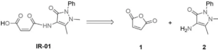
Starting with low-cost materials and a one-pot proce-dure, the reaction of maleic anhydride with the correspond-ing amine was performed uscorrespond-ing a microwave reactor but no solvent, for a cleaner methodology with high reproduci-bility and good yield, further lending validity to the method (Figure 1).
The formation of the synthetic target occurs after the attack of the 4-aminoantipyrine nitrogen on the carbonyl carbon of maleic anhydride, which provides ring opening and the subsequent formation of the acid of interest with good yield (Figure 2).
Reagents and techniques
All reagents and spectrograde solvents for synthesis and NMR measurements were purchased commercially and used without further purification.
The melting point was determined on a Quimis dry melting point apparatus, model Q340S23, and used as un-corrected data. The microwave procedure was performed in a CEM/Discover microwave reactor with sealed tube.
1H and13C NMR spectra were recorded at room
tem-perature on a Bruker 300 spectrometer (10% in deuterated dimethylsulfoxide (DMSO-d6) solutions at 298 K)
operat-ing at 300.132 and 75.476 MHz, respectively. Data
pro-cessing was conducted on a Solaris workstation. The 1H
and13C chemical shifts are reported on thedscale (ppm)
and referenced to internal DMSO-d6; coupling constantsJ
are reported in hertz (Hz). The abbreviations s, d and m rep-resent simplet, douplet and multiplet, respectively.
Synthesis process
In a sealed tube, 4-aminoantipyrine (2.0 g, 10 mmol) and maleic anhydride (1.0 g, 10 mmol) were subjected to
Figure 1- Retrosynthetic analysis for (Z )-4-((1,5-dimethyl-3-oxo-2-phe-nyl-2,3dihydro-1H-pyrazol-4-yl) amino)-4-oxobut-2-enoic acid.
microwave irradiation (150 W) at 90 °C for 10 s. The solid was washed with ethyl acetate and filtered. The remaining yellow solid was recrystallized from CH3Cl, giving IR-01
(2.79 g, 93%).1H NMR (DMSO-d6, 300 MHz)d (ppm):
2.13 (s, 3H), 3.02 (s, 3H), 6.26 (d, 1H,Jcis= 12.3 Hz), 6.48
(d, 1H,Jcis= 12.3 Hz), 7.30 (m, 3H), 7.46 (m, 2H). 13C
NMR (CDCl3, 75 MHz) d (ppm): 11.71 (CH3), 36.24
(CH3), 106.62 (C), 124.22 (CH), 126.94 (CH), 129.60
(CH), 131.54 (CH), 131.60 (CH), 135.25 (C), 152.56 (C), 161.73 (C=O), 164.58 (C=O), 167.03 (C=O). Melting point: 178.1-179.8 °C.
Chemical agents, animals and experimental design The DNA-damage-inducing agents (commercial chemotherapeutic agents) used in this study were the fol-lowing: doxorubicin (Glenmark Pharmaceuticals Ltd., Ar-gentina. MS Reg. No. 1.1013.0232.002-4, Lot #21130040) at a dose of 16 mg/kg body weight (b.w.) intraperitoneally (ip) cisplatin (Accord Pharmaceuticals Ltd., UK. MS Reg.
No. 1.5537.0002.003-7, Lot #88549) at a dose of 6 mg/kg (b.w.,ip), and cyclophosphamide (Genuxal®, Baxter Ltda.,
Germany. MS Reg. No. 1.00683.0168.003-1, Lot #F728) at
a dose of 100 mg/kg (b.w.,ip). Doxorubicin and
cyclo-phosphamide were diluted in distilled water.
IR-01 was first diluted in 5% DMSO and then in glycated serum before the drug was administered at doses of 12, 24 and 48 mg/kg (b.w.,ip).
Eighty Swiss female mice (with a mean weight of 30 g, 6-8 weeks old) were randomly distributed into 16 experi-mental groups (n = 5).
The animals were housed in individual cages lined with wood shavings on a ventilated rack (Alesco®) and pro-vided commercial feed (Nuvital®) and filtered waterad li-bitum. The experimental conditions were controlled, with a 12-hour light:12-hour dark photoperiod, mean temperature
of 22 2 °C and mean humidity of 55±10%. The
experi-ment was approved by the Animal Ethics Committee of the Federal University of Mato Grosso do Sul (Universidade Federal de Mato Grosso do Sul - UFMS) under protocol no. 399/2012 and performed according to the Declaration of Animal Rights.
For the evaluation of the IR-01 effects, the following experimental groups were established:
In lot 1, a negative control group comprised animals that received a dose of distilled water and another of 5%
DMSO in glycated serum, both at 0.1 mL/10 g (b.w.,ip).
The IR-01 groups in lot 1 comprised animals treated with IR-01 at concentrations of 12, 24 and 48 mg/kg (b.w.,ip)
and with a dose of distilled water at 0.1 mL/10 g (b.w.,ip).
To assess the effects of combining IR-01 with the commercial chemotherapeutic agents, the following exper-imental groups were established.
In lot 2, a doxorubicin group (DOX) comprised ani-mals treated with doxorubicin at a dose of 16 mg/kg (b.w.,
ip) and with 5% DMSO in glycated serum at a dose of 0.1
mL/10 g (b.w.,ip). The DOX + IR-01 groups in lot 2
com-prised animals that were treated with doxorubicin at a dose of 16 mg/kg (b.w.,ip) and IR-01 at doses of 12, 24 and 48
mg/kg (b.w.,ip).
The animals in lots 3 and 4 were treated as described in lot 2 except that doxorubicin was replaced by cisplatin (CIS and CIS + IR-01) and cyclophosphamide (CPP and CPP + IR-01) at doses of 6 and 100 mg/kg (b.w.,ip),
re-spectively.
At 24 (T1), 48 (T2) and 72 (T3) hours after the treat-ments, 20mL of peripheral blood was collected to perform a
micronucleus assay. Additionally, 20 mL of peripheral
blood was collected at T1 to perform a comet assay. A new aliquot was collected at T3 for a differential blood cell count. At the end of the experiment, at T3, the animals were euthanized by cervical dislocation to collect the spleen for a phagocytosis test and the kidney and liver for cell death analysis.
Biological assays
Peripheral blood Comet assay
The comet assay was performed according to the pro-tocol of Singhet al.(1988), with modifications by Oliveira et al. (2015a). The material was analyzed using an
epi-fluorescence microscope (Bioval®, model L 2000A) with a
40 objective, a 420-490 nm excitation filter and a 520 nm barrier filter. As described by Kobayashiet al.(1995), a
to-tal of 100 cells per treatment were inspected visually and the comets were classified as: class 0, undamaged cells showing no tail; class 1, cells with a tail size smaller than the diameter of the nucleoid; class 2, cells with a tail size 1-2 times the diameter of the nucleoid; class 3, cells with a tail size greater than two times the diameter of the nucleoid. Apoptotic cells that showed a totally fragmented nucleus were not scored. The total score was calculated as the sum of the number of cells scored for each class times that class value.
Peripheral blood Micronucleus assay
The micronucleus assay in peripheral blood was per-formed according to Hayashiet al.(1990), with
modifica-tions by Oliveiraet al.(2015a). A 20mL peripheral blood aliquot was covered with a cover slip after its deposition on
a slide precoated with 20 mL of acridine orange (1.0
mg/mL). The slide was stored in a freezer (-20 °C) for at least seven days. The analysis was performed under an epifluorescence microscope with a 40 objective (Bioval®,
model L 2000A) along with a 420-490 nm excitation filter and a 520 nm barrier filter. Two thousand cells were ana-lyzed per animal.
Cell death assay
decreasing series of ethanol concentrations (95-25%), washed with McIlvaine’s buffer for 5 min, stained with 0.01% acridine orange for 5 min and washed again with buffer. Dying cells were identified through an analysis of
the DNA fragmentation patterns, according to Carvalhoet
al.(2015) and Navarroet al.(2014).
Splenic phagocytosis assay
The spleen was macerated in saline solution. One hundred microliters of cell suspension was covered with a coverslip after its placement on a slide previously coated with 20mL of acridine orange (1.0 mg/mL). The slides were stored in a freezer until their analysis, which was performed with a fluorescence microscope (Bioval®, model L 2000A)
using a 40 objective along with a 420-490 nm filter and a 520 nm barrier filter. Two hundred cells were analyzed per animal. The presence or absence of phagocytosis was deter-mined based on the descriptions of Carvalhoet al.(2015)
and Hayashiet al.(1990).
Differential blood cell count
A 20mL aliquot of peripheral blood was used to pre-pare blood smears on glass slides. These slides were air dried and stained with a panoptic kit for 10 min. The cells were visualized under bright field microscopy using a 100 objective. A total of 100 cells per animal were analyzed and classified as lymphocytes, neutrophils, monocytes, eosino-phils and basoeosino-phils (Ishiiet al., 2011).
Calculation of percent damage reduction (%DR) and percent damage increase (%DI)
Manoharan and Banerjee (1985) and Waters et al.
(1990) proposed the calculation of percent damage reduc-tion to assess the chemopreventive ability of a substance when it is associated with a substance known to be muta-genic, such as the commercial chemotherapeutic agents used as positive control (PC). According to Oliveiraet al.
(2015a) and Navarroet al.(2014), the same calculation can
be used to estimate the increase in DNA damage. Thus, for the present study, both the percent DNA damage reduction and percent DNA damage increase were calculated using the same formula:
%DR or%DI Mean of PC (Mean of ) Mean of PC Mean of C
= - +
-IR-01 PC
ontrol
æ è
çç ö
ø ÷÷´100
Statistical Analysis
Data are reported as the mean±standard error of the
mean (SEM) and analyzed using Student’s t-test or the
Mann-Whitney test, depending on whether the data distri-bution was parametric or nonparametric, respectively, us-ing GraphPad InStat Demo version 3.6 (GraphPad Software Inc., San Diego, CA, USA). The significance level adopted wasp< 0.05.
Results
Synthesis
The product formed was characterized by1H and13C NMR, and the results described below demonstrated chem-ical shifts and integrations consistent with IR-01.
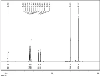
In the1H NMR spectra (Figure 3), two signals at the 6.26 and 6.48 ppm regions (J = 12.3 Hz), representing the two olefinic hydrogen doublets of the 1,4-dioxo-butenyl fragment, indicate theZconfiguration of the compound ob-tained; the signal at 9.78 ppm refers to the amidic hydrogen. In the13C NMR spectra (Figure 4), the three signals observed between 161.35 and 166.59 ppm, representing the IR-01 carbonyls, confirm the formation of the synthetic tar-get.
All other1H and13C NMR signals are in agreement with the data reported in the literature for the same com-pound (Cunhaet al., 2005).
Figure 3-1H RMN spectra of IR-01 in DMSO-d
6at 300 Mhz.
Biological assays
Assessment of genetic integrity and effects of IR-01 on DNA damage caused by commercial chemotherapeutic agents
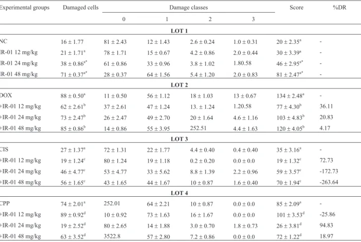
The genetic integrity assessment indicated that IR-01 can cause genomic damage (comet assay) but is unable to cause chromosomal damage (micronucleus assay). The treatments with the test compound caused an increase (p<
0.05) in the frequency of DNA damage by 2.37 and 4.44 and in the score by 2.3 and 4.05 for the 24 and 48 mg/kg doses, respectively (Table 1). The micronucleus frequency ranged from 0.6±0.24 to 3.0±0.44 in the control group
and from 3.0 ± 0.31 to 5.2 ± 0.66 in the IR-01-treated
groups (Table 2).
When associated with the commercial chemothera-peutic agents, IR-01 showed chemopreventive activity for most associations, except for the two highest doses when administered with cisplatin and evaluated in the comet as-say (Table 1) and the intermediate dose in combination with cyclophosphamide in the icronucleus assay (Table 2).
The percent damage reduction by IR-01 in the Comet assay ranged between 4.17 and 36.11% in combination
with doxorubicin and between 18.87 and 94.83% in combi-nation with cyclophosphamide and was 72.73% for the lowest IR-01 dose in combination with cisplatin. An in-crease in percent damage was observed for the two highest IR-01 doses in combination with cisplatin (172.73 and 263.64% for 24 and 48 mg/kg, respectively) and for the lowest IR-01 dose in combination with cyclophosphamide (25.86%) (Table 1).
In the micronucleus assay, the percent damage reduc-tion ranged from 65.35 to 91.43% for the combinareduc-tion with doxorubicin, from 62.50 to 86.67% for the combination with cisplatin and from 0 to 14.60% for the cyclophos-phamide combination. For the latter, an increase in DNA damage that reached 56.02% was observed at the interme-diate dose (Table 2).
Evaluation of the splenic phagocytosis potential and effects of IR-01 in combination with commercial chemotherapeutic agents
When administered alone, the two highest doses of IR-01, 24 and 48 mg/kg, increased (p < 0.05) the rate of splenic phagocytosis by 1.43 and 1.67, respectively (Table 3).
Table 1- Results of the comet assay showing the ability of IR-01 to cause or prevent genomic damage.
Experimental groups Damaged cells Damage classes Score %DR
0 1 2 3
LOT 1
NC 16±1.77 81±2.43 12±1.43 2.6±0.24 1.0±0.31 20±2.35a
-IR-01 12 mg/kg 21±1.71a 78±1.71 15±0.67 4.2±0.86 2.0±0.44 30±3.39ª
-IR-01 24 mg/kg 38±0.86a* 61±0.86 33±0.96 3.8±1.02 1.80.58 46±2.95a*
-IR-01 48 mg/kg 71±0.37a* 28±0.37 64±1.56 5.4±1.20 2.0±0.83 81±2.47a*
-LOT 2
DOX 88±0.50a 11±0.50 56±1.12 18±1.03 13±0.67 134±2.48a
-+IR-01 12 mg/kg 62±2.61b 37±2.61 47±1.24 13.±1.24 1.20.58 77±4.30b 36.11
+IR-01 24 mg/kg 73±2.47b 26±2.47 49±2.70 20±1.64 4.6±1.16 103±4.83b 20.83
+IR-01 48 mg/kg 85±0.86b 14±0.86 55±3.95 252.51 4.4±1.63 120±4.05b 4.17
LOT 3
CIS 27±1.37a 72±1.31 22±1.77 4.4±0.40 0.4±0.40 35±3.16a
-+IR-01 12 mg/kg 19±1.24c 80±1.24 19±1.18 0.2±0.20 0.0±0.0 19±1.32c 72.73
+IR-01 24 mg/kg 46±4.77c 53±4.77 33±5.62 8.8±1.39 2.2±0.96 59±3.57c -172.73
+IR-01 48 mg/kg 56±1.65c 43±1.65 44±1.67 10±0.87 1.6±0.40 70±1.94c -263.64
LOT 4
CPP 74±2.01a 252.01 64±2.21 10±0.87 0.0±0.0 85±2.09a
-+IR-01 12 mg/kg 89±0.92d 10±0.92 73±1.63 16±1.67 0.0±0.0 101±3.53d -25.86
+IR-01 24 mg/kg 19±2.52d 80±2.65 14±1.88 3.0±0.70 1.8±0.73 26±3.81d 94.83
+IR-01 48 mg/kg 63±3.52d 3522.8 57±2.80 7.2±0.86 0.0±0.0 72±1.22d 18.97
Table 2- Results of the micronucleus assay related to the ability of IR-01 to cause or prevent chromosomal damage
Experimental groups Mean±SE %DR
24 h 48 h 72 h 24 h 48 h 72 h
LOT 1
NC 3.0±0.44 1.8±0.20 0.6±0.24 - -
-IR-01 12 mg/kg 3.8±0.37a 3.0±0.31ª* 4.2±0.37ª* - -
-IR-01 24 mg/kg 4.2±0.20a 4.6±0.60ª* 4.8±0.37ª* - -
-IR-01 48 mg/kg 4.8±0.37ª* 3.4±0.50ª* 5.2±0.66ª* - -
-LOT 2
DOX 52±2.56a* 36±1.72a* 26±1.16ª* - -
-+ IR-01 12 mg/kg 7.20.58b* 5.2±0.37b* 7.2±0.86b* 91.43 90.06 74.01
+ IR-01 24 mg/kg 12±0.70b* 6.4±0.50b* 7.2±0.37b* 81.63 86.55 74.01
+ IR-01 48 mg/kg 13±1.06b* 9.4±0.40b* 9.4±0.60b* 79.59 77.78 65.35
LOT 3
CIS 27±0.50ª* 20±0.55a* 15±0.50ª* - -
-+ IR-01 12 mg/kg 6.2±0.37c* 5.6±0.24c* 4.2±0.20c* 86.67 79.12 75.00
+ IR-01 24 mg/kg 7.0±0.37c* 7.6±0.50c* 6±0.31c* 83.33 68.14 62.50
+ IR-01 48 mg/kg 8.4±0.50c* 7.4±0.60c* 5.8±0.37c* 77.50 69.23 63.89
LOT 4
CPP 41±2.17a* 50±2.34a* 28±2.80a* - -
-+ IR-01 12 mg/kg 33±2.71d 63±3.75d 24±2.16d 21.05 -26.97 14.60
+ IR-01 24 mg/kg 55±3.63d* 77±2.95d* 42±3.88d* -36.84 -56.02 -51.10
+ IR-01 48 mg/kg 42±3.85d 71±2.70d 28±2.10d -2.63 -43.57 00.00
SE: Standard error of the mean; %DR: Percent damage reduction; NC: Negative control group; DOX: Doxorubicin group; CIS: Cisplatin group; CPP: Cyclophosphamide group.(a)Statistically compared to the NC group;(b)Statistically compared to the DOX group;(c)Statistically compared to the CIS group;(d)Statistically compared to the CPP group;*statistically different (p< 0.05; Mann-Whitney test).
Table 3- Results related to splenic phagocytosis evaluation.
Experimental groups Phagocytosis
Absolute values Mean±SE
LOT 1
NC 221 44.2±0.66
IR-01 12 mg/kg 228 45.6±1.77ª
IR-01 24 mg/kg 316 63.2±1.28ª*
IR-01 48 mg/kg 369 73.8±1.15ª*
LOT 2
DOX 670 134.0±1.37ª*
+IR-01 12 mg/kg 321 64.2±1.59b*
+IR-01 24 mg/kg 405 81.0±1.30b*
+IR-01 48 mg/kg 373 74.6±1.43b*
LOT 3
CIS 269 53.8±1.35ª*
+IR-01 12 mg/kg 106 21.2±1.35c*
+IR-01 24 mg/kg 74 14.8±1.02c*
In combination with doxorubicin, IR-01 reduced (p< 0.05) the frequency of phagocytosis by 47.91, 60.45 and 55.67% at the 12, 24 and 48 mg/kg doses, respectively. In combination with cisplatin, the respective reductions were 39.40, 27.51 and 20.07%. In combination with cyclophos-phamide, the reductions were 53.40 and 61.36% for the 12 and 24 mg/kg doses, respectively (Table 3).
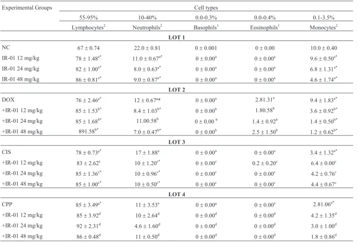
The differential blood cell count showed that at the three doses tested, IR-01 administered alone can increase (p
< 0.05) the frequency of lymphocytes and reduce (p< 0.05)
neutrophil and monocyte counts. Additionally, in the treatment with the commercial chemotherapeutic agents, an increase (p< 0.05) in the frequency of lymphocytes and
a reduction in that of monocytes occurred for doxorubicin, cisplatin and cyclophosphamide, with a reduction (p <
0.05) in neutrophils also occurring for doxorubicin (Table 4).
The following results were observed for the combina-tions of chemotherapeutic agents with IR-01: (I) for
doxo-rubicin, an increase (p < 0.05) in the frequency of
Table 4- Reference values and results related to the differential blood cell count.
Experimental Groups Cell types
55-95% 10-40% 0.0-0.3% 0.0-0.4% 0.1-3.5%
Lymphocytes2 Neutrophils2 Basophils1 Eosinophils1 Monocytes2
LOT 1
NC 67±0.74 22.0±0.81 0±0.001 0±0.00 10.0±0.40
IR-01 12 mg/kg 78±1.48a* 11.0±0.67a* 0±0.00a 0±0.00a 9.6±0.50a*
IR-01 24 mg/kg 82±1.00a* 8.0±0.63ª* 0±0.00a 0±0.00a 6.8±1.31a*
IR-01 48 mg/kg 86±0.81a* 9.0±0.87a* 0±0.00a 0±0.00a 4.6±1.74a*
LOT 2
DOX 76±2.46ª* 12±0.67ª* 0±0.00a 2.81.31a 9.4±1.83a*
+IR-01 12 mg/kg 85±1.53b* 8.4±1.03b* 0±0.00b 1.80.58b 3.6±0.92b*
+IR-01 24 mg/kg 85±1.68b* 11.00.58b
0±0.00b 1.4±0.92b 1.4±0.50b*
+IR-01 48 mg/kg 891.58b* 7.0
±0.47b* 0±0.00b 2.5±1.50b 1.2±0.62b*
LOT 3
CIS 78±0.73ª* 17±1.88a 0±0.00a 0±0.00a 3.4±1.32a*
+IR-01 12 mg/kg 83±2.62c 10±1.20c* 0±0.00c 0.2±0.20c 6.4±0.00c
+IR-01 24 mg/kg 85±1.36c* 10±0.96c* 0±0.00c 0±0.00c 4.2±0.76c
+IR-01 48 mg/kg 85±1.00c* 10±0.50c* 0±0.00c 0±0.00c 4.4±0.67c
LOT 4
CPP 85±3.49ª* 11±3.53a 0±0.00ª 0±0.00a 2.81.06a*
+IR-01 12 mg/kg 85±3.92d 10±2.64d 0±0.00d 0±0.00d 4.2±1.35d
+IR-01 24 mg/kg 92±2.31d 4.6±1.60d 0±0.00d 0±0.00d 3.0±1.00d
+IR-01 48 mg/kg 86±0.48d 11±0.50d 0±0.00d 0±0.00d 1.8±0.86d
Data is represented as mean values±standard error of the mean. Statistical tests:(1)Student’st-test(p < 0.05) and(2)Mann-Whitney test (p< 0.05). NC: Negative control group;(a)Statistically compared to the NC group;(b)Statistically compared to the DOX group;(c)Statistically compared to the CIS group; (d)Statistically compared to the CPP group;*statistically different.
LOT 4
CPP 515 103.0±2.00ª*
+IR-01 12 mg/kg 275 55.0±3.46d*
+IR-01 24 mg/kg 316 63.2±2.57d*
+IR-01 48 mg/kg 534 106.8±3.13d
SE: Standard error of the mean; NC: Negative control group; DOX: Doxorubicin group; CIS: Cisplatin group; CPP: Cyclophosphamide group.(a) Statis-tically compared to the NC group;(b)Statistically compared to the DOX group;(c)Statistically compared to the CIS group;(d)Statistically compared to the CPP group;*statistically different (p< 0.05; Student’st-test).
lymphocytes and a reduction (p< 0.05) in that of
mono-cytes for all the doses tested and a reduction (p < 0.05) in neutrophil frequency for the lowest and highest doses; (II) for cisplatin, an increase (p< 0.05) in the frequency of
lym-phocytes for the two higher doses and a reduction (p< 0.05)
in that of neutrophils for all the doses; and (III) for cyclo-phosphamide, no statistically significant change (Table 4). Neutropenia was observed in the groups treated with the two highest doses of IR-01, in DOX + IR-01 at the low-est and highlow-est doses and in CPP + IR-01 at the intermedi-ate dose. Eosinophilia was observed in the animals treintermedi-ated with cisplatin when combined with all doses of IR-01, and monocytosis occurred in the control groups treated with IR-01, in DOX, in DOX + IR-01 at the lowest dose, in CIS + IR-01 at all three doses, and in CPP + IR-01 at the lowest dose tested (Table 4).
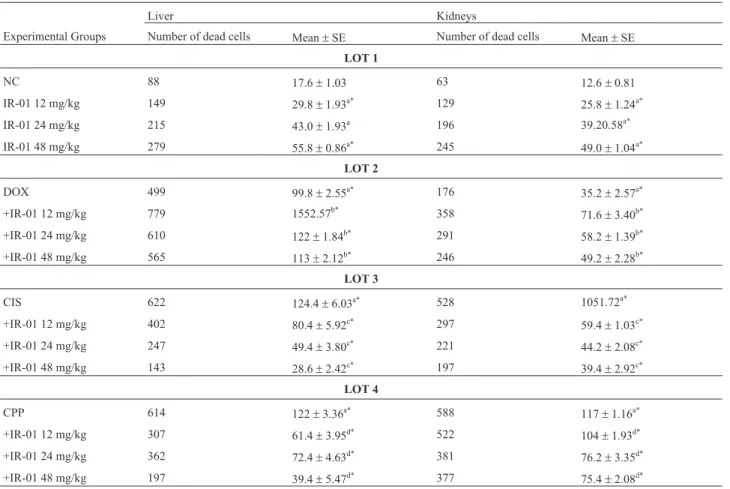
Evaluation of cell death induction and effects of IR-01 in combination with commercial chemotherapeutic agents
The administration of IR-01 increased (p< 0.05) the
frequency of dead cells in the liver by 1.69, 2.44 and 3.17 and in the kidneys by 2.05, 3.11 and 3.89 at the 12, 24 and 48 mg/kg doses, respectively (Table 5).
The commercial chemotherapeutic agents doxorubi-cin, cisplatin and cyclophosphamide caused an increase in dead cells frequency of 5.67, 7.07 and 6.98 in the liver and 2.79, 8.38 and 9.33 in the kidneys, respectively (Table 5).
The following results were observed for the chemo-therapeutic agents tested in combination with IR-01: (I) for doxorubicin, the potentiation of cell death (p < 0.05) by 155.31% in the liver and 203.41% in the kidneys for the lowest IR-01 dose tested; (II) for cisplatin, a reduction of dead cells (p < 0.05) by up to 22.99% in the liver and 37.52% in the kidneys for the highest IR-01 dose; and (III) for cyclophosphamide, reductions of 32.29 and 64.44% in the liver and kidneys, respectively, also for the highest IR-01 dose (Table 5).
Discussion
The increase in cancer incidence and the high cost of treatments motivate the search for new strategies to prevent and manage this disease (Mauroet al., 2011). An approach
with great potential is chemoprevention, which involves the use of natural and/or synthetic agents to suppress,
in-Table 5- Cell death evaluation on mice kidneys and liver.
Liver Kidneys
Experimental Groups Number of dead cells Mean±SE Number of dead cells Mean±SE
LOT 1
NC 88 17.6±1.03 63 12.6±0.81
IR-01 12 mg/kg 149 29.8±1.93a* 129 25.8±1.24a*
IR-01 24 mg/kg 215 43.0±1.93a 196 39.20.58a*
IR-01 48 mg/kg 279 55.8±0.86a* 245 49.0±1.04a*
LOT 2
DOX 499 99.8±2.55a* 176 35.2±2.57a*
+IR-01 12 mg/kg 779 1552.57b* 358 71.6±3.40b*
+IR-01 24 mg/kg 610 122±1.84b* 291 58.2±1.39b*
+IR-01 48 mg/kg 565 113±2.12b* 246 49.2±2.28b*
LOT 3
CIS 622 124.4±6.03a* 528 1051.72a*
+IR-01 12 mg/kg 402 80.4±5.92c* 297 59.4±1.03c*
+IR-01 24 mg/kg 247 49.4±3.80c* 221 44.2±2.08c*
+IR-01 48 mg/kg 143 28.6±2.42c* 197 39.4±2.92c*
LOT 4
CPP 614 122±3.36a* 588 117±1.16a*
+IR-01 12 mg/kg 307 61.4±3.95d* 522 104±1.93d*
+IR-01 24 mg/kg 362 72.4±4.63d* 381 76.2±3.35d*
+IR-01 48 mg/kg 197 39.4±5.47d* 377 75.4±2.08d*
hibit or reverse the process of carcinogenesis in its early stages (Friedman and Rasooly, 2013).
Organic synthesis has gained prominence in the search to develop more potent and less toxic molecules, and the redesign and structural modification of previously known compounds or radicals allow important advances in defining biological activities and in structure-activity stud-ies. Under this perspective, our research group designed and synthesized IR-01 (Z )-4-((1,5-dimethyl-3-oxo-2-phe-nyl-2,3dihydro-1H-pyrazol-4-yl) amino)-4-oxobut-2-enoic acid using 4-aminoantipyrine associated to the structural fragment 1,4-dioxo-2-butenyl, observing the influence of the position of the phenyl ring, the distance between frag-ments, the increased number of heteroatoms and the in-creased number of olefins, as indicated in the literature (Jha
et al., 2010).
Often, the synthesis of a biologically effective com-pound can use reagents and/or approaches of considerable environmental impact. Thus, compared with the efficient but environmently agressive synthetic procedure described in the literature (Cunhaet al., 2005), our one-pot method assisted by microwave irradiation can be a similarly effi-cient but cleaner approach for the synthesis of IR-01, with a very good yield and a reaction time of only 1% compared to that reported in the literature.
The results of the biological studies on IR-01, pre-pared using this new synthesis method, suggest that it can cause DNA damage. However, this damage does not be-come fixed in the cell genome, a hypothesis that is rein-forced by the fact that the damage evaluated by the comet assay did not result in a significant increase in
micro-nucleus frequency. According to Rundell et al. (2003),
genotoxic damage is likely to undergo repair, whereas mutagenic damage is not, with changes becoming fixed in the genetic material as mutations. Such occurrence is not uncommon in preclinical chemoprevention experiments, both with natural products (Synder and Gillies, 2002; Cu-nhaet al., 2005; Rodeiroet al., 2006; Hoshinaet al., 2013;
Mendanha da Cunhaet al., 2013; Luoet al., 2015) and with
synthetic compounds (Zhanet al., 2008; Caoet al., 2015;
Frolovaet al., 2015; de Araújoet al., 2017). Although there
are significant differences in the frequency of micronuclei between the negative control group and the groups treated with IR-01, Vazet al.(2016) describes that this isolated
fact does not necessarily imply in toxicogenetic damage. Also, according to the results observed in other
experi-ments from our research group (Oliveira et al., 2009a,
2013; Mauroet al., 2010; Pesariniet al., 2014; Navarroet al., 2015), the baseline frequency of micronuclei can be
greater than the frequency observed for the animals treated with IR-01 in the present study. For example, according to Oliveiraet al. (2015b), the baseline frequency of
micro-nuclei in Swiss female mice may reach 11.00±3.16. Based on these data, IR-01 is considered to cause genomic damage but is unable to cause chromosomal
dam-age. The genotoxic activity may occur because of the phar-macophore 1,4-dioxo-2-butenyl, which has already been described as an effective cytotoxic agent in the tumor cell
lines Molt4/C8 and CEM L1210 (Jhaet al., 2010).
How-ever, the addition of 4-aminoantipyrine may have modified this property, which is required in anticancer agents. It is important to highlight that chemotherapeutic agents gener-ally include in their mechanism of action the induction of DNA damage that causes cell death, especially that of tu-mor cells (Elsendoornet al., 2001; Nadinet al., 2005).
The chemopreventive action described may be ex-plained by IR-01, which originated from 4-aminoantipy-rine, retaining the antioxidant activity of its precursor. This hypothesis is consistent with the data from the present study because the best capacity for preventing genomic damage were observed for doxorubicin and cyclophos-phamide, chemotherapeutic agents that are capable of gen-erating free radicals that cause rather extensive genotoxic damage (Almeidaet al., 2005), triggering cell death and
thereby exerting their anticancer action. The lack of a pat-tern in the chemopreventive response of the three studied commercial chemotherapeutic agents suggests that their mechanisms of action may interfere with the response to DNA damage in the presence of IR-01.
Cisplatin is also a chemotherapeutic agent that gener-ates free radicals (Antunes and Bianchi, 2004), and this needs to be considered in its antitumor action. However, when this drug was combined with IR-01, the rate of DNA damage reduction increased, contrary to what was observed for doxorubicin and cyclophosphamide. Thus, despite the existence of the 4-aminoantipyrine radical, the antioxidant activity was undetectable, and this increase in DNA dam-age reduction could be attributed to the pharmacophore 1,4-dioxo-2-butenyl. However, further studies are needed to clarify this potentiation of the toxicogenic effects. A hy-pothesis for discussion is the reduction caused by cisplatin in the activity of antioxidant enzymes, such as superoxide dismutase, catalase, GSH peroxidase and GSH reductase (Hyppolito and Oliveira, 2005). Thus, the oxidative and consequently genotoxic capacity of cisplatin is more in-tense than that of the previously cited antineoplastic agents, which may have led to the increased genomic damage.
Regarding the micronucleus assay, the pattern of re-sponse to doxorubicin resembled that shown for the comet assay, i.e., the percent damage reduction decreased with higher dose, showing an inverse correlation. Note that over time the chemopreventive activity decreased, but the in-versely proportional response pattern was maintained. This pattern was expected, and the decrease in the chemopre-ventive activity was perhaps due mainly to the metabo-lization and elimination of IR-01. Metabometabo-lization and secretion were also observed for doxorubicin because the capacity for DNA damage induction decreased.
de-creased over the three time points. These data suggest that the increased DNA damage observed in the comet assay was not fixed into the genetic material because no chromo-somal damage occurred. This finding, in turn, suggests that the repair mechanism was effective in preventing that ge-nomic damage would be fixed as chromosomal damage.
For cyclophosphamide, increased toxicogenic activ-ity was observed over the 72 hours of the study. It occurred at the intermediate test dose despite its efficient capacity to prevent genomic damage, with reduced chemopreventive activity at the other two doses.
In addition to the ability to reduce commercial che-motherapeutics effects, IR-01 also has a pharmacophoric radical in its structure, which could increase the antioxidant defenses in non-injured cells. This hypothesis is supported by Bianchi and Antunes (1999), Albertini and Ruiz (2001),
Antunes and Bianchi (2004), and Oliveiraet al. (2013),
who reported that cells that have DNA lesions are deficient in antioxidant defenses. Therefore, these cells are more prone to suffer cytotoxic damage and undergo cell death more easily when exposed to certain cytotoxic and/or geno-toxic agents. On the other hand, when normal cells with ad-equate antioxidant enzyme activity are in contact with another antioxidant agent, there is less of a chance that DNA damage is caused by free radicals.
The splenic phagocytosis test revealed that the same toxicogenetic doses also stimulated splenic phagocytosis. Thus, cells with DNA damage were efficiently removed from the bloodstream. Other studies have shonw that the spleen has the ability to remove tumor cells and/or DNA-damaged cells that are prone to carcinogenesis from the bloodstream (Cruvinelet al., 2010; Navarro et al., 2014;
Carvalhoet al., 2015). In addition, splenic phagocytosis is
also associated with the biomonitoring of blood cell viabil-ity, and it promotes the removal of senescent leukocytes, platelets and erythrocytes (Freitaset al., 2009), as well as of
apoptotic bodies and pathogens (Huysentruyt and Seyfried, 2010).
The splenic phagocytosis analysis also showed that all the chemotherapeutic agents were able to increase splenic activity, which was expected because these agents cause DNA damage and these damaged cells tend to be se-questered. When the agents were associated with IR-01, a decrease in phagocytosis occurred in all the experimental groups. This result suggests that the absence of cells with chromosomal damage did not stimulate the spleen to in-crease splenic phagocytosis.
Considering the alterations in both leukometry and splenic phagocytosis caused by IR-01, interestingly, it was observed that this molecule was able to increase the number of circulating lymphocytes and to cause a reduction in neutrophil numbers. These data suggest that the increase in phagocytic activity may be characterized by neutrophils ex-iting the blood and migrating into the spleen to sequester cells with DNA damage. Similar findings have been
re-ported in other studies that also identified
immunostimu-latory compounds through this association (Lee et al.,
2003; Leung et al., 2005; Ishiiet al., 2011; Sang et al.,
2013).
The combination of IR-01 with doxorubicin and cis-platin also showed an increase in the number of leukocytes and a reduction in the number of neutrophils. These find-ings corroborate the increase in splenic phagocytosis ob-served and discussed for the previous assay.
No variation in the frequency of blood cells was ob-served for cyclophosphamide. However, this does not con-tradict the splenic phagocytosis observed for the two lower doses. According to Oliveiraet al.(2015a), splenic
phago-cytosis can occur efficiently even in the absence of a chan-ge in blood cell counts.
The present study also assessed cell death, given that unrepaired cells with DNA damage tend to disrupt the cell cycle and enter apoptosis (Zhou and Elledge, 2000). The re-sults showed that IR-01 is capable of increasing the fre-quency of dead cells, from now on considered apoptotic cells, in the liver and kidneys. This is an important issue be-cause IR-01 can stimulate splenic phagocytosis, increase the number of lymphocytes, and induce cell death, despite that it can cause genomic damage without causing chromo-somal damage. Such important biological activities may be required in chemopreventive compounds (De Flora and Ferguson, 2005).
Nevertheless, although IR-01 is capable of increasing apoptosis when administered alone, in combination with doxorubicin, cisplatin and cyclophosphamide, IR-01 it gen-erally reduced the frequency of apoptosis caused by these agents. The potentiation of apoptosis could be a good indi-cator of its adjuvant action in chemotherapy. However, such potentiation occurred only for doxorubicin. Therefore, the results do not encourage the use of IR-01 in combina-tion with chemotherapeutic agents in anticancer therapy.
Given the above data, IR-01 has properties that render it sufficient to be classified as a chemopreventive agent, such as the inability of causing chromosomal damage, antigenotoxic potential, and the ability to alter leukometry, increase phagocytosis and induce cell death. These proper-ties are possibly correlated with the 4-aminoantipyrine rad-ical, an important antioxidant moiety present in the IR-01 molecule and described as having anti-inflammatory, anal-gesic, and antipyretic properties (Burduleneet al., 1999; Turan-Zitouniet al., 2001; Pisoschi and Pop, 2015).
According to Fedel-Miyasatoet al.(2014) and Rocha et al.(2015) a good correlation exists between effective
caus-ing a delay or inhibition of oxidation rates (Maxwell, 1995). The latter is correlated with the desmutagenic mode of action of substances reported as potential chemopre-ventive agents (Sato et al., 1984; Ferrara et al., 2000;
Pesariniet al., 2013; Navarroet al., 2015).
If these lines of defense are still insufficient, the body may also facilitate the excretion of xenobiotics through de-toxification enzymes, making them more water soluble and, thus, assisting their elimination by the kidneys (Cor-don-Cardoet al., 1989; Hooiveldet al., 2001), while
modu-lating the DNA repair system. This last line of defense is associated with bioantimutagenesis, in which enzymes are modulated by test compounds, thus favoring the correction and integrity of the genetic material, reducing the probabil-ity of developing cancer (Oliveiraet al., 2009b; Nakamura et al., 1999; Di Giacomoet al., 2014; Leiteet al., 2015).
All of the actions that are attributed to IR-01 discour-age its use in combination with chemotherapeutic discour-agents because of the maintenance of the antioxidant activity of its precursor, 4-aminoantipyrine. This use is discouraged be-cause despite the fact that IR-01 can be-cause genomic dam-age, can increase splenic phagocytosis, lymphocyte num-ber and the frequency of cell death, all these being properties required for chemotherapeutic agents, IR-01 can interfere negatively when associated with drugs already used extensively in anticancer therapy. Such interference prevents DNA damage and apoptosis, which are the main pathways for the elimination of tumor cells.
Thus, we consider that IR-01 is not indicated for use as an adjuvant in anticancer therapy in combination with doxorubicin, cisplatin or cyclophosphamide. In this case, the properties derived from 4-aminoantipyrine, even when in combination with the 1,4-dioxo-butenyl fragment, rec-ognized as cytotoxic (Jhaet al., 2010), largely overrode the
ability to induce cell death. Corroborating this, the study by Bernoet al.(2016) states that 4-aminoantipyrine, a
dipy-rone metabolite, reduces DNA damage, apoptosis induc-tion and phagocytosi when administered in combinainduc-tion with doxorubicin, cisplatin or cyclophosphamide.
The present study is the first to propose a new syn-thetic methodology to efficiently and cleanly produce IR-01 and the first to demonstrate the chemopreventive effects of this molecule. In addition, we contraindicate the use of IR-01 as an adjuvant in anticancer therapies in combination with doxorubicin, cisplatin and cyclophosphamide because of its ability to reduce important effects of these agents.
Acknowledgments
This project was funded by Fundação de Apoio ao Desenvolvimento do Ensino, Ciência e Tecnologia do Es-tado de Mato Grosso do Sul (FUNDECT), Conselho Na-cional de Desenvolvimento Científico e Tecnológico (CNPq) and Coordenação de Aperfeiçoamento de Pessoal de Nível Superior (CAPES).
References
Albertini SM and Ruiz MA (2001) O papel da glutamina na terapia nutricional do transplante de medula óssea. Rev Bras Hematol Hemoter 23:41-47.
Almeida VL, Leitão A, Reina LDCB, Montanari CA, Donnici CL and Lopes MTP (2005) Cancer and cell cicle-specific and cell cicle nonspecific anticancer DNA-interactive agents: an introduction. Quim Nova 28:118-129.
Ames BN, Durston WE, Yamasaki E and Lee FD (1973) Carcino-gens are mutaCarcino-gens: A simple test system combining liver homogenates for activation and bacteria for detection. Proc Natl Acad Sci U S A 70:2281-2285.
Antunes LMG and Bianchi MLP (2004) Dietary antioxidants as inhibitors of cisplatin-induced nephrotoxicity. Rev Nutr 17:89-96.
Berno CR, Rós BT, da Silveira IO, Coelho HR, Antoniolli AC, Beatriz A, de Lima DP, Monreal AC, Sousa FG, da Silva Gomes R,et al.(2016) 4-Aminoantipyrine reduces toxic and genotoxic effects of doxorubicin, cisplatin, and cyclophos-phamide in male mice. Mutat Res Genet Toxicol Environ Mutagen 805:19-24.
Bianchi MLP and Antunes LMG (1999) Radicais livres e os principais antioxidantes da dieta. Rev Nutr 12:123-130. Burdulene D, Palaima A, Stumbryavichyute Z and Talaikite Z
(1999) Synthesis and antiinflammatory activity of 4-amino-antipyrine derivatives of succinamides. Pharm Chem J 33:191-193.
Cao M, Onyango EO, Williams CR, Royce DB, Gribble GW, Sporn MB and Liby KT (2015) Novel synthetic pyridyl ana-logues of CDDO-Imidazolide are useful new tools in cancer prevention. Pharmacol Res 100:135-147.
Carvalho PC, Santos EA, Schneider BU, Matuo R, Pesarini JR, Cunha-Laura AL, Monreal AC, Lima DP, Antoniolli AC and Oliveira RJ (2015) Diaryl sulfide analogs of combre-tastatin A-4: Toxicogenetic, immunomodulatory and apop-totic evaluations and prospects for use as a new chemo-therapeutic drug. Environ Toxicol Pharmacol 40:715-721. Cordon-Cardo C, O’Brien JP, Casals D, Rittman-Grauer L,
Biedler JL, Melamed MR and Bertino JR (1989) Multidrug-resistance gene (P-glycoprotein) is expressed by endothelial cells at blood-brain barrier sites. Proc Natl Acad Sci U S A 86:695-698.
Cruvinel WM, Mesquita Jr D, Araújo JAP, Catelan TT, de Souza AW, da Silva NP and Andrade LE (2010) Immune system: Part I. Fundamentals of innate immunity with emphasis on molecular and cellular mechanisms of inflammatory respon-se. Rev Bras Reumatol 50:434-461.
Cunha S, Oliveira SH, Rodrigues Jr MT, Bastos RM, Ferrari J, de Oliveira CMA, Kato L, Napolitano HB, Vencato I and Lariucci C (2005) Structural studies of 4-aminoantipyrine derivatives. J Mol Struct 752:32-39.
de Araújo FH, de Figueiredo D, Auharek S, Pesarini JR, Meza A, Gomes R, Monreal ACD, de Lima D, Antoniolli-Silva ACMB, Kassuya CA,et al.(2017)In vivochemotherapeutic insight of a novel isocoumarin (3-hexyl-5,7-dimethoxy-isochromen-1-one): Genotoxicity, cell death induction, leu-kometry and phagocytic evaluation. Genet Mol Biol 40:665-675.
Di Giacomo S, Mazzanti G, Sarpietro MG and Di Sotto A (2014)
a-Hexylcinnamaldehyde inhibits the genotoxicity of envi-ronmental pollutants in the bacterial reverse mutation assay. J Nat Prod 77:2664-2670.
Elsendoorn TJ, Weijl NI, Mithoe S, Zwinderman AH, Van Dam F, De Zwart FA, Tates AD and Osanto S (2001) Chemother-apy induced chromosomal damage in peripheral blood lym-phocytes of cancer patients supplemented with antioxidants or placebo. Mutat Res 498:145-158.
Fedel-Miyasato LES, Formagio ASN, Auharek SA, Kassuya CA, Navarro SD, Cunha-Laura AL, Monreal AC, Vieira MC and Oliveira RJ (2014) Antigenotoxic and antimutagenic effects ofSchinus terebinthifoliusRaddi inAllium cepaand Swiss mice: A comparative study. Genet Mol Res 13:3411-3425. Ferrara G, Loffredo E, Simeone R and Senesi N (2000) Evaluation
of antimutagenic and desmutagenic effects of humic and fulvic acids on root tips of Vicia faba. Environ Toxicol 15:513-517.
Freitas SH, Evêncio JN, Dória RGS, Mendonça FS, Simões MJ, Camargo LM and Sebe AA (2009) Morphologic, morpho-metric, and ultrastructural aspects of the spleen of rats after hepatic pedicle total clamping. Arq Bras Med Vet Zootec 61:1314-1321.
Friedman M and Rasooly R (2013) Review of inhibition of bio-logical activities of food-related. Toxins 5:743-775. Frolova TS, Kukina TP and Sinitsyna OI (2015) Genotoxic and
mutagenic properties of synthetic betulinic acid and betu-lonic acid. Bioorg Khim 41:462-467.
Hayashi M, Morita T, Kodama Y, Sofuny T and Ishidate M Jr. (1990) The micronucleus assay with mouse peripheral blood reticulocytes using acridine orange-coated slides. Mutat Res 245:245-249.
Hedenmaln K and Spigset O (2002) Agranulocytosis and other blood dyscrasias associated with dipyrone (metamizol). Eur J Clin Pharmacol 58:265-274.
Hooiveld GJEJ, Vam-Montfoort JE, Meijer DKF and Müller M (2001) Function and regulation of ATP-binding cassette transport proteins involved in hepatobiliary transport. Eur J Pharm Sci 12:525-543.
Hoshina MM, Santos LD and Palma MS (2013) Cytotoxic, geno-toxic/antigenotoxic and mutagenic/antimutagenic effects of the venom of the waspPolybia paulista. Toxicon 72:64-70. Huysentruyt LC and Seyfried TN (2010) Perspectives on the
mesenchymal origin of metastatic cancer. Cancer Metastasis Rev 29:695-707.
Hyppolito MA and Oliveira JÁA (2005) Ototoxycity, otopro-tection and self defense of the coclear outer hair cells. Medicina (Ribeirão Preto) 38:279-289.
Ishii PL, Prado CK, Mauro M de O, Carreira CM, Mantovani MS, Ribeiro LR, Dichi JB and Oliveira RJ (2011) Evaluation of Agaricus blazei in vivofor antigenotoxic, anticarcinogenic, phagocytic and immunomodulatory activities. Regul Toxi-col PharmaToxi-col 59:412-422.
Jha A, Mukherjee C, Prasad AK, Parmar VS, Vadaparti M, Das U, De Clercq E, Balzarini J, Stables JP, Shrivastav A, et al. (2010) Derivatives of aryl amines containing the cytotoxic 1,4-dioxo-2-butenyl pharmacophore. Bioorg Med Chem Lett 20:1510-1515.
Kim ES, Khuri FR and Hong WK (2002) Chemoprevention trials. In: Bertino JR (ed) Encyclopedia of Cancer. Academic Press, Orlando, pp 457-472.
Kobayashi H, Sugiyama C, Morikawa Y, Hayashi M and Sofuny T (1995) A comparison between manual microscopic analy-sis and computerized image analyanaly-sis in the single cell gel electrophoresis assay. MMS Commun 3:103-115.
Lee YS, Han OK, Park CW, Suh SI, Shin SW, Yang CH, Jeon TW, Lee ES, Kim KJ, Kim SH, et al. (2003) Immuno-modulatory effects of aqueous-extracted Astragali radix in methotrexate-treated mouse spleen cells. J Ethnopharmacol 84:193-198.
Leite AS, Dantas AF, Oliveira GL, Gomes Júnior AL, de Lima SG, Citó AM, de Freitas RM, Melo-Cavalcante AA and Dantas Lopes JA (2015) Evaluation of toxic, cytotoxic, mutagenic, and antimutagenic activities of natural and tech-nical cashew nut shell liquids using the Allium cepaand Artemia salinabioassays. Biomed Res Int 2015:626835. Leung KN, Leung PY, Kong LP and Leung PK (2005)
Immuno-modulatory effects of esculetin (6,7-dihydroxycoumarin) on murine lymphocytes and peritoneal macrophages. Cell Mol Immunol 2:181-188.
Luo X, Yu X, Liu S, Deng Q, Liu X, Peng S, Li H, Liu J and Cao Y (2015) The role of targeting kinase activity by natural prod-ucts in cancer chemoprevention and chemotherapy. Oncol Rep 34:547-554.
Manoharan K and Banerjee MR (1985) Beta-carotene reduces sis-ter chromatid exchanges induced by chemical carcinogens in mouse mammary cells in organ culture. Cell Biol Int Rep 9:783-789.
Mauro MO, Pesarini JR, Ishii PL, da Silva AF and Oliveira RJ (2010) Chemopreventive activity of phenylalanine against damage mutagenic prompted by the acute administration of cyclophosphamide in pregnant and non-pregnant mice using the micronucleus test. Rev Bras Farmacogn 20:334-339. Mauro MO, Sartori D, Oliveira RJ, Ishii PL, Mantovani MS and
Ribeiro LR (2011) Activity of selenium on cell proliferation, cytotoxicity, and apoptosis and on the expression of CASP9, BCL-XL and APC in intestinal adenocarcinoma cells. Mutat Res 715:7-12.
Maxwell SR (1995) Prospects for the use of antioxidant therapies. Drugs 49:345-361.
Mendanha da Cunha CR, Mendanha Neto SA, Carlos SC, Cortez AP, Gomes MN, Martins FI, Alonso A, Rezende KR, Mene-gatti R, de Magalhães MT,et al.(2013) 4-Nerolidylcatechol and its synthetic analogues: Antioxidant activity and toxicity evaluation. Eur J Med Chem 2013 62:371-378.
Nadin SB, Vargas-Roig LM, Drago G, Ibarra J and Ciocca DR (2005) DNA damage and repair in peripheral blood lympho-cytes from health individuals and cancer patients: A pilot study on the implications in the clinical response to chemo-therapy. Cancer Lett 21:11-14.
Nakamura Y, Suganuma E, Matsuo T, Okamoto S, Sato K and Ohtsuki K (1999) 2,4-Nonadienal and benzaldehyde bioan-timutagens in Fushimi Sweet Pepper (Fushimi-Togarashi). J Agric Food Chem 47:544-549.
Navarro SD, Mauro MO, Pesarini JR, Ogo FM and Oliveira RJ (2015) Resistant starch: A functional food that prevents DNA damage and chemical carcinogenesis. Genet Mol Res 14:1679-1691.
Oliveira RJ, Salles MJ, Da Silva AF, Kanno TY, Lourenço AC, Freiria GA, Matiazi HJ, Ribeiro LR and Mantovani MS (2009a). Effects of the polysaccharide b-glucan on clasto-genicity and teratoclasto-genicity caused by acute exposure to cyclophosphamide in mice, Regul Toxicol Pharmacol 53:164-173.
Oliveira RJ, Baise E, Mauro MO, Pesarini JR, Matuo R, Silva AF, Ribeiro LR and Mantovani MS (2009b). Evaluation of che-mopreventive activity of glutamine by the comet and the micronucleus assay in mice’s peripheral blood. Environ Toxicol Pharmacol 28:120-124.
Oliveira RJ, Sassaki ES, Monreal AC, Monreal MT, Pesarini JR, Mauro MO, Matuo R, Silva AF, Zobiole NN, Siqueira JM,et al. (2013) Pre-treatment with glutamine reduces genetic damage due to cancer treatment with cisplatin. Genet Mol Res 12:6040-6051.
Oliveira RJ, Navarro SD, Lima DP, Mauro MO, da Silva AF, Souza TR and Ribeiro LR (2015a) A novel cytosporone 3-Heptyl-4,6-dihydroxy-3H-isobenzofuran-1-one: Synthe-sis; toxicological, apoptotic and immunomodulatory proper-ties; and potentiation of mutagenic damage. BMC Cancer 15:561.
Oliveira RJ, Mantovani MS, Pesarini JR, Mauro MO, da Silva AF, Souza TR and Ribeiro LR (2015b) 6-Dimethylaminopurine and cyclohexamide are mutagenic and alter reproductive performance and intrauterine development in vivo. Genet Mol Res 14:834-849.
Pathak SK, Sharma RA and Mellon JK (2003) Chemoprevention of prostate cancer by diet-derived antioxidant agents and hormonal manipulation. Int J Oncol 22:5-13.
Pesarini JR, Zaninetti PT, Mauro MO, Carreira CM, Dichi JB, Ribeiro LR, Mantovani MS and Oliveira RJ (2013) Anti-mutagenic and anticarcinogenic effects of wheat bran in vivo. Genet Mol Res 12:1646-1659.
Pesarini JR, Victorelli SG, Vicentini AP, Ferreira LK, Mauro MO, Matuo R, Oliveira JR, Antoniolli AC, Mantovani MS and Oliveira RJ (2014) Antigenotoxic and antimutagenic ef-fects of glutamine supplementation on mice treated with cisplatin. Genet Mol Res 13:4820-4830.
Pisoschi AM and Pop A (2015) The role of antioxidants in the chemistry of oxidative stress: A Review. Eur J Med Chem 15:55-74.
Rocha RS, Kassuya CAL, Formagio ASN, Mauro MO, Andrade-Silva M, Monreal AC, Cunha-Laura AL, Vieira MC and Oliveira RJ (2015) Analysis of the anti-inflammatory and chemopreventive potential and description of the
antimuta-genic mode of action of theAnnona crassifloramethanolic extract. Pharm Biol 54:35-47.
Rodeiro I, Cancino L and González JE (2006) Evaluation of the genotoxic potential of Mangifera indica L. extract (Vi-mang), a new natural product with antioxidant activity. Food Chem Toxicol 44:1707-1713.
Rundell MS, Wagner ED and Plewa MJ (2003) The comet assay: Genotoxic damage or nuclear fragmentation?. Environ Mol Mutagen 42:61-67.
Salgado P, Suarez-de-La-Rica A, Maseda E, Maggi G, Hernán-dez-Gancedo C, Lopez-Tofiño A, Palacios E, Ruiz E and Gilsanz F (2015) Severe Mucor necrotizing fasciitis associ-ated to dipyrone-induced agranulocytosis. Rev Esp Qui-mioter 28:58-60.
Sang X, Fei M, Sheng L, Zhao X, Yu X, Hong J, Ze Y, Gui S, Sun Q, Ze X, et al.(2013) Immunomodulatory effects in the spleen-injured mice following exposure to titanium dioxide nanoparticles. J Biomed Mater Res A 102:3562-3572. Sato T, Suzuki Y, Ose Y and Ishikawa T (1984) Desmutagenic
substance in water extract of grass-wrack pondweed (Potamogeton oxyphylusMiquel). Mutat Res 129:33-38. Singh NP, McCoy MT, Tice RR and Schneider EL (1988) A
sim-ple technique for quantitation of low levels of DNA damage in individual cells. Exp Cell Res 175:184-191.
Steward WP and Brown K (2013) Cancer chemoprevention: A rapidly evolving field. Br J Cancer 109:1-7.
Synder D and Gillies PJ (2002) Evaluation of the clastogenic DNA intercalative and topoisomerase II interactive proper-ties of bioflavonoids in Chinese hamster V79 cells. Environ Mol Mutagen 40:266-276.
Turan-Zitouni G, Sivaci M, Kiliç FS and Erol K (2001) Synthesis of some triazolyl-antipyrine derivatives and investigation of analgesic activity. Eur J Med Chem Chemistry 36:685-689. Vaz MS, Vaz da Silva MS, Oliveira RJ, da Silva Mota J, Brait DR,
de Carvalho LN, Vani JM, Berno CR, Araújo FH and de Barros ME (2016) Evaluation of the toxicokinetics and apoptotic potential of ethanol extract from Echinodorus macrophyllus leaves in vivo. Regul Toxicol Pharmacol 82:32-38.
Waters MD, Brady AL, Stack HF and Brockman HE (1990) Antimutagenicity profiles for some model compounds. Mutat Res 238:57-85.
Zhan Y, Du X, Chen H, Liu J, Zhao B, Huang D, Li G, Xu Q, Zhang M, Weimer BC,et al.(2008) Cytosporone B is an ag-onist for nuclear orphan receptor Nur77. Nat Chem Biol 4:548-556.
Zhou BB and Elledge SJ (2000) The DNA damage response: putt-ing checkpoints in perspective. Nature 408:433-439.
Associate Editor: Daisy Maria Fávero Salvadori