Printed version ISSN 0001-3765 / Online version ISSN 1678-2690 http://dx.doi.org/10.1590/0001-3765201820170736
www.scielo.br/aabc | www.fb.com/aabcjournal
UV induced surface modification on improving the
cytocompatibility of metallocene polyethylene
SARAVANA K. JAGANATHAN1,2,3
and MANI M. PRASATH4
1
Department for Management of Science and Technology Development, Ton Duc Thang University, Ho Chi Minh City, Vietnam 2
Faculty of Applied Sciences, Ton Duc Thang University, Ho Chi Minh City, Vietnam 3
IJN-UTM Cardiovascular Engineering Centre, Department of Clinical Sciences, Faculty of Biosciences and Medical Engineering, Universiti Teknologi Malaysia,
81300 Skudai, Johor, Malaysia 4
Faculty of Biosciences and Medical Engineering, Universiti Teknologi Malaysia, 81300 Skudai, Johor, Malaysia
Manuscript received on September 19, 2017; accepted for publication on December 25, 2017
ABSTRACT
Demand for medical implants is rising day by day as the world becomes the place for more diseased and older people. Accordingly, in this research, metallocene polyethylene (mPE), a commonly used polymer was treated with UV rays for improving its biocompatibility. Scanning electron microscopy (SEM) images confirmed the formation of crests and troughs, which depicts the improvement of surface roughness of mPE substrates caused by UV etching. Accordingly, the contact angle measurements revealed that the wettability of mPE-2.5 J/cm2
(68.09º) and mPE-5 J/cm2
(57.93º) samples were found to be increased compared to untreated mPE (86.84º) indicating better hydrophilicity. Further, the UV treated surface exhibited enhanced blood compatibility as determined in APTT (untreated mPE- 55.3 ± 2.5 s, mPE-2.5 J/ cm2
- 76.7 ± 4.1 s and mPE-5 J/cm2
- 112.3 ± 2 s) and PT (untreated mPE - 24.7 ± 1.5 s, mPE- 2.5 J/cm2 - 34.3 ± 1.1 s and mPE-5 J/cm2
- 43 ± 2 s) assay. Moreover, the treated mPE-2.5 J/cm2
(4.88%) and mPE-5 J/cm2 (1.79%) showed decreased hemolytic percentage compared to untreated mPE (15.40%) indicating better safety to red blood cells. Interestingly, the changes in physicochemical properties of mPE are directly proportional to the dosage of the UV rays. UV modified mPE surfaces were found to be more compatible as identified through MTT assay, photomicrograph and SEM images of the seeded 3T3 cell population. Hence UV-modified surface of mPE may be successfully exploited for medical implants.
Key words: metallocene polyethylene, UV treatment, surface properties, blood compatibility, Cytotoxic-ity properties, Biocompatible polymers.
Correspondence to: SaravanaKumarJaganathan E-mail: [email protected]
INTRODUCTION
Biomaterials are the promising choice of materials for an extensive range of applications in both diagnostic and therapeutic industries (Hollinger
2011, Bhat and Kumar 2013). It has a well-defined
reputation in the field of tissue engineering,
clinical devices, drug delivery, medical implants,
biosensors, cosmetics and food industries
(Poncin-Epaillard and Legeay 2003, Yoruc and Sener 2012).
biomaterial-based industries is anticipated to exceed $88.4 billion by 2017 from the current value of $58.1 billion. Every year, USA alone spend 7-8% of its total global healthcare outgoings exclusively for biomaterial-related usages (Global materials market). Meanwhile, in coming years the demand for promising biomaterials is anticipated to surge radically due to an increasing number of diseased population. It insinuates the need for more research toward improving the properties of existing
materials using simple and feasible modification
techniques. So, biomaterials have a significant
future in both research and commercial fields. In general, biomaterials can be classified into
three groups based on their origin and applications as (1) synthetic materials, (2) naturally derived, and (3) semisynthetic or hybrid materials. Among the above, synthetic materials like metals, ceramics, polymers and composites are most commonly used for various biomedical applications. The exceptional mechanical properties of metals and their alloys such as tensile strength, elasticity
coefficient and fatigue life make them attractive
materials for many load-bearing biomedical systems. Some of the examples include wires,
screws, etc., to fracture fixation plates and artificial
joints. Nevertheless, metallic materials are highly prone to corrosion and tend to release harmful side products in the form of ions, chemical compounds and insoluble components which will cause adverse biological reactions. In the meantime, ceramics emerged as desirable biomaterials because of its captivating bioactive, bioinert and biodegradable properties. They have been used
in several applications in the dental field; though
the poor mechanical characteristics like brittleness and low strength, made them unsuitable for wide exploitation. Later, polymers gained greater attention than other materials because of their versatility and easy to tailor nature. Presently, polymers are reported to be the most promising type of biomaterials (Jaganathan et al. 2014).
Common biological substances fall under the second category like collagen, heparin, proteins, peptides, carbohydrates, bio-ceramics, etc., are utilized for both surface coating and material synthesis. Though materials completely made of natural substances possess fascinating biocompatible properties they fail in several aspects because of poor physicochemical and mechanical properties. To avoid that complication, natural materials are coupled with synthetic substances and it falls under the third category (Balaji et al. 2015). The longevity of an implant/biomaterial inside the human body is dependent on its ability to avoid any adverse reaction or damage to the
surrounding environment which chiefly relies on
the biocompatibility of materials used. But this
crucial property is greatly influenced by its physical,
chemical, mechanical and biological characteristics. If analyzed deeply, the existence of interconnections between all these essential properties and the durability of a biomaterial can be inferred (John et al. 2015). So, the presence of appropriate surface, mechanical and biological properties will ensure desired function and longevity of an implant. Several methods employing radiation were employed to improve the surface properties and biocompatibility of the polymer. Among them, UV is commonly employed due to its advantages like portability and
cost-effectiveness (Jaganathan et al. 2015).
With the growth in the polymer technology, the family of metallocene single-site catalyst
produced a new brand of polyolefins and they were
reported to have better performance properties like enhanced toughness, sealability, clarity, and elasticity. Metallocene with general formula M (C5H5)2 have two cyclopentadienyl anions (Cp,) which are attached to the metal center (M) with the oxidation state II. Among these, metallocene
polyethylene (mPE) finds wide spread applications
ethanol followed by rinsing with distilled water
to get rid of impurities (if any) presents on the
surface. Then, the samples were subjected to two
different doses 2.5 J/cm2
and 5 J/cm2 respectively.
After the completion of UV treatment, the samples
were washed with distilled water and dried at room
temperature overnight before performing any
characterization studies. The untreated sample was
named as mPE whereas the UV treated samples
were named as mPE-2.5 J/cm2 and mPE-5 J/cm2.
PHYSICOCHEMICAL CHARACTERISATION
Contact Angle Assay
Wettability is an important feature which highly
influences the interaction of the material surface
with the biological environment in our body.
Through the contact angle test, we can infer whether
a particular material is hydrophilic or hydrophobic
by measuring the angle of water droplet placed on
the surface of the sample. Here, the measurement
was taken using Dynamic Contact Angle Analyzer
(FTA200—First Ten Angstroms). A water droplet
of 1 µL was used in both cases and the photographs
were taken in the ultrafast mode within 30 seconds.
Then, the degree of the angle formed was analyzed
using a computer interfaced software. The Contact
angle experiment was repeated three times using
different substrates.
SEM Micrographs
The SEM micrographs were utilized to illustrate
the effect of UV treatment on the mPE surface.
For that, mPE, mPE-2.5 J/cm2 and mPE-5 J/cm2
samples were sputter coated with gold and imaged
through Hitachi Tabletop Microscope (TM3000)
analysis system, at a magnification of 4000x. At
least three images were taken for each sample. and resistance towards ammonia and water, it
suffers lack of blood compatibility to make it ideal
candidate for clinical applications (Mohandas et al. 2013). In clinical applications, the assessment of the blood compatibility is one of the main factors which decides the application of the developed biomaterials. When the material contacts with the blood, the first process that occurs is the rapid absorption of plasma proteins which facilitates the platelet surface interaction. The platelet surface interaction might cause the formation of the thrombus which leads to the failure of the developed material (Mao et al. 2004). In this article, UV-induced blood compatibility changes and cytocompatibility of the metallocene polyethylene (mPE) were investigated.
EXPERIMENT DETAILS
Materials
The Enable™ 20-10 Series metallocene polyeth-ylene beads were purchased from ExxonMobil and cast into sheets of 1 mm thickness at Rubber Technology centre, Indian Institute of Technology, Kharagpur, India. The melt index of mPE is 1.1 g/10 min and density is about 0.919 g/cm3. The reagents
used in the coagulation assay such as rabbit brain activated cephaloplastin, calcium chloride (0.025 M), and thromboplastin (Factor III) were obtained from Diagnostic Enterprises, India. The Roswell Park Memorial Institute medium (RPMI-1640) cell culture medium, fetal bovine serum (FBS), addi-tional sources like sodium pyruvate, nonessential amino acids, L-glutamine, vitamin solution, peni-cillin and streptomycin were purchased from Life Technologies, Inc., Grand Island, United States.
UV-TREATMENT OF MPE
Coagulation Assays
The polymer induced abnormalities in the blood clotting cascade were measured by coagulation
assays. The onset of fibrin clot was determined
by the end-point of these assays once the platelet-poor plasma comes in contact with the control and treated surfaces. The damage to the red blood cells was performed by hemolysis assay.
Prothrombin Test (PT)
The interdiction of the extrinsic pathway was investigated by the measurement of prothrombin time. To begin the test, 50 µL of platelet poor plasma (PPP) incubated at 37°C was applied on tested samples followed by adding 50 µL of NaCl-thromboplastin (Factor III) containing Ca2+ ions. Then, the activation of the clot was measured using a stopwatch and a steel hook (Ayyar et al. 2017).
Activated Partial Thromoplastin Test (APTT)
Similarly, the interdiction of the intrinsic pathway was investigated by the measurement of APTT. To begin the test, the samples to be tested were added to the rabbit brain encephalin and preincubated with 50 µL of PPP at 37°C. Then, the mixture was added to 0.025 M of calcium chloride and the activation of the blood clot was detected using a steel hook and stopwatch (Ayyar et al. 2017).
CYTOCOMPATIBILITY STUDIES
Hemolysis Test
The hemolysis assay was performed to investigate the damage to the red blood cells. To begin, the samples to be tested were equilibrated with 0.9% w/v of physiologic saline for 30 min at 37°C. Then, the equilibrated silane was added with 3 mL of diluted blood and incubated for 60 min at 37°C. The positive control was the complete hemolysis prepared by mixing blood and distilled water in the
ratio of 4 : 5 by volume and the negative control was the physiological saline solution. Then, the mixtures were incubated at 37°C for 60 min and were taken out and centrifuged at 1000 rpm for 5 min. Finally, the absorbance of the supernatant was measured at 542 nm which represents red blood cell (RBC) damage. The percentage of hemolysis or hemolytic index was calculated as described in previous work (Ayyar et al. 2017).
3-(4,5-Dimethylthiazol-2-Yl)-2,5-Diphenyltetrazolium Bromide (MTT) Assay
3T3 fibroblast cells were maintained as monolayer
cultures in DMEM F-12 supplemented with 10% fetal calf serum, 1% glutamine and 1% each of penicillin and streptomycin. The cells were
cultured in a T-25 tissue culture flask for passage
and experiments. Before seeding the cultured cells, the untreated and treated mPE samples were cut into small discs and were properly washed with 70% alcohol and stored in the sterile environment. Then, 3T3 cells were counted (10,000) and seeded on the surface of polymers. Polymer discs seeded with cells were carefully incubated with 5% CO2 incubator for 24 h. For positive control, Nunc 96-well cell tissue culture plate (Thermo Fischer
Scientific, Waltham, MA, USA) was used. After
24 h incubation, MTT reagent was added to the wells after aspirating the medium. Finally, DMSO was added to dissolve the accumulated dye in the mitochondria to evaluate the percentage viability by measuring the optical density (OD) at 542 nm. 100% cell viability is the maximum OD under the positive control. Also, photomicrograph pictures are made using an inverted microscope.
Cell Morphology
samples were dehydrated using serial dilutions of alcohol (10%, 20%, 40%, 60% and 80% respectively). Then, the slides were treated with hexamethyl disilazane (HMDS) for few seconds and gold plated before analyzing using SEM.
Statistical Analysis
All experiments were conducted thrice independently. Unpaired t-test was used to
determine the statistical significance. The results
obtained from all experiments are expressed as mean ± SD. In the case of qualitative experiments, a representative of three images is shown.
RESULTS AND DISCUSSION
Surface properties of a material play an important role in creating a favourable environment for the cells to regenerate and in averting the damages of blood native components. Biomaterials based researches performed over the years to create better understanding of required properties and its role in controlling the biocompatibility of a material has revealed few essential characteristics. The properties such as surface wettability, energy, roughness charge,
chemistry, and reactivity are identified to be most
important. And to attain better biocompatibility, a material should possess above characters to some appropriate extent (Anderson et al. 2001).Among
various properties, the surface roughness is a vital factor which greatly determines the material relationship with biomolecules and biological environment. Previous studies have reported that surfaces with better roughness will greatly encourage cell adhesion, proliferation and migration in both in vitro and in vivo conditions. Moreover, the micro-rough surfaces were noted to stimulate
cells for further differentiation and modulates the
biological response of tissues in contact when compared to smooth surfaces. However, the extent of roughness varied depends on the application, for instance, large cells like osteoblasts were reported to proliferate better on surfaces with roughness in micro levels. In contrast, for neural and endothelial cells regeneration nanoscale roughness was noted to be more suitable. (Chang and Wang 2011, Vagaska et al. 2010, Kim et al. 2007).
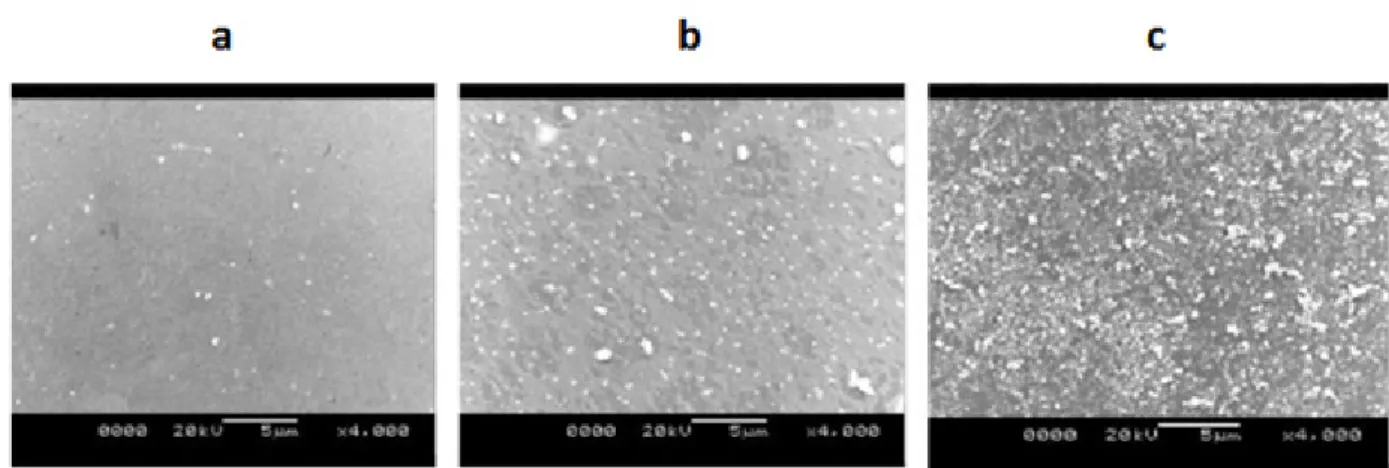
SEM ANALYSIS
To demonstrate the presence of required roughness, the SEM micrographs recorded. Fig. 1 clearly shows the formation of pits in mPE-2.5 J/cm2 when
compared to the untreated mPE because of the etching of polymer surface by the UV rays. Whilst, the quantity and size of the pits observed to increase in mPE-5 J/cm2 substrates because of prolonged etching. Therefore, the characteristic changes visibly express a significant increase in surface
Figure 1 - Representative SEM images of the a) mPE, b) mPE-2.5 J/cm2
and c) mPE-5 J/cm2
roughness of mPE samples after UV treatment.
So, the surface modified samples are anticipated
to establish a platform for friendly relation with biological substances.
WETTABILITY
Despite surface roughness, wettability is also an important surface parameter which plays a pivotal role in controlling biomolecule adsorption and cellular interaction. In general, a substrate with less wettability and low surface energy interacts poorly with a native environment which often results
in the activation and aggregation of undesired biomolecules. In this study, the mean contact angle of P-mPE was calculated as 86.84º, whereas in mPE-2.5 J/cm2 and mPE-5 J/cm2 samples it was reduced to 68.09º and 57.93º respectively as shown in Table I. So, from the results, we can clearly infer that after UV treatment the mPE surfaces become hydrophilic because the contact angle recorded is well below 70º. Basically, the hydrophilic surfaces are reported to facilitate the bioadhesion, i.e., formation of uniform biofilm. This property is particularly important in long-term implants such as cardiovascular and orthopaedic grafts since it is reported to enhance cells incorporation and biomechanical characteristics (Jaganathan et al. 2014, Maitz et al. 2015).
COAGULATION ASSAYS
Blood coagulation assays of the surface treated samples were indicated in the Fig. 2a, b. It was observed that the both PT and APTT time of treated samples were observed to be enhanced
TABLE I
Mean contact angles of the mPE, mPE-2.5 J/cm2
and
mPE-5 J/cm2 treated samples (*mean differences were
significant compared with untreated mPE (p< 0.05)).
S.No Samples Contact angle (mean ± SD)
1 (untreated mPE)Control 86.84 ± 1.65 2 mPE- 2.5 J/cm
2 68.09 ± 0.94* 3 mPE- 5 J/cm2
57.93 ± 2.05*
Figure 2 -a) APTT and b) PT assessment of mPE, mPE-2.5 J/cm2
and mPE-5 J/cm2
treated samples. The mean of the mPE and mPE-2.5 J/cm2
and mPE-5 J/cm2
treated samples along with their standard deviation
is indicated from three individual experiments. Further mean differences between the mPE and the mPE-2.5
J/cm2
when compared with the untreated surface. The APTT of mPE-2.5 J/cm2 and mPE-5 J/cm2 treated
samples were found to be 76.7 ± 4.1 s and 112.3 ± 2 s and for the untreated surface, the APPT time was observed to be only 55.3 ± 2.5 s as shown in Fig. 2a. Similarly, the value of PT for mPE-2.5 J/cm2 and mPE-5 J/cm2 treated samples were
observed to be 34.3 ± 1.1 s and 43 ± 2 s and for the untreated surfaces, the value of PT was found to be 24.7 ± 1.5 s as shown in Fig. 2b. In the recent
study, Mohandas et al investigated the effect of microwave induced surface modification of mPE.
Their results showed enhanced blood compatibility and of mPE and they ascribed it to the improved physicochemical properties like wettability and surface roughness (Mohandas et al. 2013). In this work, it was observed an increase in wettability and improved surface roughness and hence, the improved blood compatibility of UV- treated surface samples must be linked to its improved physicochemical properties.
CYTOCOMPATIBILITY STUDIES
The surfaces with higher wettability allow the displacement of undesired plasma proteins (like
fibrinogen) by cell adhesive serum proteins, such
as fibronectin and vitronectin, due to
protein-specific affinity (Balaji et al. 2015). This property
of controlling and translating protein adsorption is greatly needed to avoid the complication of blood component activation. Moreover, the obtained contact angle falls in the optimum range of 70º-30º which has previously been reported to be suitable
for regeneration of cells like fibroblasts, HUVECs,
osteoblasts and cardiomyocytes (Anderson 2001).
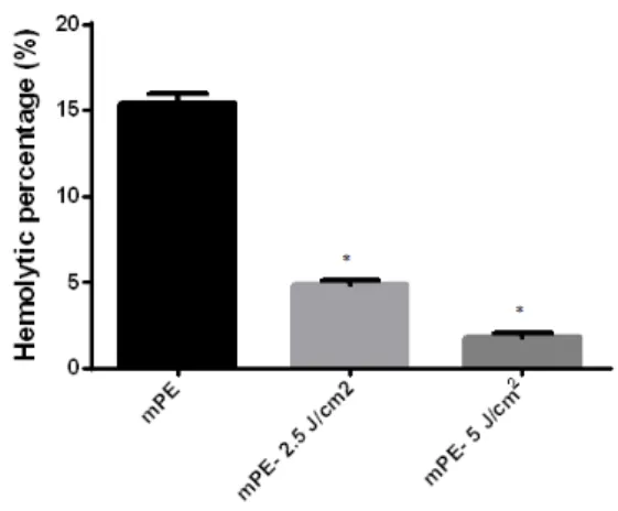
The above discussed effect of improved surface
roughness and wettability was clearly observed in the cytocompatibility tests. The hemolysis assay of the surface treated samples are indicated in Fig. 3. The results of hemolysis assay showed that untreated sample exhibited 15.40% hemolysis,
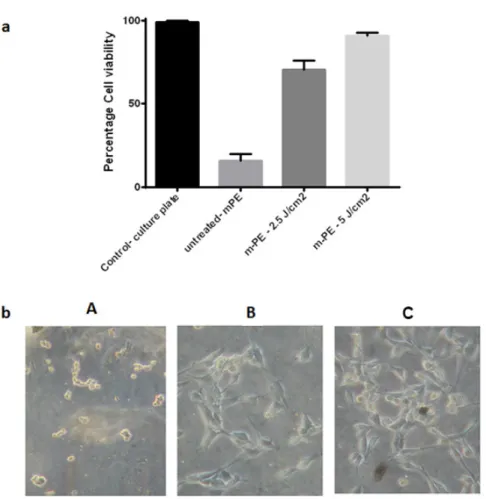
whereas, the surface showed only treated samples mPE-2.5 J/cm2 and mPE-5 J/cm2 showed 4.88% and 1.79% hemolysis. It was clearly evident that the treated surfaces showed decreased hemolytic index indicating high safety to red blood cells. Further, MTT assay showed that the cultured 3T3 cells adhered well on the UV treated surface when compared to untreated mPE as shown in Fig. 4a, b. The mean percentage cell viability of the untreated surface was found to be 15.67. The treated mPE-2.5 J/cm2 and mPE-5 J/cm2 samples expressed a cell
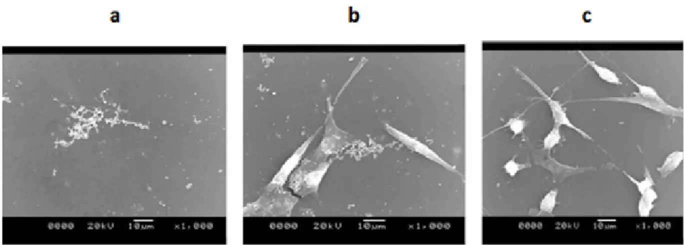
viability percentage of 70.67 and 91 respectively as shown in Fig. 4a. In the photomicrographs on mPE-5 J/cm2 surface, the cells seem to adhere at a much higher rate as shown in Fig. 4b. Moreover, the presence of spindle structure indicates a suitable environment for the proliferation of the 3T3 cells (Zhou et al. 2016). Further, it is also evident from the
SEM images that the cells seeded on the modified
surface are more viable than on untreated samples as shown in Fig. 5. It was observed that the mPE-2.5 J/cm2 treated mPE seems to be non-toxic surface, whereas, mPE-5 J/cm2 treated mPE displayed
similar cell viability percentage as the tissue
Figure 3 - Hemolysis assay of mPE, mPE-2.5 J/cm2
and mPE-5 J/cm2
treated samples. The mean of the mPE and mPE-2.5 J/ cm2
and mPE-5 J/cm2
treated samples along with their standard deviation is indicated from three individual experiments.
Further mean differences between the mPE and the mPE-2.5 J/
cm2
culture plates. The commercial polymers used for manufacturing the cell culture plates were styrene and propylene (Coassin et al. 2001). Generally, they are hydrophobic which limits the cell adherence and proliferation (Dowling et al. 2011). To improve the cell adherence and proliferation, the biocoatings with variety of surface treatment were utilized to improve the hydrophilic nature in order to enhance the cell adherence and proliferation (Mandracci et al. 2016). In our study, the UV treated mPE improved the hydrophilic nature which facilitates the enhanced cell adherence and proliferations.
Further, the UV treatment was cost effective and
the UV treated mPE might be used as an alternate candidate for the manufacturing of the cell culture plates.
CONCLUSIONS
Technologies existing in this modern era paves a way for the exploration of several advanced materials, however, the availability of those materials for medical use will take a few decades of time as it has to undergo a series of clinical trials. So, in addition to discovering new substitutes, more researches need to be carried out on improving the quality of materials which have been utilized for
Figure 4 - (a) Cell viability of mPE, mPE-2.5 J/cm2
and mPE-5 J/cm2
treated samples after 24 h using MTT assay (b) Photomicrograph of the mPE, mPE-2.5 J/cm2
and mPE-5 J/cm2
treated samples seeded with 3T3 fibroblast cells after 24 h. The mean of the mPE and
mPE-2.5 J/cm2
and mPE-5 J/cm2
various medical applications. This not only ensures
timely implementation of the resultant product
for clinical usage, but also help us to combat
the future demand for reliable and cost-effective
implants. Hence, this work shows that following
UV treatment, the surface roughness, wettability
and blood compatibility of mPE samples enhanced
notably which also depended on the UV dose.
The cytocompatibility of mPE also improved as
evidently seen from the photomicrograph and
SEM images. The improved blood compatibility
of the UV exposed mPE may be utilized for blood
contacting devices. Further, the cell viability results
of mPE-5 J/cm2 was similar with the existing
cell culture plates. Hence, it might be a plausible
candidate for manufacturing the cell culture plates.
However, it has to be further validated in other
cell lines and animal model to justify its proposed
applications.
ACKNOWLEDGMENTS
This work was supported by the Ministry of
Higher Education Malaysia with the Grant no.
Q.J130000.2545.17H00.
REFERENCES
ANDERSON JM. 2001. Biological responses to materials. Annual Rev Mater Res 31(1): 81-110.
AYYAR M, MANI MP, JAGANATHAN SK AND RATHANASAMY R. 2017. Preparation, characterization and blood compatibility assessment of a novel electrospun nanocomposite comprising polyurethane and ayurvedic-indhulekha oil for tissue engineering applications. Biomed Eng/Biomedizinische Technik: 1-9.
BALAJI A, JAGANATHAN SK, VELLAYAPPAN MV, JOHN AA, SUBRAMANIAN AP, SELVAKUMAR M, MOHANDAS H AND SUPRIYANTO E. 2015. Prospects of common biomolecules as coating substances for polymeric biomaterials. RSC Adv 5(85): 69660-69779. BHAT S AND KUMAR A. 2013. Biomaterials and
bioengineering tomorrow’s healthcare. Biomater 3(3): e24717.
CHANG HI AND WANG Y. 2011. Cells and Biomaterials Cell Responses to Surface and Architecture of Tissue Engineering Scaffolds. Regenerative Medicine and Tissue Engineering, Intech Publications, 27 p.
COASSIN PJ, HAROOTUNIAN AT, TSIEN RY AND PHAM
AA. 2001. Inventors; Aurora Biosciences Corporation,
assignee. Low background multi-well plates with greater than 864 wells for spectroscopic measurements. United States patent US 6: 229-603.
DOWLING DP, MILLER IS, ARDHAOUI M AND GALLAGHER WM. 2011. Effect of surface wettability and topography on the adhesion of osteosarcoma cells on plasma-modified polystyrene. J Biomater App 26(3): 327-347.
HOLLINGER JO. 2011. An Introduction to Biomaterials. CRC Press, 2nd
ed., 644 p. http://www.marketsandmarkets.com/
Figure 5 - SEM micrographs of a) mPE, b) mPE-2.5 J/cm2
PressReleases/globalbiomaterials-market-worth-US58.1-Billion-by-2014.asp.
JAGANATHAN SK, SUPRIYANTO E, SELVAKUMAR M, BALAJI A AND ASOKAN MK. 2014. Biomaterials in Cardiovascular Research: Applications and Clinical Implications. BioMed Res Int: 1-11.
JAGANATHAN SK, BALAJI A, VELLAYAPPAN MV, SUBRAMANIAN AP, JOHN AA, ASOKAN MK AND SUPRIYANTO E. 2015. Radiation-induced surface modification of polymers for biomaterial application. J Mater Sci 50(5): 2007-2018.
JOHN AA, SUBRAMANIAN AP, VELLAYAPPAN MV, BALAJI A, JAGANATHAN SK, MOHANDAS H, PARAMALINGGAM T, SUPRIYANTO E AND YUSOF M. 2015. Physico-chemical modification as a versatile strategy for the biocompatibility enhancement of biomaterials. RSC Adv 5(49): 39232-39244.
KIM MH, KINO-OKA M, KAWASE M, YAGI K AND TAYA M. 2007. Response of human epithelial cells to culture surfaces with varied roughnesses prepared by immobilizing dendrimers with/without d-glucose display. J Biosci Bioeng 103(2): 192-199.
MAITZ MF. 2015. Applications of synthetic polymers in clinical medicine. Biosurf Biotribol 1(3): 161-176.
MANDRACCI P, MUSSANO F, RIVOLO P AND CAROSSA S. 2016. Surface treatments and functional coatings for biocompatibility improvement and bacterial adhesion reduction in dental implantology. Coatings 6(1): 1-22. MAO C, QIU Y, SANG H, MEI H, ZHU A, SHEN J AND
LIN S. 2004. Various approaches to modify biomaterial surfaces for improving hemocompatibility. Advances in colloid and interface science. 110(1): 5-17.
MOHANDAS H, SIVAKUMAR G, KASI P, JAGANATHAN SK AND SUPRIYANTO E. 2013. Microwave-assisted surface modification of metallocene polyethylene for improving blood compatibility. BioMed Res Int 253473: 1-7.
PONCIN-EPAILLARD FA AND LEGEAY G. 2003. Surface engineering of biomaterials with plasma techniques. J Biomater Sci Polym Ed 14(10): 1005-1028.
VAGASKA BA, BACAKOVA L, FILOVA EL AND BALIK KA. 2010. Osteogenic cells on bio-inspired materials for bone tissue engineering. Physiol Res 59(3): 309-322. YORUC ABH AND SENER BC. 2012. Biomaterials, A
Roadmap of Biomedical Engineers and Milestones. Intech Publications, p. 1-240.