http://dx.doi.org/10.1590/s2175-97902018000117347
Article
*Correspondence: Pál Perjési, PhD. Institute of Pharmaceutical Chem-istry, University of Pécs, Rókus u. 2, H-7624 Pécs, Hungary. E-mail: pal.perjesi@gytk.pte.hu
Changes in hepatic metabolic enzyme activities and biliary
excretion of 4-nitrophenol in streptozotocin induced diabetic rats
Attila Almási
1*, Évora da Ibéria Leite Nogueira Pinto
4, Noémi-Piroska Kovács
3, Tamás Fischer
2,
Zoltán Markovics
1, Emil Fischer
2, Pál Perjési
1,*1Institute of Pharmaceutical Chemistry, University of Pécs, Pécs, Hungary, 2Institute of Pharmacology and Pharmacotherapy, University of Pécs, Pécs, Hungary, 3Farmacie Selena, Târgu Mureş, Romania, 4Department of Pharmaceutical Sciences,
Federal University of Pernambuco, Recife, PE, Brazil
Activity of hepatic metabolic enzymes of glucuronidation and sulfation of 4-nitrophenol (PNP) and biliary excretion of its glucuronide (PNP-G) and sulfate (PNP-S) conjugates have been investigated in control and streptozotocin (STZ)-induced diabetic rats. 500 μM PNP solution was luminally perfused in a cannulated jejunal loop for 90 minutes. It was found that biliary excretion of PNP-G was significantly decreased in the diabetic rats. This effect of STZ could be completely reversed by administration of rapid-acting insulin. Activity of hepatic UDP-glucuronyltransferase and β-glucuronidase was also depressed by the STZ pretreatment. Administration of insulin antagonized the inhibitory action of STZ on UDP-glucuronyltransferase, but the reduced activity of β-glucuronidase was not reversed. Biliary excretion of PNP-S was also depressed in the diabetic rats. Whereas, different effects of insulin administration were observed. Namely, the lower biliary excretion rate of PNP-S was not changed after administration of insulin. Activity of the sulfotransferase and the arylsulfatase enzymes was not altered either by STZ pretreatment or by insulin administration. Biliary excretion of PNP was also significantly depressed by STZ and this depression was not changed after insulin administration. The results call attention to hepatobiliary circulation of low molecular weight xenobiotics and their glucuronide and sulfate conjugates.
Keywords: 4-Nitrophenol. Hepatic excretion. Glucuronide conjugate. Sulfate conjugate. Experimental diabetes.
INTRODUCTION
Orally administered drugs, before they reach the systemic blood circulation, can be metabolized in the intestinal tract and the liver. Biotransformation of drugs before entering the systemic circulation is referred to as first pass metabolism. Numerous (Phase I. and Phase II.) reactions are involved in biotransformation of xenobiotics in the liver and small intestine (Linn, Chiba, Baillie, 1999; Parkinson et al., 2013). Phenolic compounds are generally metabolized by Phase II reactions. In the present experiments 4-nitrophenol (PNP) was used as a model substance, because it is known to be metabolized almost exclusively by glucuronidation and sulfation (Kuhn, Rost, Müller, 2001) producing 4-nitrophenol-glucuronide
(PNP-G) and 4-nitrophenol-sulfate (PNP-S). These reactions are catalyzed by UDP- glucuronyltransferases (Tukey, Strassburg, 2000; Parkinson et al., 2013) and sulfotransferases (Capiello et al., 1989; Maiti et al., 2004; Parkinson et al., 2013) in the liver and the small intestine. Esterases (β-glucuronidase and arylsulfatase) play also an important role in biotransformation of drugs, because these enzymes can hydrolyze the glucuronide and the sulfate conjugates (Danovitch, Laster, 1969; Parkinson
et al., 2013).
Several factors, e.g. drug interactions, nutrient deficiencies, pathological alterations, diseases including diabetes can influence metabolism of drugs (Liu et al., 2012; Tormo et al., 1995; Watkins, Dykstra, 1987).
drug metabolism and hepatic and intestinal transport of drugs, (Fischer, Lauterbach, 1984; Tomkin, Owens, 2015). Little definitive data are available, however, concerning the effects of diabetes on the excretory function of the liver, the metabolic enzyme activities and the role of insulin in these processes and alterations.
In our previous experiments effect of diabetes on the excretory function of the small intestine and the activities of the related enzymes were investigated (Fischer et al., 2015). The results indicated that experimental diabetes increased intestinal glucuronidation of PNP but did not influence sulfate conjugation.
The present experiments were designed to study the effects of STZ-induced experimental diabetes on the hepatic elimination of PNP during its luminal perfusion in a jejunal loop of rats. For this purpose, biliary excretion of PNP-G and PNP-S as well as activities of hepatic UDP-glucuronyltransferase, β-glucuronidase, sulfotransferase and arylsulfatase enzymes were determined to get information about the relationship between the biliary excretion rates and the respective enzyme activities. The prompt effect of the insulin was also studied on biliary excretion of the PNP metabolites and the enzyme activities in the diabetic rats.
MATERIAL AND METHODS
Chemicals
4-nitrophenol (PNP), 4-nitrophenol-ß-glucuronide (PNP-G), 4-nitrophenol-sulfate (PNP-S), 4-ethylphenol (ETP), streptozotocin (STZ), tetrabutyl ammonium bromide (TBAB), phenolphthalein-ß-glucuronide, uridine 5’-diphosphoglucuronic acid (UDPGA), 3’-phosphoadenosine-5-phosphosulphate (PAPS), nitrocatechol (NC)-sulfate, 4-naftol, Brij56®, HEPES, DL-dithiothreithol were obtained from Sigma Aldrich Company (Budapest, Hungary). All other chemicals and reagents were analytical or HPLC grade. The standard isotonic perfusion medium had the following composition (mmol/l): NaCl 96.4, KCl 7.0, CaCl2 3.0, MgSO4 1.0, sodium phosphate buffer (pH 7.4) 0.9, TRIS buffer (pH 7.4) 29.5, glucose 14.0, mannitol 14.0.
Animals and experimental procedure
Male Wistar rats (weighing 220-250 g) were randomly separated into three groups of seven rats each. In group I and II experimental diabetes was induced by i.v. administration of STZ (65 mg/kg bw). Control animals (group III) were treated with saline. Experimental
procedure was performed after one week of the STZ-treatment. Rats of group II were treated with rapid-acting insulin (Actrapid) (1 IU/kg bw, i.v.) just before the anesthesia. Liver and bile samples were obtained from the same rats of the three different groups of rats (control, STZ-pretreated with or without insulin).
The animals were anesthetized with urethane (1.2 g/kg bw, i.p.). The abdomen was opened by a mid-line incision and a jejunal loop (length about 10 cm) was isolated and cannulated in vivo. Body temperature was
maintained at 37 °C with a heat lamp. The lumen of the jejunal loop was gently flushed with warmed isotonic solution to remove digesta and food residues and then blown empty with 4-5 mL of air. Perfusion through the lumen of the jejunal loop with isotonic medium containing 500 µM PNP was carried out at a rate of 13 ml/min in a recirculation mode for 90 minutes. The initial volume was 15 mL. The temperature of perfusion medium was maintained constant at 37 °C. Bile was collected in 15 minute-periods into tared Eppendorf tubes placed in ice. The collected samples were stored in refrigerator (-20 oC) until analysis. Bile flow was measured gravimetrically assuming a specific gravity of 1.0. Biliary excretion was calculated as the product of the concentration in bile and bile flow.
Analytical conditions, instrumentation
Bile samples were analyzed by a UV-Vis RP-HPLC method, as it was developed and published earlier (Almási
et al., 2011). Briefly, 50 µL of bile samples was mixed
and vortexed with cold methanol in a total volume of containing 2.5 mM ETP as internal standard. A mixture of 0.01 M citrate buffer (pH 6.2) and methanol (47:53 v/v%) containing 0.03 M tetrabutyl ammonium bromide was used as a mobile phase. The flow rate of the mobile phase was 1.0 mL.min-1.
The HPLC system consisted of a Varian 2010 pump, a Rheodyne 7724i injection valve and a UV-Detector 308 (Labor MIM, Budapest, Hungary). Data were recorded and evaluated by a PowerChrom 280 data module and software (ADInstruments, Sydney, Australia). Separation of compounds was performed on a Nucleosyl 100 C18 reversed phase analytical column (250 mm x 4.6 mm I.D., 10 µm particle size) with a TR-C-160K1 ODS guard column. UV measurements were performed on a Pye Unicam (Philips) PU 8800 UV-Vis spectrophotometer (Philips, Cambridge, UK) at ambient temperature. Injection volume was 20 μL.
Hungary) were used to adjust the pH of the electrolyte solutions.
Enzyme assays
UDP-glucuronyltransferase and ß-glucuronidase assays
UDP-glucuronytransferase assay
Liver obtained from control (isotonic medium) and perfused (500 μM PNP) rats were gently dissected and immediately frozen and stored at – 80 °C temperature. 250 – 500 mg liver samples were dissected and placed into 2.5-5 mL of cold (4 °C) homogenization buffer (250 mM sucrose, 1 mM DL-dithiothreithol, 10 mM Hepes-Tris buffer pH 8.0) and disrupted using an Ultra-Turrax and Potter-Elvehjem homogenizers.
After disruption, the homogenate was diluted with the homogenization buffer to have a protein concentration of 1 mg/ml. Quantification of protein was performed in each enzyme assay by the biuret method (van Norman, 1909).
UGT enzyme activity assays were carried out in an incubation mixture of 100 μL of liver homogenate (1 mg/ ml protein) and 100 μL of incubation buffer (200 mM Tris-HCl, 10 mM MgCl2, 0.1 % Brij56® pH 7.4) containing 2 mM PNP. Half of the incubates contained 5 mM UDPGA in the total volume. After 30 min incubation at 37 °C, the reaction was quenched by adding 3.8 ml of 0.1 M ice cold NaOH and the absorbance of the unconjugated PNP was measured at 405 nm. Specific UGT activity was calculated on the base of metabolized PNP and expressed in μmol PNP/mg/min (Capiello, Giuliani, Pacifici, 1991).
β-Glucuronidase assay
Dissection, storage and homogenization of liver samples were the same as described for the UDP-glucuronyltransferase assay. ß-Glucuronidase activity assays were carried out in an incubation mixture of 100 μL of liver homogenate (1 mg/mL protein) and 150 μL of 70 mM pH 5.0 acetate buffer. Half of the incubates contained 1 mM phenolphthalein-β-glucuronide in the total volume. After 60 min incubation at 37 °C the reaction was quenched by adding 250 μL of ice cold 5% trichloroacetic acid. Samples were neutralized by adding 0.22 M glycine solution of pH 12.9. The absorbance was measured at 540 nm. β-Glucuronidase activity was calculated on the base of the metabolized phenolphthalein and it is expressed in the amount of the formed free phenolphthalein: µmol/mg/min. The mixture of the acetate buffer, the glycine solution and distilled water was used as a reference (Fishman, 1974).
Sulfotransferase and arylsulfatase assays
• Sulfotransferase assay
Dissection, storage and homogenization of liver samples were the same as described for the UDP-glucuronyltransferase assay. Liver was quickly dissected and immediately frozen and stored at -80 °C until analysis. Homogenization was performed in cold (4 °C) homogenization buffer (250 mM sucrose, 1 mM DL-dithiothreithol, 10 mM Hepes-Tris buffer pH 8.0).
Determination of sulfotransferase activity was carried out with incubation of 100 μL of liver homogenate and 400 μl of incubation buffer (62.5 mM Tris buffer, pH 6.2) containing 25 μM PAPS, 125 μM 2-naftol, and 6.25 mM 4-nitrophenol sulfate. The reaction was started by addition of the homogenate to the incubation buffer.
After 30 min incubation at 37 °C the reaction was quenched by adding 500 μL of 0.25 M Tris buffer, pH 8.7. Absorption of the formed 4-nitrophenol was measured at 401 nm. Reference solution contained homogenization buffer without organ homogenate (Maiti et al., 2004). The advantage of this assay is that the sulfotransferases also catalyze the sulfuryl group transfer from PNP-S to regenerate PAP-S and the forming PNP can be easily detected by UV-Vis spectrophotometry.
• Arylsulfatase assay
Dissection, storage and homogenization of liver samples were the same as described for the UDP-glucuronyltransferase assay.
Determination of arylsulfatase activity was carried out with incubation of 50 μL of liver homogenate and 400 μL of incubation buffer (in a concentration of 0.25 M sodium-acetate buffer pH 5.5) containing 0.01 M nitrocatechol sulfate. The reaction was started by adding the homogenate to the incubation buffer. After 30 min incubation at 37 °C the reaction was quenched by adding 500 uL of 5 m/v% trichloroacetic acid. Thereafter the mixture was centrifuged at 1600 x g for 10 minutes. Then 500 μL of supernatant was added to 500 μL of 5 M NaOH solution and absorption of the formed nitrocatechol was measured at 520 nm. Reference solution contained homogenization buffer instead of the homogenate (Danovitch, Laster, 1969).
Calculations, statistical analysis
The biliary excretion rate of PNP and the metabolites were calculated on the base of their concentration in the bile and the volume of the biliary flow. Data show the mean ± S.E. of seven experiments.
Data were analyzed by one-way ANOVA, the difference among groups was determined by post hoc Tukey test and (p<0.05) was significant.
Ethical approval
All procedures were carried out on animals according to the Hungarian Animal Protection Act (Hungarian Act XXVIII, 1998). The study was approved by the Ethics
Committee on Animal Research of the University of Pécs.
RESULTS
The blood glucose level of the experimental animals was 8.61 ± 1.59 mmol/L in the control, 27.1 ± 4.04 mmol/L in the diabetic, and 9.49 ± 3.88 mmol/L in the diabetic rats with insulin treatment.
The bile flow was determined for each 15 minute-period. The obtained data are summarized in Table I. As it shown STZ treatment statistically did not change the bile flow rate. On the contrary, insulin treatment resulted in statistically significant increase of the bile flow rate in comparison to both the nontreated and the STZ-treated groups.
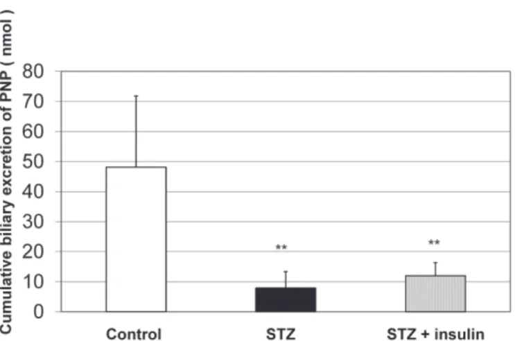
Figure 1 shows the cumulative biliary excretion (sum of the excreted amounts in 90 minutes) of PNP-G at the luminal perfusion of 500 μM PNP. Significantly lower amount of PNP-G was excreted into the bile in diabetic rats than in the controls. Depression of biliary excretion of PNP-G was compensated by rapid-acting insulin administration.
Figure 2 and Figure 3 summarize activities of the enzymes which play important role in synthesis (UDP-glucuronyltransferase) and hydrolysis (β-glucuronidase) of PNP-G in the liver. As Figure 2 shows, activity of UDP-glucuronyltransferase was significantly decreased in the diabetic rats. The depression was antagonized by insulin administration. The results show parallelism with alterations of biliary excretion of PNP-G in the respective groups.
Similarly to UDP-glucuronyltransferase, a decreased activity of β-glucuronidase was observed in the diabetic rats (Figure 3). This decrease, however, was less pronounced than that measured for the corresponding transferase. The depressed activity was not reversed by insulin administration.
TABLE I - The 15-minute period bile flows (µL/kg/min) in the various experimental groups
Time period
(min) 500 µM PNP
500 µM PNP+STZ
500 µM PNP+STZ+Insulin
0 – 15 63.1 ± 4.4 54.8 ± 8.7 81.6 ± 7.5#
15 – 30 60.5 ± 3.7 54.8 ± 6.6 103.1 ± 7.8**,##
30 – 45 60.6 ± 4.2 49.6 ± 5.7 96.7 ± 12.7*,##
45 – 60 58.7 ± 4.1 58.6 ± 7.8 96.1 ± 4.0**,##
60 – 75 59.6 ± 3.5 52.7 ± 6.1 93.3 ± 6.2**,##
75 – 90 57.4 ± 3.2 47.1 ± 6.9 90.2 ± 8.1**,##
The 15-minute period bile flows (µL/kg/min) over the 90-min luminal perfusion of 500 µM PNP in control (PNP) and diabetic rats without (PNP+STZ) or with insulin (PNP+STZ+Insulin) administration. Data represent the mean of ± S.E. of seven rats. Significant difference from the control value: ** p<0.01, * p<0.05. Significant difference from the value of STZ-diabetic rats without insulin
administration: ## p< 0.01, # p < 0.05.
FIGURE 1 - Cumulative biliary excretion of PNP-G after the 90-min luminal perfusion of 500 µM PNP in control and diabetic
The cumulative biliary excretion of PNP-S is shown in Figure 4. STZ-pretreatment significantly decreased biliary excretion of PNP-S in comparison to the control values. These results indicate that alteration in the biliary excretion of PNP-S shows the same tendency observed for PNP-G. Insulin administration, however, could not reverse the depressed biliary excretion rate of PNP-S in the diabetic rats. No significant difference was found between the values of the STZ-pretreated rats with or without insulin administration.
Sulfotransferase activity of the liver was not changed either by STZ or insulin treatment (Figure 5). No significant difference was observed between the values of the control and the diabetic rats without or with insulin treatment.
Figure 6 illustrates hepatic activity of arylsulfatase. No significant difference was observed between the values of control and STZ-pretreated rats without or with insulin. These finding shows similarity to that of the corresponding change of sulfotransferase activity.
Data of Figure 7 shows that experimental diabetes produced by STZ significantly decreased biliary excretion of the unconjugated PNP. It can also be seen that insulin was unable to antagonize the decreasing effect of STZ pretreatment on the biliary excretion rate of PNP.
DISCUSSION
The effect of STZ-induced experimental diabetes
FIGURE 2 - Activity of UDP-glucuronyltransferase of the
liver in control and diabetic rats without or with insulin administration. Values represent the mean ± S.E. of seven rats. Significant difference from the control value: ** p<0.01.
Significant difference from the value of STZ-diabetic rats
without insulin administration: ## p<0.01. The enzyme activity
is expressed in μmol PNP/mg/min, which was conjugated in 1
minute calculated for 1 mg protein of the liver.
FIGURE 3 - Activity of β-glucuronidase of the liver in control
and diabetic rats without or with insulin administration. Values represent the mean ± S.E. of seven rats. Significant difference from the control value: ** p<0.01. Significant difference from the value of STZ-diabetic rats without insulin administration:
## p<0.01. The columns show the amount of phenolphthalein formed from its glucuronide in 1 minute calculated for 1 mg liver protein.
FIGURE 4 - Cumulative biliary excretion of PNP-S after the 90-min lu90-minal perfusion of 500 µM PNP in control and diabetic
rats without or with insulin administration. Data represent the mean of ± S.E. of seven rats. Significant difference from the control value: ** p<0.01. No significant difference was found between the values of STZ-diabetic rats without or with insulin
has been investigated on biliary excretion of PNP and its conjugated metabolites (PNP-G, PNP-S) as well as on activity of the respective metabolic enzymes, while PNP was luminally perfused in the jejunal loop of anesthetized rats. The pathological state induced by STZ administration, due to its specific B-cell cytotoxicity, is considered to closely resemble that of insulin-dependent diabetes mellitus (Agarwal, 1980). STZ-doses of 50-65 mg/kg bw lead to hyperglycemia (20-30 mmol/L) but severe ketosis does not develop even if insulin is not administered (Wei et al, 2003). Such a treatment is also useful as a model of phrenic nerve neuropathy in the rats
(Rodrigues Filho, Fazan, 2006). The experiments were performed 1 week after the STZ treatment. By that time the expected hyperglycemia (27 mmol/L) has been developed.
Earlier results showed that bile flow rate in STZ-treated rats is a function of time after administration of STZ: bile flow is diminished immediately for several days after STZ injection, whereas the flow is normal one month after STZ (Stone et al., 1997; Boyer, 2013). This might
be the reason why some authors have reported decreased (Andrews, Griffiths, 1984; Carnovale, Rodriguez-Garay, 1984) whereas others have reported normal bile flow or even choleresis (Watkins, Dyktsra, 1987; Watkins, Sherman, 1992). In long term diabetes, however, several other factors - change in steady state volume of distribution and in some pharmacokinetic parameters (transport proteins, bile duct contractility) - may also influence hepatic disposition of xenobiotics (Tormo et al., 1995; Liu
et al., 2010; Zhang et al., 2011). In the present experiments bile flow rate was not affected by the 7-day STZ-treatment (Table I). The bile flow rate values are in accord with the expected normal choleretic values (Boyer, 2013).
It was found that STZ treatment depressed biliary excretion of PNP-G, which could be compensated by short-acting insulin administered prior to the experiment (Figure 1). Activity of both liver UDP-glucuronyltransferase and β-glucuronidase was also lower in the diabetic rats than those in the controls (Figures 2 and 3). Insulin antagonized the depression of UDP-glucuronyltransferase, whereas the decreased activity of β-glucuronidase was not influenced by administration of rapid-acting insulin.
FIGURE 5 - Activity of sulfotransferase of the liver in control
and diabetic rats without or with insulin administration. Values
represent the mean ± S.E. of seven rats. The sulfotransferase
activity is expressed in nmol PNP-S, which was produced in 1
minute calculated for 1 mg liver protein.
FIGURE 6 - Activity of arylsulfatase of the liver in control and
diabetic rats without or with insulin administration. Values
represent the mean ± S.E. of seven rats. Data expressed in nmol
of 4-nitrocatechol (4-NC), which was produced from 4-NC
sulfate in 1 minute calculated for 1 mg liver protein.
FIGURE 7 - Cumulative biliary excretion of PNP after the 90-min lu90-minal perfusion of 500 µM PNP in control and diabetic
rats without or with insulin administration. Data represent the mean of ± S.E. of seven rats. Significant difference from the control value: ** p<0.01. No significant differences were found between the values of STZ-diabetic rats without or with insulin
There have been several studies demonstrating that conjugation reactions, including conjugation of PNP, are sensitive to the level of carbohydrate reserves and cofactor supply (Reinke et al., 1979; Thurman et al., 1981; Qu, Kauffman, Thurman, 1995). Increased production of UDP-GA can augment the capacity of liver to form glucuronide even without a detectable increase of glucuronyltransferase activity (Miettinen, Leskinen, 1970). The background of the increased production of UDP-GA is that the flux of glucose in tissues of diabetic rats is shunted to insulin insensitive pathways when flow through insulin-sensitive routes is inhibited (Watkins, Dyktsra, 1987).
Diabetes is known to be a major disorder in which oxidative stress and free radical production have been implicated through several lines of evidence (Hinokio
et al., 1999; Suzuki et al., 1999; Brownlee, 2001) In the
liver, involvement of reactive oxygen radicals has been suggested in apoptotic cell death of hepatocytes and endothelial cells (Jaeschke, 2000). It was demonstrating that insulin treatment impairs the increase of hydroxyl radical (.OH) production generated by the STZ-induced
diabetic state in the rat liver (Frances et al., 2010). According to these earlier observations, the enhanced glucuronide excretion caused by insulin administration is supposed to be the consequence of relief of the oxidative stress resulting in increased availability of UDP-GA and regenerated enzyme activity.
Biliary excretion of PNP-S was decreased in diabetic rats similarly to that of PNP-G, whereas, insulin was unable to compensate the depressed biliary excretion of PNP-S (Figures 4-6). No change was found, however, either in sulfotransferase or in arylsulfatase activities in diabetic rats regardless they were treated with insulin or not. Sulfate conjugation in hepatocytes of insulin-treated diabetic rats was also found to be less intense than that of in the control rats (Eacho, Sweeny, Weiner, 1981). Decrease in sulfation of PNP and in biliary excretion of PNP-S can be explained by depletion of ATP, because it is required in synthesis of PAPS. Hyperglycemia induced oxidative stress depletes ATP, which is subsequently utilized for synthesis of PAPS, the cofactor required for sulfation. Similar mechanism is considered for reduced availability of UDP-GA (Li et al., 2015).
STZ-induced diabetes significantly decreased biliary excretion of the unconjugated PNP (Figure 7). Administration of insulin doesn’t antagonize the decreasing effect. In intact rats anions with molecular weight lower than 350 mainly excreted by the kidney (Lehman-McKeeman, 2013). Our experiment shows that some PNP can be excreted into the bile, which – in intact
rats - can be reabsorbed from the small intestine. These experimental findings call attention to hepatobiliary circulation of law molecular weight xenobiotics as well as their sulfate and glucuronide conjugates. Further mechanistic studies are needed to identify the molecular mechanism of hyperglycemia-induced decrease of the three (PNP, PNP-G and PNP-S) ionizable compounds into the bile.
Comparison the above results with those obtained in our previous work shows different patterns of change in enzyme activities and the amount of the respective metabolites. Thus, the STZ-initiated diabetes raised the luminal appearance of PNP-G and the increase could be completely reversed by rapid-acting insulin. The changes were in parallel with alterations in the respective enzyme activities (Fischer et al., 2015). The differences
can be explained by the different effects determining the amount of the excreted metabolites in the two organs. Whereas, glucuronidation in the small intestine is directly related to the actual blood glucose level, in the hepatic processes direct action of insulin has also to be taken into considerations. Insulin has been reported to increase the bile flow and biliary excretion of drugs in experimental animals (Garberoglio et al., 1983; Thomsen, Larsen, 1982). Neither the luminal appearances of PNP-S nor the related intestinal enzyme activities, however, were significantly influenced by the experimental hyperglycemia (Fischer
et al., 2015).
CONCLUSION
These experiments show importance of hyperglycemia in hepatic excretion of PNP as well as its glucuronic acid and sulfate conjugates in the rats. The changes in the biliary excretion can be partly explained by the altered activities of the metabolic enzymes involved in synthesis of the two conjugates. No direct correlation was found between alterations of the enzyme activities and appearance of the respective metabolites in the bile. Accordingly, other factors (e.g. transport processes, availability of cofactors, bile acid production) should also be important in the effects of experimental diabetes on hepatic elimination of drugs.
ACKNOWLEDGEMENT
express their special thanks to Reiszné Horváth Mária for her excellent technical assistance.
REFERENCES
Almási A, Fischer E, Perjési P. Isocratic ion-pair HPLC method
for quantitation of 4-nitrophenol and it’s conjugated metabolites
from bile. Sci Pharm. 2011;79(4):837-847.
Agarwal MK. Streptozotocin: mechanisms of action. FEBS
Lett. 1980;120(1):1-3.
Andrews SM, Griffiths LA. The metabolism and disposition of
[2-14C]diazepam in the streptozotocin-diabetic rat. Xenobiotica. 1984;14(10):751-760.
Boyer JL. Bile formation and secretion. Compr Physiol.
2013;3(3):1035-1078.
Brownlee M. Biochemistry and molecular cell biology of
diabetic complications. Nature. 2001;414(6865):813-820.
Capiello M, Franchi M, Giuliani L, Pacifici GM. Distribution
of 2-naphthol sulfotransferase and its substrate adenosine 3-phosphate 5-phosphosulfate in human tissues. Eur J Clin Pharmacol. 1989;37(3):317-320.
Capiello M, Giuliani L, Pacifici GM. Distribution of
UDP-glucuronyltransferase and its endogenous substrate uridine 5-diphosphoglucuronic acid in human tissues. Eur J Pharmacol. 1991;41(4):345-350.
Carnovale CE, Rodriguez-Garay EA. Reversible impairment of hepatobiliary function induced by streptozotocin in the rat. Experientia 1984;40(3):248-250.
Danovitch SH, Laster R. The development of arylsulfatase in
the small intestine of the rat. Biochem J. 1969;114(2):343-350.
Eacho PI, Sweeny D, Weiner M. Conjugation of p-nitroanisole
and p-nitrophenol in hepatocytes isolated from streptozotocin diabetic rats. J Pharmacol Exp Ther. 1981;218(1):34-40.
Fischer E, Almási A, Bojcsev S, Fischer T, Kovács NP, Perjési P. Effect of experimental diabetes and insulin replacement on
intestinal metabolism and excretion of 4-nitrophenol in rats. Can J Physiol Pharmacol. 2015;93(6):459-464.
Fischer E, Lauterbach F. Effect of hyperglycaemia on sugar transport in the isolated mucosa of guinea-pig small intestine. J Physiol. 1984;355:367-386.
Fishman WH. β-Glucuronidase. In: Bergmeyer HU (Ed). Methods of Enzymatic Analysis New York: Academic Press;
1974; p. 929-943.
Frances DE, Ronco MT, Monti JA, Ingaramo PI, Pisani GB, Parody JP, Pellegrino JM, Sanz PM, Carrillo MC, Carnovale
CE. Hyperglycemia induces apoptosis in rat liver through the
increase of hydroxyl radical: new insights into the insulin effect.
J Endocrinol. 2010;205(2):187-200.
Garberoglio CA, Richter HM3rd, Henarejos A, Moossa AR, Baker AL. Pharmacological and physiological doses of insulin and determinants of bile flow in dogs. Am J Physiol.
1983;245(1):G157-173.
Hinokio Y, Suzuki S, Hirai M, Chiba M, Hirai A, Toyota T. Oxidative DNA damage in diabetes mellitus: its association with
diabetic complications. Diabetol. 1999;42(8):995-998.
Hungarian Act 1998 XXVIII on Protection and Careful Treatment of Animals.
Jaeschke H. Reactive oxygen and mechanisms of inflammatory
liver injury. J Gastroenterol Hepatol. 2000;15(7):718-724.
Kuhn MD, Rost M, Müller D. Para-nitrophenol glucuronidation
and sulphatation in rat and human slices. Exp Toxic Pathol. 2001;53(1):81-87.
Li S, Tan H-Y, Wang N, Zhang Z-J, Lao L, Wong C-W, Feng Y.
The role of oxidative stress and antioxidants in liver diseases.
Int J Mol Sci. 2015;16(11):26087-26124.
Linn LH, Chiba M, Baillie TA. Is the role of the small intestine
in first–pass metabolism overemphasized? Pharm Rev. 1999;51(2):135-151.
Liu C, Su H, Wang Y, Tung T, Chou P, Chou Y, Liu J, Chen J. Reduced bile duct contractile function in rats with chronic
hyperglycemia. Health. 2010;2(9):1072-1077.
Liu H, Wu B, Pan G, He I, Li Z, Fan M, Jian L, Chen M, Wang K, Huang C. Metabolism and pharmacokinetics of mangiferin
in conventional rats, pseudo-germ free rats, and
streptozotocin-induced diabetic rats. Drug Metab Dispos.
2012;40(11):2109-2118.
Lehman-McKeeman LD. Absorption, distribution and excretion
of toxicants. In: Casarett and Doull’s toxicology: the basic science of poisons. 8th Ed. (Klaassen CD, Ed.). New York:
Changes in hepatic metabolic enzyme activities and biliary excretion of 4-nitrophenol in streptozotocin induced diabetic rats
Maiti S, Grant S, Baker SM, Karanth S, Pope CN, Chen G. Stress regulation of sulfotransferases in male rat liver. Biochem Biophys Res Commun. 2004;323(1):235-241.
Miettinen TA, Leskinen E. Glucuronic acid pathway. In: Fishman WH (Ed.) Metabolic conjugation and metabolic hydrolysis. New York: Academic Press; 1970; p.157-238.
Parkinson A, Ogilvie BW; Buckley DB, Kazmi F, Czerwinski M, Parkinson O. Biotransformation of Xenobiotics. In: Casarett and Doull’s toxicology: the basic science of poisons, 8th Ed. (Klaassen CD, Ed.). New York: McGraw-Hill Education; 2013; Chap. 6; p. 185-366.
Qu W, Kauffman FC, Thurman RG. Food restriction stimulates
conjugation of p-nitrophenol ion perfused rat liver. Arch
Biochem Biophys. 1995; 319(2):451-456.
Reinke LA, Kauffman FC, Evans RK, Belinsky SA and Thurman
RG. p-Nitrophenol conjugation in perfused livers from normal
and phenobarbital-treated rats: Influence of nutritional state.
Res Commun Chem Pathol Pharmacol. 1979;23(1):185-193.
Rodrigues Filho OA, Fazan VPS. Streptozotocin induced
diabetes as a model of phrenic nerve neuropathy in rats. J
Neurosci Meth. 2006;151(2):131-138.
Stone JL, Braunstein JB, Beaty TM, Sanders RA, Watkins JB
III. Hepatobiliary excretion of bile acids and rose bengal in streptozotocin-induced and genetic diabetic rats. J Pharmacol Exp Ther. 1997;281(1):412-419.
Suzuki S, Hinokio Y, Komatu K, Ohtomo M, Onoda M, Hirai S, Hirai M, Hirai A, Chiba M, Kasuga S, Akai H,
Toyota T. Oxidative damage to mitochondrial DNA and its relationship to diabetic complications. Diabetes Res Clin Pract. 1999;45(2-3):161-168.
Thomsen OO, Larsen JA. Interaction of insulin, glucagon, and
DBcAMP on bile acid-independent bile production in the rat.
Scand J Gastroenterol. 1982;17(5):687-693.
Thurman RG, Reinke LA, Belinsky S, Evans RK, Kauffman FC.
Co-regulation of the mixed-function oxidation of nitroanisole and glucuronidation of p-nitrophenol in the perfused rat
liver by carbohydrate reserves. Arch Biochem Biophys.
1981;209(1):137-142.
Tomkin GH, Owens D. Dyslipidaemia of diabetes and the intestine. World J Diabetes. 2015;6(7):970-977.
Tormo MA, Gómez-Zubeldia MA, Roppero F, Muñoz-Casillas
M, Moreno JC, Campillo JE. Experimental
streptozotocin-reduced diabetes and intestinal glucose metabolism in the rat, in vivo and in vitro. Acta Diabetol. 1995;32(3):182-186.
Tukey RH, Strassburg CP. Human UDP-glucuronosyltransferases: metabolism, expression and disease. Ann Rev Pharmacol Toxicol. 2000;40:581-616.
Van Norman KH. The biuret reaction and the cold nitric acid test in the recognition of protein. Biochem J. 1909;4(3-4):127-35.
Watkins JBIII, Dykstra TP. Alterations in biliary excretory
function by streptozotocin-induced diabetes. Drug Metab
Dispos. 1987;15(2):177-183.
Watkins JBIII, Sherman SE. Long-term diabetes alters the
hepatobiliary clearance of acetaminophen, bilirubin and digoxin. J Pharmacol Exp Ther. 1992;260(3):1337-1343.
Wei M, Ong L, Smith MT, Ross FB, Schmid K, Hoey AJ, Burstow D, Brown L. The streptozotocin-diabetic rat as a model
of the chronic complications of human diabetes. Heart Lung Circulation. 2003;12(1):44-50.
Zhang L, Liang L, Jin S, Jing X, Yao D, Hu N, Liu L, Duan R, Liu X, Wang G, Xie L. Tissue specific alterations in expression
and function of P-gp in STZ induced diabetic rats. Acta Pharmacol Sin. 2011;32(7):956-966.