RESUMO.- [Bactérias multirresistentes isoladas de ar-trite séptica equina.] Artrite séptica é uma artropatia infecciosa debilitante de equinos, que requer diagnóstico
precoce e intervenção terapêutica imediata, com intuito de evitar a degeneração de a cartilagem articular e a perda da capacidade atlética e de trabalho dos animais. Poucos
estu-Multidrug resistant bacteria isolated from septic arthritis
in horses
1Rodrigo G. Motta2,3*, Lorrayne S.A. Martins3, Igor G. Motta4, Simony T. Guerra2,
Carolina L. de Paula2, Carmen Alicia Daza Bolanos2,5, Rodrigo Costa da Silva6
and Marcio Garcia Ribeiro2
ABSTRACT.- Motta R.G., Martins L.S.A., Motta I.G., Guerra S.T., De Paula C.L., Bolanos C.A.D., Silva R.C. & Ribeiro M.G. 2017. Multidrug resistant bacteria isolated from septic arthritis in horses.Pesquisa Veterinária Brasileira 37(4):325-330.Departamento de Higiene Veteriná-ria e Saúde Pública, Faculdade de Medicina VeterináVeteriná-ria e Zootecnia, Universidade Estadual Paulista, Cx. Postal 560, Botucatu, SP 18618-681, Brazil. E-mail: [email protected]
Septic arthritis is a debilitating joint infectious disease of equines that requires early diagnosis and immediate therapeutic intervention to prevent degenerative effects on the articular cartilage, as well as loss of athletic ability and work performance of the animals. Few studies have investigated the etiological complexity of this disease, as well as multi-drug resistance of isolates. In this study, 60 horses with arthritis had synovial fluid sam -ples aseptically collected, and tested by microbiological culture and in vitro susceptibility test (disk diffusion) using nine antimicrobials belonging to six different pharmacological groups. Bacteria were isolated in 45 (75.0%) samples, as follows: Streptococcus equi subsp.
equi (11=18.3%), Escherichia coli (9=15.0%), Staphylococcus aureus (6=10.0%), Strepto-coccus equi subsp. zooepidemicus (5=8.3%), Staphylococcus intermedius (2=3.3%), Prote-us vulgaris (2=3.3%), Trueperella pyogenes (2=3.3%), Pseudomonas aeruginosa (2=3.3%),
Klebsiella pneumoniae (1=1.7%), Rhodococcus equi (1=1.7%), Staphylococcus epidermidis
(1=1.7%), Klebsiella oxytoca (1=1.7%), Nocardia asteroides (1=1.7%), and Enterobacter clo-acae (1=1.7%). Ceftiofur was the most effective drug (>70% efficacy) against the pathogens
in the disk diffusion test. In contrast, high resistance rate (>70% resistance) was observed to penicillin (42.2%), enrofloxacin (33.3%), and amikacin (31.2%). Eleven (24.4%) isolates were resistant to three or more different pharmacological groups and were considered multidrug resistant strains. The present study emphasizes the etiological complexity of equine septic arthritis, and highlights the need to institute treatment based on the in vitro
susceptibility pattern, due to the multidrug resistance of isolates. According to the available literature, this is the first report in Brazil on the investigation of the etiology. of the septic arthritis in a great number of horses associated with multidrug resistance of the isolates. INDEX TERMS: Joint infectious diseases, horses, etiology, antimicrobial resistance, arthritis.
1 Received on June 11, 2016. Accepted on December 16, 2016.
2 Departamento de Higiene Veterinária e Saúde Pública, Faculdade de Medicina Veterinária e Zootecnia (FMVZ), Universidade Estadual Paulista (Unesp), Cx. Postal 560, Botucatu, SP 18618-681, Brazil. *Corresponding author: [email protected]
3 Departamento de Clínica Médica de Grandes Animais, Universidade de Rio Verde (UniRV), Cx. Postal 104, Rio Verde, GO 75901-970, Brazil.
4 Programa de Pós-Graduação em Reprodução Animal, Faculdade de Me-dicina Veterinária e Zootecnia (FMVZ), Universidade de São Pulo, (USP), Cx. Postal 225, Pirassununga, SP 13635-900, Brazil.
5 Médica Veterinária Autônoma, Av. Panamericana, Carrera 13, Barrio Ciudad Jardín, Manzana D, casa 15, San Juan de Pasto, Colômbia.
dos têm investigado a complexidade etiológica da afecção, bem como a presença de multirresistência dos isolados aos antimicrobianos. Foram investigados 60 equinos portado-res de artrite, submetidos à colheita asséptica de líquido sinovial para a realização de cultivo microbiológico e teste de sensibilidade microbiana in vitro (difusão com discos) com nove antimicrobianos pertencentes a seis diferentes grupos farmacológicos. Foi obtido isolamento microbia-no em 45 (75,0%) amostras, como segue: Streptococcus equi subsp. equi (11=18,3%), Escherichia coli (9=15,0%),
Staphylococcus aureus (6=10,0%), Streptococcus zooepi-demicus (5=8,3%), Staphylococcus intermedius (2=3,3%),
Proteus vulgaris (2=3,3%), Trueperella pyogenes (2=3,3%), Pseudomonas aeruginosa (2=3,3%), Klebsiella pneumoniae
(1=1,7%), Rhodococcus equi (1=1,7%), Staphylococcus epi-dermidis (1=1,7%), Klebsiella oxytoca (1=1,7%), Nocardia asteroides (1=1,7%)e Enterobacter cloacae (1=1,7%). Cef-tiofur foi o antimicrobiano mais efetivo (>70% eficácia) in vitro diante dos patógenos. Em contraste, alta resistência dos isolados (>70% de resistência) foi observada para peni-cilina (42,2%), enrofloxacino (33,3%) e amicacina (31,2%). Onze (24,4%) isolados foram resistentes a três ou mais diferentes grupamentos de fármacos e considerados com resistência múltipla aos antimicrobianos. O presente estu-do enaltece a complexidade etiológica envolvida na artrite séptica em equinos e ressalta a necessidade de instituir o tratamento dos animais com respaldo de testes de sensibili-dade microbiana in vitro em virtude da resistência múltipla dos isolados. De acordo com a literatura consultada, esta é a primeira descrição no país da etiologia da artrite séptica em grande número de equinos associada a multirresistên-cia dos isolados aos fármacos testados.
TERMOS DE INDEXAÇÃO:Artropatia infecciosa, cavalos, etiologia, multirresistência aos antimicrobianos, artrite.
INTRODUTION
Equines are in contact with humans for thousands years, being used for transport, work, leisure, food, entertainment, sports and, more recently, to recovery of adults and chil-dren with cognitive or psychomotor problems (Thomassian 2005). Brazil has the fourth largest herd of equines of the world, with about 8,000,000 animals, and it only behind of China, Mexico, and EUA. Horse breeding and raising genera-te 3.2 million direct and indirect jobs, moving US$7.3 billion with equine rearing, and US$4.4 million with the exports of animals. In addition, Brazil ranks eighth in worldwide horse meat exports, selling the product to Belgium, the Netherlan-ds, Italy, Japan, France, and the United States (Brasil 2016).
Joint diseases are problematic conditions for horses and can lead to permanent lameness, functional incapa-city, or death due to septic shock in severe cases (Beccati et al. 2015). Septic arthritis is an inflammatory process of the joints caused by bacteria, fungi, and viruses (Taylor et al. 2010). It is possible that all joint structures are affected, which may cause clinical manifestations according to the location of the lesion (Bertone 2003, Olive et al. 2014). Sep-tic arthritis is usually caused by bacteria. Pathogens reach the joint via blood, local invasion (traumatic inoculation),
or by iatrogenic route, a consequence of local contamina-tion after direct infiltracontamina-tion of a drug into the joint (Colahan et al. 2000, Hall et al. 2012).
The clinical signs of the septic arthritis in equines inclu-de lameness of variable intensity, einclu-dema, local hyperther-mia, effusion, and marked sensitivity to pain at palpation (Thomassian 2005, Steel et al. 2013). In its initial course, diagnosis is based on clinical signs, rigorous clinical exami-nation of the affected joints and limbs, and identification of potential risk factors in the anamnesis (recent traumas, intra-articular injections, systemic diseases, and immuno-suppression) (Olive et al. 2014). Diagnosis is confirmed by arthrocentesis, followed by laboratory analysis of the syno-vial fluid, microbial culture, in vitro susceptibility test, and imaging tests of the affected joint (Taylor et al. 2010, Hall et al. 2012). In septic arthritis, the synovial fluid can be turbid, with purulent or hemorrhagic aspect, and may present alte-red viscosity. The liquid shows increased inflammatory cell counts and protein concentration (Haerdi-Landerer et al. 2010). Early diagnosis of the causative agent is necessary, because many animals evolve rapidly to chronic and debili-tating lesions, and the animal may not support its weight on the affected limb. Consequently, the animal will gradually lose joint function, tending to decubitus and leading to a poor prognosis (Forresu et al. 2006, Olive et al. 2014).
Treatment focuses on the use of antimicrobial drugs and articular drainage, and aims at the elimination of the causative agent, and removal of cell debris and fibrin from the join, which may damage the articular cartilage (Haerdi--Landerer et al. 2010). Microbial culture, associated with the in vitro susceptibility tests, increases therapy efficiency
(Morton 2005, Taylor et al. 2010).
Different antimicrobial drugs are used to treat septic ar-thritis, as monotherapy or in association, via intra-articular and/or parenteral injection (Haerdi-Landerer et al. 2010, Beccati et al. 2015), including b-lactams, aminoglycosides, sulfonamides, fluoroquinolones, macrolides, rapamycin, and amphenicols (Morton 2005, Giguère et al. 2010, Hall et al. 2012). Broad spectrum antimicrobials and those that re-ach high intra-articular concentrations should be preferred (Schneider et al. 2002, Haerdi-Landerer et al. 2010).
The multidrug resistance of bacterial species is an emer-gent global concern included in the “One Health” concept (Ribeiro et al. 2015), and poses a major threat to control and treatment strategies of many infectious diseases that affect domestic animals (Giguère et al. 2010). Nevertheless, to date, the prevalence of drug resistance or the degree of multidrug resistance of pathogens isolated from septic equine arthritis is unclear or unnoticed.
etiology of septic arthritis in 60 horses, as well as the oc-currence of multidrug resistance of the isolates to the main antimicrobials used to treat the disease in this species.
MATERIAL AND METHODS
Sixty horses with septic arthritis, 28 (46.7%) males and 32 (53.3%) females, were studied from 2013 to 2015. The animals showed the following distribution according to breed: Quar-ter Mile Horse (n=17; 28.3%), Mangalarga Marchador (n=10; 16.7%), Criollo (n=6; 10%), Appaloosa (n=3; 5%), Paint Horse (n=8; 13.3%), and mixed breed horses (n=16; 26.7%). Age ranged from 2 to 11 years, with 20 (33.3%) animals between 2-3 years; 31 (51.7%) between 4-5 years; 3 (5.0%) animals between 6-7 years, 2 (3.3%) animals between 8-9 years and 4 (6.7%) >10 ye-ars. Animals were used for tie-down roping (n=16; 26.7%), riding (n=19; 31.7%), barrel racing (n=14, 23.3%), and marching (n=11; 18.3%), and came from different farms of Rio Verde, GO, Brazil (17°47’53”S, 50°55’41”W), and its surrounding areas.
Horses with lameness (variable intensity), increased articular volume, occasionally effusion in severe or chronic cases, hyper-thermia, joint enlargement, and/or marked local pain sensibility were considered highly suggestive of having septic arthritis. Once signs were compatible with the disease, local antisepsis of the affected joint(s) was performed with 2% povidone iodine solu-tion, and 1mL synovial fluid was aseptically drawn using a 5-mL disposable syringe and 30x7 disposable needles. The following joints were sampled: carpal (n=22; 36.7%), tarsal (n=16; 26.7%), femoro-tibio-patellar (n=11; 18.3%), proximal interphalange-al (n=5; 8.3%), metacarpinterphalange-al/metatarsinterphalange-al-phinterphalange-alangeinterphalange-al (n=4; 6.7%), and distal interphalangeal (n=2; 3.3%). Samples were kept at 4°C into sterile flasks and sent to the Veterinary Microbiology Lab, University of Rio Verde, GO, for microbiological culture, and in vi-tro susceptibility testing. No antimicrobials were used in horses sampled.
All the samples were plated simultaneously in defibrinated sheep blood agar (5%) and MacConkey agar, and incubated aerobi-cally at 37oC for 72 hours. Isolated microorganisms were identified
based on conventional phenotypic methods (Songer & Post 2005, Quinn et al. 2011). All isolates were subjected to an in vitro antimi-crobial diffusion disk test, according to the Clinical and Laboratory Standards Institute (CLSI) guidelines (CLSI 2014), previously the National Committee for Clinical Laboratory Standards (NCCLS). Nine commercially available antimicrobials, used in livestock ve-terinary practice and/or indicated for equine septic arthritis were tested, totalizing six different antimicrobials groups, as follows: b−
-lactam (ceftiofur, 30µg; penicillin, 10 IU), aminoglycosides (amika-cin, 30µg; gentami(amika-cin, 10µg), analog of thiamphenicol (florpheni -col, 30µg), fluoroquinolones (enrofloxacin, 5µg; norfloxacin, 10µg), sulfonamides (trimethoprim-sulfamethoxazole, 1.25/23.7µg), and macrolides (azithromycin, 15µg). Multidrug resistance was consi-dered when one isolate showed simultaneous resistance to three or more antimicrobials (Schwarz et al. 2010, Girardini et al. 2013).
Considering a population of 11,000 equines in the studied region, and prevalence of septic arthritis ranging from 1 to 2%, the minimum number of animals with clinical signs to be sampled was estimated in 47 horses. Associations between the frequency of isolated microorganisms and the variables (anatomic location of the joints, age, breed, gender, and use of the animals) were tes-ted using the Chi-square and/or Fisher exact test, considering P<0.05 as significant (Triola 2005). The present study was appro -ved by the Committee of Ethics and Animal Experimentation of University of Rio Verde (UniRV), GO, Brazil (Protocol #01/2015).
RESULTS
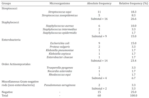
Bacteria were isolated in 45/60 (75%) synovial fluid sam -ples from horses with arthritis. The most prevalent micro-organisms were streptococci and enterobacteria, followed by staphylococci, order Actinomycetales, and Pseudomonas aeruginosa (Table 1).
Table 1. Groups and microorganisms isolated from 60 samples of synovial fluid from horses with septic arthritis. Rio Verde, GO, Brazil, 2013-2015
Groups Microorganisms Absolute frequency Relative frequency (%)
Streptococci
Streptococcus equi 11 18.3
Streptococcus zooepidemicus 5 8.3
Subtotal = 16 26.6
Staphylococci
Staphylococcus aureus 6 10.0
Staphylococcus intermedius 2 3.3
Staphylococcus epidermidis 1 1.7
Subtotal = 9 15.0
Enterobacteria
Escherichia coli 9 15.0
Proteus vulgaris 2 3.3
Klebsiella pneumoniae 1 1.7
Klebsiella oxytoca 1 1.7
Enterobacter cloacae 1 1.7
Subtotal = 14 23.4
Order Actinomycetales
Trueperella pyogenes 2 3.3
Nocardia asteroides 1 1.7
Rhodococcus equi 1 1.7
Subtotal = 4 6.7
Miscellaneous Gram-negative
rods (non-enterobacteria) Pseudomonas aeruginosa 2 3.3
Subtotal = 2 3.3
Negative - 15 25.0
Total - 60 100.0
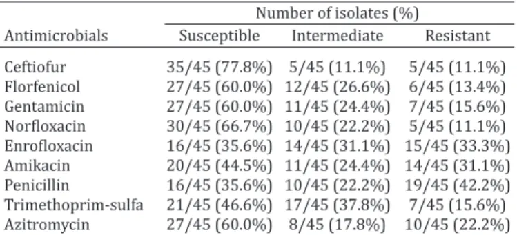
The results on the most relevant antimicrobial suscepti-bility patterns for the isolates are show in Table 2. The most effective drug (>70% efficacy) was ceftiofur (77.8%). High resistance rates (>30% inefficacy) were observed for peni -cillin (35.6%), enrofloxacin (33.3%), and amikacin (31.2%) (Table 2). Simultaneous drug resistance to three or more groups of drugs was observed in 11 (24.4%) isolates.
No significant association (P>0.05) was observed be -tween the frequency of isolated microorganisms and the anatomical location of the sampled joints, age, gender, bre-ed, and use of the animals.
DISCUSSION
We highlight in this study the complexity of groups and pa-thogens identified in septic arthritis of horses, as well as the multidrug resistance of the isolates to conventional an-timicrobials recommended for the treatment of infectious diseases of the joints.
Streptococcus equi subsp. equi and Streptococcus equi
subsp. zooepidemicus were, the most prevalent bacterial group observed in this study. This group can be found in the skin, conjunctival and mucous membranes (mainly upper respiratory tract) of the animals, causing opportunistic infections in different organs and tissues (Songer & Post 2005). Particularly in horses, these pathogens are linked to the occurrence of strangles, a highly prevalent disease of the upper respiratory tract (Waller et al. 2014). The wide occur-rence of these pathogens causing septic arthritis in horses can be justified by the presence of this group of microor -ganisms on the skin of animals, making it easy for trauma-tic inoculation into the artrauma-ticular region, contamination of intra-articular administration of drugs (iatrogenic rou-te) (Steel et al. 2013), or systemic dissemination from the upper respiratory tract (Songer & Post 2005, Orsini 2006). In addition, Pelkonen et al. (2013) warned about the zoo-notic potential of Streptococcus equi subsp. zooepidemicus
in Finland as a causative agent of an emerging disease with severe evolution in humans, given the risk of transmission to humans that are in close contact with infected horses.
Staphylococci were also isolated from many animals in this study. Similar to the streptococci group, these micro-organisms are found on the skin, conjunctival and mucous membranes, and may cause opportunistic infections in the joint by traumatic inoculation, iatrogenic or systemic
Table 2. In vitro antimicrobial susceptibility profile (disk diffusion method) of microorganisms from equines with
septic arthritis. Rio Verde, GO, Brazil, 2013-2015
Number of isolates (%)
Antimicrobials Susceptible Intermediate Resistant Ceftiofur 35/45 (77.8%) 5/45 (11.1%) 5/45 (11.1%) Florfenicol 27/45 (60.0%) 12/45 (26.6%) 6/45 (13.4%) Gentamicin 27/45 (60.0%) 11/45 (24.4%) 7/45 (15.6%)
Norfloxacin 30/45 (66.7%) 10/45 (22.2%) 5/45 (11.1%)
Enrofloxacin 16/45 (35.6%) 14/45 (31.1%) 15/45 (33.3%)
Amikacin 20/45 (44.5%) 11/45 (24.4%) 14/45 (31.1%) Penicillin 16/45 (35.6%) 10/45 (22.2%) 19/45 (42.2%) Trimethoprim-sulfa 21/45 (46.6%) 17/45 (37.8%) 7/45 (15.6%) Azitromycin 27/45 (60.0%) 8/45 (17.8%) 10/45 (22.2%) Trimethoprim-sulfa = Trimethoprim-sulfamethoxazole.
routes (Benites et al. 2016). Moreover, Taylor et al. (2010) referred to the refractoriness to treatment of equine sep-tic arthritis caused by Staphylococcus aureus, as well as its poor prognosis, in Great Britain.
Enterobacteria presented high rates of isolation in the sampled horses. This complex group belongs to enteric mi-crobiota of animals. These bacteria are also found in the en-vironment, feces, water, organic material, or contaminated materials for general use in animal raising (Songer & Post 2005). They are responsible by enteric and extra-enteric opportunistic infections in animals, and have a wide and diverse set of virulence factors (Quinn et al. 2011). The high occurrence of enterobacteria in the synovial fluid sam -ples of horses is in agreement with similar studies in other countries (Schneider et al. 2002, Bertone 2003), and pro-bably is a consequence of traumatic contamination of the joint by environmental microorganisms, as well as being secondary to systemic dissemination (Steel et al. 2013).
Pseudomonas aeruginosa is a Gram-negative bacterium that affects humans and animals, and usually causes wa-terborne disease. It is found in the environment, intestinal tract of animals, or contaminated surgical materials. It cau-ses opportunistic disease, and leads to many clinical signs in livestock (Songer & Post 2005, Quinn et al. 2011). In this study, Pseudomonas aeruginosa has been isolated in two animals with septic arthritis, probably secondary to con-tamination with water, needles during intra-articular ino-culation of drugs, or feces from the environment (Steel et al. 2013). In these situations, prognosis is usually poor due to the high refractoriness of the pathogen to the conventio-nal antimicrobials used to treat the disease (Songer & Post 2005, Quinn et al. 2011).
The order Actinomycetales is a heterogeneous group of microorganisms relevant to animal and human health, once they are related to high-impact livestock diseases (i.e. tu-berculosis, rhodococcosis, nocardiosis, corynebacteriosis, and dermatophilosis), besides the risks these pathogens pose to public health (Songer & Post 2005). Nocardia aste-roides and Trueperella pyogenes are aerobic actinomycetes related to opportunistic pyogranulomatous infectious in livestock (Quinn et al. 2011). Different species of Nocardia
are found on the ground, particularly in sites where soil, organic material, and high humidity are found. Trueperella pyogenes is found on the skin and mucous membranes of the animals, and is usually transmitted by flies (Ribeiro et al. 2015). Although the occasional occurrence of this micro-organism in horses, as observed in this study, lesions cau-sed by these pathogens are intensely pyogenic, of difficult tissue repair, and usually refractory to the treatment with conventional antimicrobials, which frequently leads to chronic processes with poor prognosis (Quinn et al. 2011).
dis-seminated infection from the lung or intestines. However, rhodococcosis is rare in adult horses, and usually occurs in weak animals or those co-infected with immunosuppressi-ve agents (Ribeiro & Vargas 2016).
From 60 sampled horses, 15 (25 %) presented negati-ve results in microbiological culture. In spite of the criteria used in the sampling collection, equine joint diseases may also be caused by traumatic, generative or immune-media-ted processes (Thomassian 2005, Madigan & House 2006). In addition, some uncommon microorganisms in horses may occasionally cause joint, ligament, and bursa lesions secondary to systemic infections, i.e., Brucella abortus and Mycoplasma sp. These microorganisms need selective me-dia or optimal oxygen tension conditions to be isolated, which were not used in the present study. Furthermore, according to what was mentioned above, in immune-me-diated conditions caused by Rhodococcus equi, the articular lesion is caused by the deposition of immune complexes (antigen-antibody) and is not possible the isolation of the microorganism (Ribeiro & Vargas 2016). All these factors may justify, in part, the negative isolation of the microorga-nisms in 15 samples of synovial fluid.
No statistical association was observed between the isolated pathogens and age, breed, gender, use of the ani-mals, and anatomical location of the sampled joints. These results agree with similar studies from other countries, in which no influence from the same variables were observed in septic joint diseases in horses (Carstanjen et al. 2010, Steel et al. 2013). However, in the current study, grea-ter occurrence of equine septic arthritis was observed in Quarter Miles, Mangalarga Marchador, Paint Horse, and mi-xed breeds ranging from 2-5 years, with lesions on tarsal, carpal, and femoro-tibio-patellar joints. The bias of septic arthropathies in these breeds and age can be attributed to the use of these animals in the studied region, particularly for entertainment and sport (tie-down roping, riding, barrel racing, and marching); which require intense use, exposing and predisposing the animals to tarsal and carpal lesions, as well as lesions in patella and fetlock.
b-lactams (penicillin, cephalosporins), aminoglycosides (Morton 2005), and fluoroquinolones (Haerdi-Landerer et al. 2010) are the antimicrobials of choice for intra-articular and/or systemic therapy of equine septic arthritis. In this study, only ceftiofur revealed in vitro sensitivity >70% to all isolates. The high efficacy of ceftiofur may be credited to the fact that intensive use of this cephalosporin in equine prac-tice therapy in Brazil took place only in the last decade (An-drade & Giuffrida 2008), reducing the selection pressure for multidrug resistance (Giguère et al. 2010). In contrast, high resistance rates (>30%) were registered for penicillin (42.2%), enrofloxacin (33.3%), and amikacin (31.2%). In -terestingly, penicillin, enrofloxacin, and amikacin are the antimicrobials of choice in the treatment of equine septic arthritis (Morton 2005, Haerdi-Landerer et al. 2010). In addition, 11 (24.4%) isolates presented simultaneous re-sistance to three or more groups of antimicrobials tested, which is a phenotypic evidence for the presence of isolates harboring genes related to multidrug resistance (Schwarz et al. 2010).
The inappropriate or empiric use of antimicrobials agents increases the selection rate for multidrug resistant bacteria, which is an emergent global threat (Giguère et al. 2010). Based on these findings, the selection of first-choice antimicrobial therapy should be based on regional in vitro resistance profile. In fact, the responsible use of antimi -crobials for animals is an emergent “One Health” concern (Ribeiro et al. 2015) in order to preserve these drugs for human therapy approaches.
CONCLUSIONS
This study highlights the complexity of bacterial agents involved in equine septic arthritis, with predominance of streptococci and enterobacteria groups, and reinforces the importance of in vitro susceptibility patterns of isolated pa-thogens to improve the success of therapy protocols.
This is the first report of septic arthritis in a large num -ber of affected horses in Brazil, showing diversity of the etiology and multidrug resistance of the isolates.
Acknowledgements.- We thank Ana Carolina Alves by support in micro-biological procedures.
REFERENCES
Andrade S.F. & Giuffrida R. 2008. Quimioterápicos antimicrobianos e an-tibióticos, p.25-72. In: Andrade S.F. (Ed.), Manual de Terapêutica Veteri-nária. 3ª ed. Roca, São Paulo.
Beccati F., Gialletti R., Passamonti F., Nannarone S., Di Meo A. & Pepe M.
2015. Ultrasonographic findings in 38 horses with septic arthritis/ te
-nosynovitis. Vet. Radiol. Ultrasound 56:68-76.
Benites N.R., Melville P.A. & Ribeiro M.G. 2016. Estafilococcias, p.300-314. In: Megid J., Ribeiro M.G. & Paes A.C. (Eds), Doenças Infecciosas em Ani-mais de Produção e de Companhia. Roca, Rio de Janeiro.
Bertone A.L. 2003. Infe ctious arthritis, p.598-604. In: Ross M.W. & Dyson S.J. (Eds), Diagnosis Management of Lameness in the Horse. 2nd ed. B.W. Saunders, Philadelphia.
Brasil 2016. Espécies. Equídeos. Ministério da Agricultura, Pecuária e Abastecimento. Disponível em <http://www.agricultura.gov.br/ani-mal/especies/equideos> Accessed on May 21, 2016.
Carstanjen B., Boehart S. & Cislakova M. 2010. Septic arthritis in adult horses. Pol. J Vet. Sci. 13:201-212.
CLSI 2014. Performance Standards for Antimicrobial Susceptibility Test-ing. 24th Information Suplement, Clinical and Laboratory Standards Institute (NCCLS).
Colahan T.B., Merritt A.M., Moore J.N. & Mayhew I.G. 2000. Equine Medi-cine and Surgery. 5th ed. Mosby, St Louis, Missouri. 207p.
Forresu D., Lepage O.M. & Cauvin E. 2006. Septic bicipital bursitis, ten-donitis and arthritis of the scapulohumeral joint in a mare. Vet. Rec. 159:352-354.
Giguère S., Prescott J.F., Baggot J.D., Walker R.D. & Dowling P.M. 2010. Te-rapia antimicrobiana em medicina veterinária. 4ª ed. Roca, São Paulo. 683p.
Girardini L.K., Gressler L.T., Costa M.M., Botton S.A., Pellegrini D.C.P. & Var-gas A.C. 2013. Perfil de suscetibilidade antimicrobiana e presença do gene vapA em Rhodococcus equi de origem humana, ambiental e equina. Pesq. Vet. Bras. 33:735-740.
Haerdi-Landerer M.C., Habermacher J., Wenger B., Suter M.M. & Steiner A. 2010. Slow release antibiotics for treatment of septic arthritis in large animals. Vet. J. 184:14-20.
Hall M.S., Pollock P.J. & Russell T. 2012. Surgical treatment of septic physi-tis in 17 foals. Aust Vet J. 90:479-484.
p.366-368. In: Smith B.P. (Ed.), Medicina Interna de Grandes Animais. 3ª ed. Manole, São Paulo.
Morton A.J. 2005. Diagnosis and treatment of septic arthritis. Vet. Clin. North Am., Equine Pract. 21:627-649.
Olive J., Lambert N., Bubeck K.A., Beauchamp G. & Laverty S. 2014 Compar-ison between palpation and ultrasonography for evaluation of experi-mentally induced effusion in the distal interphalangeal joint of horses. Am. J. Vet. Res. 75:34-40.
Orsini J.A. 2006. Artrite séptica (Artrite infecciosa), p.1096-1099. In: Smi-th B.P. (Ed.), Medicina Interna de Grandes Animais. 3ª ed. Manole, São Paulo.
Pelkonen S., Lindahl S.B., Suomala P., Karhukorpi S., Karhukorpi J., Vuorine S., Koivula I., Vaisanen T., Pentikainen J., Autio T. & Tuuminen T. 2013. Transmission of Streptococcus equi subspecies zooepidemicus infection from horses to humans. Emerg. Infect. Dis. 19:1041-1048.
Quinn P.J., Markey B.K., Leonard F.C., Fitzpatrick E.S., Fanning S. & Hartigan P.J. 2011. Veterinary Microbiology and Microbial Disease.2nd ed. Bla-ckwell Science Ltd, Oxford.1231p.
Ribeiro M.G., Risseti R.M., Bolaños C.A.D., Caffaro K.A., Morais A.C.B., Lara G.H.B., Zamprogna T.O., Paes A.C., Listoni F.J.P. & Franco M.M.J. 2015. Trueperella pyogenes multispecies infections in domestic animals: a ret-rospective study of 144 cases (2002-2012). Vet. Quart. 35:1-6. Ribeiro M.G. & Vargas A.C. 2016. Rodococose, p.458-477. In: Megid J.,
Ri-beiro M.G. & Paes A.C. (Eds), Doenças Infecciosas em Animais de Produ-ção e de Companhia. Roca, Rio de Janeiro.
Schneider R.K., Bramlage L.R. & Moore R.M. 2002. A retrospective study of 192 horses affected with septic arthritis/tenosynovitis. Equine Vet. J. 24:435.
Schwarz S., Silley P., Simjee S., Woodford N., Van Duijkeren E., Johnson A.P. & Gaastra W. 2010. Assessing the antimicrobial susceptibility of bacteria obtained from animals. Vet. Microbiol. 141:1-4.
Songer J.G. & Post K.W. 2005. Veterinary Microbiology: bacterial and fun-gal agents of animal disease. St Louis. Elsevier Saunders, St Louis. 687p. Steel C.M., Pannirselvam R.R. & Anderson GA. 2013. Risk of septic arthritis after intra-articular medication: a study of 16,624 injections in Thor-oughbred racehorses. Aust. Vet. J. 91:268-73.
Taylor A.H., Mair T.S., Smith L.J. & Perkins J.D. 2010. Bacterial culture of septic synovial strutures of horses: does a positive bacterial culture in-fluence progno sis. Equine Vet. J. 42(3):213-218.
Thomassian A. 2005. Afecções do Aparelho Locomotor (osso e articula-ções), p.97-136. In: Thomassian A. (Ed.), Enfermidades dos Cavalos. 4ª ed. Varela, São Paulo.
Triola M.F. 2005. Introdução à Estatística. 9ª ed. LTC, Rio de Janeiro. 682p. Waller A.S., Sellon D.C. & Sweeney C.R. 2014. Streptococcal infections,