In Situ Formation of Hydroxyapatite During Powder Metallurgy Preparation of Porous Ti/
HA Nano Composite: A Candidate for Dental Implants
Golsa Mousavia , Rasoul Sarraf-Mamoorya*
Received: October 29, 2017; Revised: February 21, 2018; Accepted: March 26, 2018
Porous Ti/HA nano-composite was prepared for implant fixtures and in situ formation of Hydroxyapatite. At first, Ca(OH)2 nanoparticles were synthesized by precipitation. Raw materials included Ca(NO3)2.4H2O, NaOH, a mixture of deionized water and ethanol. Synthesized Ca(OH)2 nano powder was mixed with (NH4)2HPO4 and the mixture was calcined at 700 ºC for 30 min. After calcination, Ca2P2O7 and CaPO3(OH).2H2O were synthesized with imperfect hexagonal morphology. The resulting powder was mixed with pure titanium powder and pressed under 300, 500 and 700 MPa. The samples were sintered at 1000 ºC for an hour under pure argon. Final composition was Ti/HA with hexagonal morphology. XRD and SEM showed the in situ formation of nano hydroxyapatite besides titanium phases during the sintering procedure. The density and open porosity of the 700 MPa pressed-samples were 3.07 g/cm3 and 28.2%, respectively. In general, porous Ti/HA nano-composite with suitable porosity could be successfully synthesized by co-precipitation procedure.
Keywords: Hydroxyapatite, Titanium, In situ synthesis, Calcium hydroxide, Nano-composite.
*e-mail: [email protected]
1. Introduction
Calcium hydroxide, a mineral with chemical formula of Ca(OH)2, has many applications in chemical substances, industries, environments, medications, and constructions1,2. One of the most important applications of Ca(OH)2 is to use it as raw material for synthesis of hydroxyapatite 3. Hydroxyapatite, with chemical formula of Ca10(PO4)6(OH)2, is a ceramic substance. HA is broadly used in medicine and oral biology due to the apatite-like structure of enamel, dentine and human bones. So, it exhibits biological affinity to nearest host tissues in the body and has a great compatibility with body. Thus, it can be used as a basic implant raw material4,5,6.
2. Literature Review
Bouyer et al.3 was the first one who introduced synthesis of hydroxyapatite using Ca(OH)2. In their study, Ca(OH)2 and ortho-phosphoric acid (H3PO4) react according to reaction (1). This reaction leads to formation of hydroxyapatite:
(1)
Santos et al.6, chose ammonium hydrogen phosphate [(NH4)2HPO4] and calcium hydroxide as raw materials. According to reaction (2), these two substances react and produce hydroxyapatite.
(2)
Also, they selected calcium hydrogen phosphate [Ca(H2PO4)2.H2O] and Ca(OH)2 as the raw materials, to achieve hydroxyapatite, according to reaction (3):
(3)
Another way to synthesize hydroxyapatite material is to apply a milling system. Alqap et al.7 chose (NH
4)2HPO 4 and calcium hydroxide as raw materials.
Since hydroxyapatite has poor mechanical properties in applications such as dental implants, metallic titanium has been added to produce a composite with excellent mechanical and biological properties that can endure forces to jawbone8. Thian et al.9 used powder injection metallurgy to produce Ti/HA composite. They prepared Ti6Al4V and hydroxyapatite powder with poly vinyl alcohol as a binder in slurry form, and mixed them for a specific time.
Karanjai et al.10 produced titanium based biocomposite with in situ formation of tricalcium-phosphate using powder metallurgy. They used titanium hydride and different ratios of calcium-phosphate compounds which contains calcium carbonate and diammonium hydrogen phosphate. After sintering, in situ formation of hydroxyapatite, calcium titanite, and tricalcium phosphate phases were observed.
In addition to high strength, a dental implant should be a porous surface-structured design. Although, the porous structure of the implant reduces the Young's modulus, it increases the rate of osteointegration. Moreover, feature of porous structure plays an important role in bone reconstruction due to the link between cavities which causes
aCeramic Group, Department of Materials Engineering, Tarbiat Modares University, Tehran, Iran
( )
( ) ( )
Ca OH H PO Ca
PO OH H O
10 6
18
2 3 4 10
4 6 2 2
" +
+
( ) ( )
( ) ( )
Ca OH NH HPO Ca
PO OH H O NH OH
10 6
6 12
2 4 2 4 10
4 6 2 2 4
" +
+ +
( ) ( ) .
( ) ( )
Ca OH Ca H PO H O
Ca PO OH H O
7 3
15
2 2 4 2 2
10 4 6 2 2
"
+
the flow of blood and ions into the structure and encourages the osteointegration. There is no standard for size of the porosities so, pore size can be determined regarding to the required mechanical properties. To have a porous dental implant, powder metallurgy method can be used. In this method, by controlling particle size, compaction pressure, and sintering temperature, pore size can be controlled11.
If the precursor raw materials for hydroxyapatite formation are in nanometer sizes, they can react faster and at a lower temperature. It can produce nano scale size hydroxyapatite material due to high surface area of reactants12.
Webster et al.13, reported that nano structured materials, the grain size of less than 100 nm, increase the adhesion of osteoblast and improve bone growth and amendment. Also, making Ti/HA composite in nano scale has benefits such as increased in hardness, Young's modulus, and corrosion resistance.
In this project, nano scale precursors were synthesized (i.e. Ca(OH)2) and applied to manufacture dental implants. The main objective of the present study was to prepare porous Ti/ HA nano-composite for implant fixtures and in situ formation of Hydroxyapatite during powder metallurgy processing.
3. Experimental Procedures
In the first step, Ca(OH)2 was synthesized by using hydrated calcium nitrate powder [Ca(NO3)2.4H2O] (Ghatran Shimi, 99%), sodium hydroxide powder (NaOH) (Merck, 99.9%), and ethanol as raw materials. Ca(OH)2 was precipitated according to reaction (4).
(4)
After synthesis and characterization of nano Ca(OH)2, this powder was mixed with (NH4)2HPO4 (DAP) and milled in a mortar for 10 min. Then, the mixture was screened by a 120 micrometer sieve and was calcined to prepare hydroxyapatite, according to reaction (5).
(5)
Calcination temperature was selected on the decomposition temperature of the raw materials. The mixture of powder was heated to 250 ºC and held at this temperature for 30 minutes to complete decomposition of (NH4)2HPO4, then the temperature increased to 700 ºC and kept for 30 minutes to form calcium phosphates of biological interest, and then cooled down slowly.
After calcination, the mixture of powder was again mixed with pure titanium powder (with the aspect of 1:4 ratio) and screened to have a homogenized sample. Then, the mixture was pressed as cylinder parts with height and diameter of 1 cm, with compaction pressures of 300, 500, and 700 MPa and sintered at 1000 ºC for an hour under pure argon atmosphere.
4. Results and Discussion
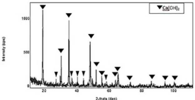
To study the formation of phases in the samples, synthesized by precipitation method, the X-ray diffraction technique was used. Figure 1 shows the XRD diffraction pattern of the calcium hydroxide. According to this Figure, it can be seen that the reaction (5) has been completely performed and there is not any unreacted materials in this condition of precipitation. It proves that the duration times for the experiments were suitable.
( ) .
( )
Ca NO H O NaOH NaNO Ca OH H O
4 2
2 4
3 2 2
3 2 2
" +
+ +
( ) ( )
( )
Ca OH NH HPO
Ca PO OH H O NH
5 3
9 6
2 4 2 4
5 4 3 2 3
" +
+ +
Figure 1. The XRD diffraction pattern of calcium hydroxide
Figure 2 shows the scanning electron microscopy (SEM) of synthesized calcium hydroxide. As it shows the calcium hydroxide particles have hexagonal plate like morphology with very fine thicknesses, which results in the highest specific surface area and highest reactivity.
Figure 2. SEM image of synthesized calcium hydroxide
(6)
The temperature of 700 ºC was selected because it was the nearest temperature to HA formation.
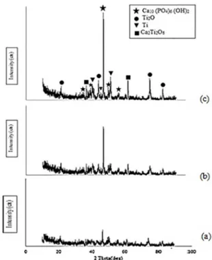
calcium phosphate compounds. These hydroxyl groups can cause synthesis of hydroxyapatite and partialy oxidation of titanium. Therefore, according to the primary objective of this project, hydroxyapatite was synthesized next to the titanium and the composite of Ti/HA was formed. On the other hand, the presentation of titanium oxide and calcium titanate in dental implants will not be harmful and will not affect its function due to this fact that these compounds are inert and they will not react with titanium and hydroxyapatite.
Also, Fig. 5 shows that the pattern of the sample, pressed with the pressure of 700 MPa, is more sharper than the other samples. In other words, the sharpness of the main peak corresponds to perfect crystallization of HA in this sample. The reason is that at higher pressure, the reactants of Ca2P2O7 and CaPO3 (OH) .2H2O are closer to each other and hence affects the reaction kinetics and diffusion process. .
CaPO OH H O Ca P O
Ca PO OH H O P O
2 2 4
4 2
3 2 2 2 7
10 4 6 2 2 2 5
"
+
+ +
Q
QQ
V
V V
Figure 3. The XRD diffraction pattern of the calcined materials in 700 ºC
Fig. 4 shows the SEM image of the calcined Ca(OH)2 and DAP sample in 700 ºC.
As it can be seen, the particle sizes of the calcium phosphates are about 1 to 5 microns and the morphology of calcined particles look like imperfect hexagonal. As we know these particles will be converted into hydroxyapatite with hexagonal morphology at 1000 ºC. This morphology is a confirmation of hydroxyapatite as well.
Figure 4. SEM image of calcined materials in 700 ºC
Figure 5 shows the XRD pattern of samples, pressed under pressure of 300, 500 and 700 MPa and sintered in 1000 ºC for 1 hour. As it can be seen, in all pressure conditions, hydroxyapatite, calcium titanate, and titanium oxide have been synthesized in addition to Ti phase. The reason of titanium oxidation is probably the early presence of titanium oxide on the surface of titanium powder and releasing the hydroxyl groups in the decomposition of
Figure 5. The XRD diffraction pattern of pressed samples under pressure of a) 300, b) 500 and c) 700 MPa and sintered in 1000ºC
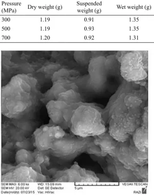
Table 1 shows that while the compaction pressure have increased; the apparent and bulk densities have also increased. In addition, it shows that open porosities in samples have decreased with increasing the compaction pressure. According to the purpose of this research, which is preparation of dental implant with adequate open porosities, the morphology and porosity distributions are the most important parameters, so the assessment of compaction pressure should be studied.
As these figures show, with the increasing compaction pressure of 300 to 700 MPa the amount of open porosity extremely reduced because the particles have become closer and the spaces between them have reduced. On the other hand, with increasing the compaction pressure, the grains with regular geometric shape have increased and their distribution throughout the sample is quite evident. These grains which have hexagonal-like sections are marked in Figures 7 and 8. We have these particles as a result of the reaction between Ca2P2O7 and CaPO3(OH).2H2O which led to hydroxyapatite. The images indirectly confirmed the results of XRD that the grains in pressed samples at a compaction pressure of 700 MPa, have shown more perfect crystallization.
5. Conclusions
1. According to the results of XRD, it was found that after calcination of mixed Ca(OH)2 and DAP at 700 ºC, Ca2P2O7 and CaPO3(OH).2H2O were obtained. SEM results showed that the particle size is in the range of 1 to 5 microns and morphology of these particles are imperfect hexagonal.
2. Sintering of mixed titanium and calcined powders at 1000 ºC, led to the formation of composites of titanium, hydroxyapatite, titanium oxide, and calcium titanate.
3. Compaction pressure of 700 MPa caused perfect crystallization during sintering. Therefore, it was much better than the other compaction pressures. Also, in 700 MPa, bulk density and the percentage of open porosities were 3.07 g/cm3 and 28.2%, respectively. Moreover, with increasing the compaction pressure, open porosities decreased and the number of grains with regular geometric shapes increased.
6. References
1. Rodriguez-Navarro C, Ruiz-Agudo E, Ortega-Huertas M, Hansen E. Nanostructure and irreversible colloidal behavior of Ca(OH)2: Implications in cultural heritage
conservation. Langmuir. 2005;21(24):10948-10957.
2. Mohammadi Z, Dummer PMH. Properties and applications of calcium hydroxide in endodontics and dental traumatology.
International Endodontic Journal. 2011;44(8):697-730.
3. Bouyer E, Gitzhofer F, Boulos MI. Morphological study of hydroxyapatite nanocrystal suspension. Journal of Materials Science: Materials in Medicine. 2000;11(8):523-531.
Table 1. Apparent density, Bulk density and Open porosity for three samples pressed under 3 different pressures.
Pressure
(MPa) Dry weight (g) Suspended weight (g) Wet weight (g) Volume (cm3) density (g/cmApparent 3)
Bulk density (g/cm3)
Open porosity
(%)
300 1.19 0.91 1.35 0.43 2.77 2.71 36.36
500 1.19 0.93 1.35 0.42 2.83 2.83 38.09
700 1.20 0.92 1.31 0.40 3.00 3.07 28.20
Figure 6. SEM image of pressed sample under pressure of 300 MPa
Figure 7. SEM image of pressed sample under pressure of 500 MPa
Figure 8. SEM image of pressed sample under compaction pressure
4. Misch CE. Contemporary Implant Dentistry. 3rd ed. Saint Louis: Mosby; 2008.
5. Kim W, Zhang Q, Saito F. Mechanochemical synthesis of hydroxyapatite from Ca(OH)2-P2O5 and CaO-Ca(OH)2-P2O5 mixtures. Journal of Materials Science. 2000;35(21):5401-5405.
6. Santos MH, Oliveira M, de Freitas Souza LP, Mansur HS, Vasconcelos WL. Synthesis control and characterization of hydroxyapatite prepared by wet precipitation process. Materials Research. 2004;7(4):625-630.
7. Alqap ASF, Adzila S, Sopyan I, Hamdi M, Ramesh S. Thermal analysis on hydroxyapatite synthesis through mechanochemical method. In: Osman NAA, Abas WABW, Wahab AKA, Ting HN, eds. 5th Kuala Lumpur International Conference on
Biomedical Engineering 2011. IFMBE Proceedings, vol 35.
Berlin, Heidelberg: Springer; 2011.
8. Thian ES, Loh NH, Khor KA, Tor SB. Processing of biocomposite Ti-6Al-4V/HA powder. Journal of Materials Science Letters.
2003;22(10):775-778.
9. Yura K, Fredrikson KC, Matijevic E. Preparation and properties of uniform colloidal indium compounds of different morphologies.
Colloids and Surfaces. 1990;50:281-293.
10. Karanjai M, Sundaresan R, Rao GVN, Mohan TRR, Kashyap BP. Development of titanium based biocomposite by powder metallurgy processing with in situ forming of Ca-P phases.
Materials Science and Engineering: A.
2007;447(1-2):19-26.
11. Arifin A, Sulong AB, Muhamad N, Syarif J, Ramli MI. Material processing of hydroxyapatite and titanium alloy (HA/Ti) composite as implant materials using powder metallurgy: A review. Materials & Design. 2014;55:165-175.
12. German RM. Intermediate Stage Processes: Solution -
Reprecipitation. In: German RM. Liquid Phase Sintering.
Chapter 5. New York: Springer; 1985.
13. Webster TJ, Ejiofor JU. Increased osteoblast adhesion on
nanophase metals: Ti, Ti6Al4V, and CoCrMo. Biomaterials.