Surface, microstructural, and adhesion strength investigations of a bioactive
hydroxyapatite-titanium oxide ceramic coating applied to Ti-6Al-4V alloys by plasma
thermal spraying
Renan Carreiro Rochaa, André Gustavo de Sousa Galdinob* , Sidnei Nicodemos da Silvac, Marcelo Lucas Pereira Machadob
Received: December 26, 2017; Revised: March 31, 2018; Accepted: April 15, 2018
Ti-6Al-4V alloy is employed in implants because of its good mechanical strength, excellent biocompatibility, and good resistance to corrosion in biological environments. Herein, a composite (HAp-TiO2, 50% by volume of both components) coating surface employing Ti-6Al-4V was characterized. Samples were analyzed post fabrication and post heat treatment to analyze the coating recrystallization, phases, crystallinity, porosity, and roughness. The coating showed rutile crystalline and amorphous Hap phases with crystallinity, porosity, and roughness of 55.6%, 13.6 ± 1.0%, and
4.2 ± 0.6 µm; whereas, after heat treatment, it showed a rutile hydroxyapatite phase and β-TCP
with values 75.6%, 13.9 ± 1.9%, and 3.8 ± 0.2 µm, respectively. The composite exhibited 874 ± 26 HV100 hardness and 30 ± 2 MPa adhesion strength after heat treatment, which agree with previously reported data on other bioactive coatings. Therefore, this composite becomes much more crystalline
after heating at 750 °C for 1 h.
Keywords: Hydroxyapatite-titanium oxide coating, Ti-6Al-4V alloy, plasma thermal spraying, surface characterization.
* e-mail: [email protected]
1. Introduction
Metallic materials have been used in implants to replace or repair human body parts over the last few decades. This is mainly due to two factors: higher life expectancy and a larger number of accidents involving means of transportation and extreme sports1.
Titanium alloys have been used as implant materials
because of their desirable characteristics such as a low specific
mass (as compared to stainless steel and cobalt-chromium alloys), high resistance to biocorrosion, biocompatibility, and outstanding mechanical properties. Titanium and its alloys stand out among the main metallic alloys typically used in implants, mainly the alpha-beta alloys with high mechanical strength such as Ti-6Al-4V (ASTM F67 and F136 or Ti-6Al-4V ELI). These alloys are now widely used for a number of clinical applications2,3.
One of the limitations of this alloy is the potential release of Al and V ions into the human body, because these ions can eventually cause long-term health issues1-3. Recent research has focused on finding ways to prevent the release of these
alloying elements and to increase the osseointegration of these metal alloys3-6. Coating these alloys with bioactive ceramics is a common practice in orthopedics and dentistry, because it combines high mechanical strength, corrosion resistance, and ease of manufacturing metal implants with enhanced biocompatibility associated with bioactive ceramic
films such as hydroxyapatite (HAp)1-3.
Coatings are applied to metal substrates through various
methods such as sol-gel7, biomimetic1, electrolytic1, sputtering ion coating1, physical vapor deposition8, and plasma thermal spray methods9-11. The plasma spray process has the best chemical control, resistance to biocorrosion, and process
efficiency among the aforementioned methods9-11.
The main concerns regarding the use of hydroxyapatite as a coating are related to substrate/coating interface instability (adhesion) and coating longevity in the physiological
environment; studies have only assessed the coating efficiency
over a short term (maximum of three years)10,12.
Therefore, recent studies have sought to develop bioactive ceramic coatings with improved adhesion between the coating and metal implants to thus increase the service life of implants6,9,10,11-14.
aInstituto Federal de Educação, Ciência e Tecnologia do Espírito Santo, Rua Governador José Sete,
S/N, Itacibá, 29150-410, Cariacica, ES, Brasil
bInstituto Federal de Educação, Ciência e Tecnologia do Espírito Santo, Av. Vitória, 1729, Jucutuquara,
29040-780, Vitória, ES, Brasil
cCentro Federal de Educação Tecnológica de Minas Gerais, Av. Amazonas, 5253, Nova Suíça,
The aim of the present study was to characterize the surface of 50% - 50% by volume HAp-TiO2 coatings applied to the Ti-6Al-4V alloy by plasma thermal spraying.
2. Materials and Methods
2.1 Hydroxyapatite Powder (HAp)
The hydroxyapatite (HAp) used in this work was kindly provided by Inside Materiais Avançados Ltda., Belo Horizonte
County, Minas Gerais State, Brazil. X-ray fluorescence assays (XRF) were conducted to identify the chemical composition in the aforementioned powder. The XRF assay was performed with the Rigaku RIX 3100 X-ray fluorescence spectrometer
in the Materials Engineering Department (DEMA) at the
Unicamp Mechanical Engineering School (FEM) - Campinas County, São Paulo State, Brazil. The XRF assay results are
presented in Table 1.
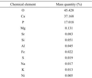
Table 1. XRF data of the as-received hydroxyapatite powder.
Chemical element Mass quantity (%)
O 45.428
Ca 37.168
P 17.018
Mg 0.131
Sr 0.083
Si 0.051
Al 0.045
Fe 0.022
S 0.019
Na 0.017
K 0.013
Ni 0.005
The hydroxyapatite used in this work had a mean particle diameter of 8.67 ± 0.14 µm and D90 = 17.34 ± 0.58 µm.
2.2 Titanium oxide (TiO
2)
The titanium oxide used was Metco 102 (Sulzer Metco), which was kindly provided by the Mechanical Engineering School of Unicamp. This titanium oxide is intended for plasma spraying applications, and its particle size ranges from 7.8 to 88 µm (nominal value). The TiO2 was characterized
by XRF, particle size analysis, morphology, and scanning
electron microscopy (SEM) prior to plasma spraying. The observed characteristics of TiO2 are presented in Table 2.
Powder granulometry is one of many variables that influences the quality of the plasma-sprayed coating due to
the high heat extraction that is inherent to plasma thermal
spraying. Very fine particles, of the order of 106 ºC/s, can solidify before they touch the substrate. These particles are
Table 2. Characteristics of the titanium oxide powder (Metco 102).
Characteristic Quantity/Quality
TiO2chemical composition 99%
Particle size 27.7 ± 0.89 µm (on average)
D90= 47.97 ± 0.41 µm
Morphology Angular
then incorporated and encapsulated by the lamellar structure of the coating after they solidify, leading to a possible drop in the adhesion strength of the coating11. Therefore, it is recommended to use powders with a particle size ranging from 20 to 100 µm16. However, issues concerning the incorporation of solid particles by the coating can be addressed by adjusting operational variables such as the distance from the torch to the substrate, primary/secondary
gas flow, and electric current in the plasma.
2.3 Ti-6Al-4V alloy
The Ti-6Al-4V alloy substrate was manufactured by the National Institute of Science and Technology in Biofabrication (BIOFABRIS - Brazil) by direct metal laser sintering (DMLS). The substrate had a diameter of 25.4 mm and height of 4.0 mm.
The chemical composition data obtained by XRF were
provided by the alloy powder supplier as well as the ASTM
F136 standard (which specifies the Ti-6Al-4V composition
for application in surgical implants), and are presented in Table 3.
Table 3. Chemical composition of Ti-6Al-4V.
Source Chemical composition (%)
Ti Al V
ASTM F136 Balance 5.5 - 6.75 3.5 - 4.5
XRF Balance 5.9 4.2
2.4 Determination of the composition of the
HAp-TiO
2composite
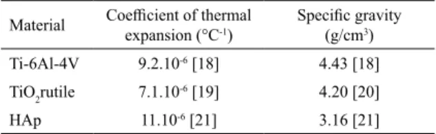
Initially, the composition HAp-TiO2 was chosen because it was expected to adhere well to the metallic substrate. The ratio between Hap and TiO2 took into account the variation
between the coefficient of thermal expansion of the coating
and that of the substrate. Equations (6.8) and (6.9) by Ashby and Jones were applied for the elasticity modulus, whereas
Equations [1] and [2] by Garmong and Shepard were applied
for the elasticity modulus17:
(1)
(2)
f
f
cu
$
HAp$
TiO2a
=
a
+
a
f
f
1
cl
HAp TiO2
a
a
a
=
+
where α is the coefficient of thermal expansion, indices
cu and cl represent the upper and lower limits of the composite, HAp represents hydroxyapatite, TiO2 represents titanium oxide, and f represents the volume fraction of the
assessed material. The coefficient of thermal expansion
and density of hydroxyapatite and titanium oxide are presented in Table 4.
sanding with silicon carbide (SiC) meshes with granulates
ranging from 220 to 1200. Next, mechanical polishing was performed using a nylon cloth soaked in 1 µm granulation diamond paste, followed by washing with acetone in an ultrasonic bath for 20 min. Finally, the Keller's reaction
was performed. The diffractograms presented herein were
collected with a voltage of 40 kV and a current of 10 mA in
steps of 0.05° over an observation range of 20° ≤ 2θ ≤ 90°.
The crystallinity of the deposited coatings was assessed with Difract Suite Eva® 2010 software, version 1.3, which automatically calculated the crystallinity by simply selecting the boundary between the typical broad peak of an amorphous
pattern and the discrete diffraction peaks. The crystallinity
index that was calculated by the software is based on Equation (3), wherein Acris is the area of the crystalline peaks
of the coating, because the XRD background represents the
amorphous pattern area.
(3)
The coating roughness and porosity were evaluated
to assess the physical properties of the coating. Porosity
images were recorded with OM and captured with a digital camera coupled to the microscope. Next, these images were
edited in the Adobe Photoshop® CS2 software to increase the contrast between the coating and pores. The porosity
was assessed with a GSA (Semi-Automatic Granulometer)
Image Analyzer. This software operates by counting pores at the intersection of lines that are pre-established by the analyzer.
2.7 Hardness tests
Coating hardness was assessed with the micro-hardness
method according to the Vickers scale. A Shimadzu hardness tester, model HMV-2000, was used for the analysis at a 100-g load for 15 s. Five measurements were performed per sample, and the mean coating hardness of these measurements was considered.
2.8 Adhesion tests
The adhesion test was performed according to the
ASTM C633-08 standard. Ten cylindrical specimens with
a diameter of 25.4 mm and a length of 25.4 mm, as well as a self-aligning device, were produced. The self-aligning device had two degrees of freedom in order to avoid shear stress during the test. The device shown in Figure 1 was
manufactured according to ASTM C633-08 recommendations.
High-adhesion Scotch-Weld 2214 3M epoxy resin was used to bond the two metallic cylindrical pieces.
The set was drawn into an EMIC conventional tensile
testing machine with a 100 kN capacity. A 100 kN load cell was used in the study and the deformation rate was found to be 0.02 mm/s.
Table 4. Thermal expansion coefficient and density of hydroxyapatite and titanium oxide.
Material Coefficient of thermal expansion (°C-1)
Specific gravity
(g/cm3)
Ti-6Al-4V 9.2.10-6 [18] 4.43 [18]
TiO2rutile 7.1.10-6 [19] 4.20 [20]
HAp 11.10-6 [21] 3.16 [21]
2.5 Thermal plasma spraying process
The HAp-TiO2 composite was mixed by tumbling. Prior to coating, the substrate specimens were prepared by grinding with coarse alumina (Al2O3) particles (grain size of 60 mesh at 75 psi pressure, 90° angle, and grinding time 30 s) in order to improve substrate roughness, eliminate possible oxides
that can influence coating adhesion, and facilitate mechanical
anchoring of the HAp-TiO2 composite. The specimens were cleaned with compressed air after blasting to remove loose alumina from the blasted surface and to prepare them for thermal spraying.
The composite was sprayed onto the Ti-6Al-4V substrate
using a plasma thermal spray gun (9MBII METCO), and the
coatings were manually applied in passes. This pistol model used herein has a radial dust feed, works at low to medium power levels, which can reach a maximum of 40 kW (500 A and 80 V). The operational deposition conditions that were used are presented in Table 5. After the aspersion thermal
process, some samples underwent heat treatment at 750 °C
for one hour, in order to recrystallize and release stress.
Table 5. Parameters in the thermal plasma spray process.
Variable Value
Current (A) 400
Voltage (V) 74
H2flow (L/min) 18
Air flow (L/min) 90
Powder flow g/min (L/min) 10
Gun to substrate distance (mm) 150
2.6 Coating characterization
Optical microscopy (OM), SEM, and X-ray diffraction (XRD) were used to characterize the coating. Samples were
prepared by sanding, polishing, and etching, as well as
%
I
cA
A
A
100
cris amor cris
$
=
+
Titanium oxide, on the other hand, had an angular morphology
with a well-defined shape and size.
3.3 Microstructural assessment and coating
crystallinity
Microscopical analysis of the coatings formed through
thermal spraying allowed for verification of several important
structural features such as thickness and coating adhesion to the substrate. The thickness of the deposited coating was uniform and observed to be 66.7 ± 1.8 µm.
Complete overlap between the parts was observed in the
substrate-coating interaction, without voids or cracks on the
interface. There were fine lines parallel to the substrate in
the structure, which indicates that the coating was formed by
lamellar deposition, and also signifies the possible presence of diffusion processes that resulted in better coating adhesion
(Figure 4 (a)).
Figure 4 (b) shows the microstructure formed by the
flattened, solidified HAp-TiO2 droplets which were arranged in successive layers forming the coating. It also shows the addition of well-scattered lamellae, which suggests a higher composite plasticity state at the moment they reach
the surface. The mechanism responsible for film formation
is triggered by the accumulation of successive layers of bioceramic powder droplets that existed in the plasma torch environment. Finally, these droplets were launched against the substrate (at a speed in excess of 400 m/s) which resulted in
the liquid coating. The coating was flattened and experienced
partial elastic recoil at the moment of impact, subsequently solidifying to form the lamellar microstructure15.
The coating phases are shown in Figure 5 (a). Only peaks characteristic of the high-crystallinity rutile phase were observed before heat treatment. There were no expected Hap peaks or peaks in phases derived from hydroxyapatite decomposition. Amorphous poplars at angles 29°, 32°, 33°, 34°, and 39° were observed, and these angles correspond with the angles where Hap peaks are expected; this indicates a low crystallinity index in the Hap portion of the composite. The distinct behavior that was observed in the two ceramics after the thermal process in relation to crystallinity seems
to be associated with the difference in the lattice parameter of the HA phase (a = 9.4 Å), which was significantly higher
than that of TiO2 (4.6 Å). Since TiO2 has lower network parameter, it also has a lower tendency than HAp to become amorphous, due to the higher network parameter of HAp as compared with that of titanium oxide.
However, in addition to β-TCP formation and the presence
of rutile, hydroxyapatite recrystallization was observed after the thermal treatment (Figure 4 (b)). No phase was formed from the reaction between HAp and TiO2.
Thus, there was a greater amount of the amorphous phase after deposition without heat treatment. In quantitative terms, the crystallinity under the deposited condition was 55.6%, whereas that after heat treatment was 75.6%.
Figure 1. Fixing device used in the adhesion test. Adapted from
ASTM C633-08.
3. Results and Discussion
3.1 Determination of volumetric composition of
the composite
The plot in Figure 2 illustrates the influence of titanium oxide addition on the thermal expansion coefficient of the
formed composite by applying the values shown in Table 3 to Equations (1) and (2).
Figure 2. Influence of TiO2 on the thermal expansion coefficient
of the HAp-TiO2 composite.
Figure 2 shows that the addition of titanium oxide to
hydroxyapatite reduced the thermal expansion coefficient of
the formed composite. The range of TiO2 volume varied from
47% to 57% near the thermal expansion coefficient of the
substrate (Ti-6Al-4V), which was 9.2.10-6 °C-1. A composition with a higher volumetric fraction of hydroxyapatite was chosen
for better composite biocompatibility and lesser difference in
thermal expansion between the substrate and coating. This is because hydroxyapatite is a bio-active material whereas titanium oxide is bioinert14,22.
3.2 Characterization of ceramic powder before
deposition
Figure 3 shows the morphology of the ceramic powders used in the present study, which exhibits granulometric dispersion as well as natural agglomeration of the particles.
Figure 3. Ceramic morphology assessment. (a) Titanium oxide powder; and (b) Hydroxyapatite powder.
Figure 4. Micrographs of Ti-6Al-4V alloy (a) substrate/coating interaction; amorphous and crystalline HAp-TiO2 phases after plasma
spraying (500X), without etching (optical microscopy); (b) SEM image of the morphological aspect of the coating surface with splash (white arrows), and the resulting lamellae as a result of molten projection or semi-cast droplets (1000X), without etching.
Figure 5. Diffractogram of HAp-TiO2 composite showing (a) the
phases before the heat treatment; (b) the phases after the heat treatment.
Table 6. Surface roughness of substrate and ceramic coating
Material Roughness RA (µm)
Substrate after blasting 4.2 ± 0.6 µm
Coating 3.8 ± 0.2 µm
3.4 Physical properties of coating
The rough surface on Ti-6Al-4V was necessary to impart good mechanical anchoring of HAp-TiO2 to the substrate. Roughness was generated by an abrasive alumina blasting process. Table 6 presents the surface roughness of the substrate and ceramic coating.
The substrate and coating roughness were similar, because the ceramic-composite deposition resulted in successive lamellar layers. Apart from promoting good mechanical coating anchoring, the substrate roughness promoted roughness in
the coating. Surface roughness is known to have a significant positive influence on bone growth24.
Our results corroborate preliminary studies25; 1) An HAp coating roughness of 3.8 ± 0.4 µm was recorded for a blasted surface and 4.23 ± 0.46 µm for the coatings, 2) a TiO2 film roughness of 2.8 ± 0.6 µm was recorded for a blasted surface, and 3) 4.6 ± 0.4 µm for the coatings. All the cited authors used plasma thermal spraying for coating deposition26.
property for coatings, because low porosity is known to minimize toxic metal ion dissolution and release from the substrate into the human body27.
Table 7. Coating porosity data.
Material Porosity (%)
As deposited 13.6 ± 1.0
After heat treatment 13.9 ± 1.9
Table 8. Hardness of HAp-TiO2 compared to values reported in the literature.
Coating material Hardness (HV) Reference
HAp rich coating 234.5 [27]
TiO2rich coating 363.9 [27]
HAp coating 366 [29]
HAp coating 320 [30]
Porosity levels close to those recorded in this study have
been reported in the literature. Khor et al.29 studied HAp coatings on Ti-6Al-4V alloys, and reported a coating porosity of 19%. Sun et al.30 investigated the microstructure, structure, and phases of different HA coatings, and obtained coating at a
porosity of up to 12%. The aforementioned authors attributed this result to the low power of the plasma equipment used in the study (27.5 kW). The power of the equipment was increased to 42.0 kW, which reduced the porosity level to 7%. The explanation for the porosity reduction to 7% is related to the incorporation of unfused particles, which decreases the coating porosity level. A plasma power of 28.8 kW was used in the present study and may have contributed to the observed increase in porosity.
3.5 Hardness
The developed composite exhibited a hardness of 523 ± 10 HV100 before and 874 ± 26 HV100 after the heat treatment.
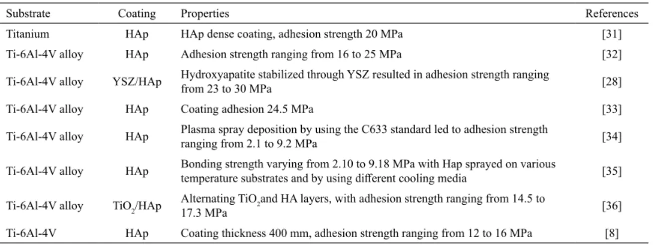
Table 9. Adhesion strength of HAp-TiO2 compared to those reported in the literature
Substrate Coating Properties References
Titanium HAp HAp dense coating, adhesion strength 20 MPa [31]
Ti-6Al-4V alloy HAp Adhesion strength ranging from 16 to 25 MPa [32]
Ti-6Al-4V alloy YSZ/HAp Hydroxyapatite stabilized through YSZ resulted in adhesion strength ranging from 23 to 30 MPa [28]
Ti-6Al-4V alloy HAp Coating adhesion 24.5 MPa [33]
Ti-6Al-4V alloy HAp Plasma spray deposition by using the C633 standard led to adhesion strength ranging from 2.1 to 9.2 MPa [34]
Ti-6Al-4V alloy HAp Bonding strength varying from 2.10 to 9.18 MPa with Hap sprayed on various temperature substrates and by using different cooling media [35]
Ti-6Al-4V alloy TiO2/HAp
Alternating TiO2and HA layers, with adhesion strength ranging from 14.5 to
17.3 MPa [36]
Ti-6Al-4V HAp Coating thickness 400 mm, adhesion strength ranging from 12 to 16 MPa [8]
The higher hardness recorded after heat treatment is due to the enhanced crystallinity of the coating. A comparison with previous studies showed that the addition of titanium oxide
to hydroxyapatite provided a significant enhancement in
coating hardness, as shown in Table 8. The authors therein used deposition variables similar to those used in the present study, including the same metallic substrate.
3.6 Adhesion strength
Tensile tests were performed to analyze the adhesion strength of the coating. The composite showed adhesion
strength of 16.6 ± 2.0 MPa prior to heat treatment and 30.0 ± 2.0 MPa after thermal treatment. The coating
herein presented better adherence than previously reported coatings which contained only hydroxyapatite. Table 9 presents the results from preliminary studies conducted over the last ten years.
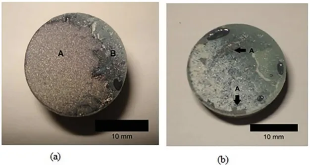
Figure 6. (a) Non-heat-treated coating after failure; (b) Heat-treated coating after failure.
4. Conclusions
The best range for the HAp-TiO2 composition is between 47% and 57% for the addition of TiO2 to hydroxyapatite. This
is so that the coefficient of linear thermal expansion of the
composite is close to that of the Ti-6Al-4V alloy substrate. Thus, the HAp-TiO2 composite with 50% by volume of both components was chosen because it displayed the crystalline phase of amorphous TiO2 and HAp after spraying. Moreover,
the rutile, recrystallized hydroxyapatite and β-TCP phases
after thermal treatment showed no phases resulting from the reaction between hydroxyapatite and titanium oxide. Without heat treatment, 55.6% crystallinity was observed in the as-deposited specimen, whereas 75.6% crystallinity was observed after the heat treatment. Thus, heat treatment enabled an approximately 36% increase in crystallinity in the
composite. There was no significant variation in roughness
and porosity, which indicates that these properties were not
influenced by the heat treatment. The composite had a HV100 hardness of 874 ± 26 and adhesion strength of 30.0 ± 2.0
MPa after the heat treatment. Both these values are higher
than those described in the literature.
5. Acknowledgements
The authors would like to thank INCT-Biofabris for the
manufacture of the test specimens through rapid prototyping, to Inside Ltda. for providing the hydroxyapatite, Labiomec/
DEMA/FEM/UNICAMP for providing the titania, and CEFET-MG for the scanning electron microscope that was
used in the microstructural analysis and Federal Institute of
Espirito Santo (IFES) by the financial contribution intended
for the revision of the article.
6. References
1. Harun WSW, Asri RIM, Alias J, Zulkifli FH, Kadirgama K, Ghani SAC, Shariffuddin JHM. A comprehensive review of
hydroxyapatite-based coatings adhesion on metallic biomaterials.
Ceramics International. 2018;44(2):1250-1268.
2. Ho WF. Effect of omega phase on mechanical properties of
Ti-Mo alloys for medicals applications. Journal of Medical and Biological Engineering. 2007;28:47-51.
3. Chen Q, Thouas GA. Metallic implant biomaterials. Materials
Science and Engineering. 2015;R 87:1-57.
4. Höhn S, Virtanen S. Effect of inflammatory conditions and H2O2on bare and coatedTi-6Al-4V surfaces: Corrosion behavior, metal ion release and Ca-Pformation under long-term
immersion in DMEM. Applied Surface Science. 2015;357:101-111. doi:10.1016/j.apsusc.2015.08.261.
5. Oliveira DP, Palmieri A, Carinci F, Bolfarini C. Gene expression
of human osteoblasts cells on chemically treated surfaces of Ti-6Al-4V-ELI. Materials Science and Engineering: C. 2015;51:248-255. doi:10.1016/j.msec.2015.03.011.
6. Ma Z, Ren L, Liu R, Yang K, Zhang Y, Liao Z, Liu W, Qi
M, Misra RDK. Effect of heat treatment on Cu distribution, antibacterial performance and cytotoxicity of Ti-6Al-4V-5Cu
alloy. Journal of Materials Science and Technology. 2015;31:723-732. doi:10.1016/j.jmst.2015.04.002.
7. Milella E, Cosentino F, Licciulli A, Massaro C. Preparation and
8. Rahmati B, Sarhan AAD, Zalnezhad E, Kamiab Z, Dabbagh
A, Choudhury D, Abas WABW. Development of tantalum oxide (Ta-O) thin film coating on biomedical Ti-6Al-4V
alloy to enhance mechanical properties and biocompatibility.
Ceramics International. 2015;42:466-480. doi:10.1016/j. ceramint.2015.08.133.
9. Tsui YC, Doyle C, Clyne TW. Plasma sprayed hydroxyapatite coatings on titanium substrates. Part 1: Mechanical properties
and residual stress levels. Biomaterials. 1998;19:2015-2029. doi:10.1016/S0142-9612(98)00103-3.
10. Lu YP, Li MS, Li ST, Wang ZG, Zhu RF. Plasma-sprayed
hydroxyapatite+titania composite bond coat for hydroxyapatite coating on titanium substrate. Biomaterials. 2004;25:4393-4403. doi:10.1016/j.biomaterials.2003.10.092.
11. Chou B, Chang E. Interface investigation of plasma-sprayed
hydroxyapatite coating on titanium alloy with ZrO2 intermediate layer as bond coat. Scripta Materialia. 2001;45:487-493.
12. Morris WF. Hydroxyapatite-coated implants: a case for their use.
Journal of Oral and Maxillofacial Surgery. 1998;56:1303-1311.
13. Chalisgaonkar V, Das M, Balla VK. Laser processing of Ti
composite coatings reinforced with hydroxyapatite and bioglass.
Additive Manufacturing. 2018;20:134-143.
14. Li H, Khor KA, Cheang P. Thermal sprayed hydroxyapatite
splats: nanostructures, pore formation mechanisms and TEM characterization. Biomaterials. 2004;25:3463-3471. Doi:10.1016/j. biomaterials.2003.10.051
15. Höhn S, Virtanen S. Biocorrosion of TiO2 nanoparticle coating
of Ti-6Al-4V in DMEM under specific in vitro conditions.
Applied Surface Science. 2015;329:356-362. doi:10.1016/j. apsusc.2014.12.114.
16. McPherson R. The relationship between the mechanism of
formation, microstructure and properties of plasma-sprayed coatings. Thin Solid Films. 1981;83:297-310. doi:10.1016/0040-6090(81)90633-7.
17. Garmong G, Shepard LA. Matrix Strengthening Mechanisms of an Iron Fiber-Copper Matrix Composite as a Function of Fiber
Size and Spacing. Metallurgical Transactions. 1971;2(1):175-180.
18. Lu X, Lin X, Chiumenti M, Cervera M, Li J, Ma L, Wei L, Hu
Y, Huang W. Finite element analysis and experimental validation of the thermomechanical behavior in laser solid forming of Ti-6Al-4V. Additive Manufacturing. 2018;21:30-40.
19. Sheppard LM. Surge in electronic materials continues. American Ceramic Society Bulletin. 1992;70(9):1467.
20. Guo HL, Zhao XP. Preparation of a kind of red encapsulated
electrophoretic ink. Optical Materials. 2004;26:297-300.
21. Castañeda L, Alonso JC, Ortiz A, Andrade E, Saniger JM, Bañuelos JG. Spray pyrolysis deposition and characterization of titanium oxide thin films. Materials Chemistry and Physics. 2002;77:938-944. doi:10.1002/crat.200710918.
22. Willmann G, Richter H, Wimmer M. Rotational bending test of
hydroxyapatite plasma-coated models of Ti-6Al-4V. Biomedical Engineering/Biomedizinische Technik. 1993;38:6-14.
23. Warashina H, Sakano S, Kitamura S, Yamauchi KI, Yamaguchi J, Ishiguro N, Hasegawa Y. Biological reaction to alumina, zirconia, titanium and polyethylene particles implanted onto
murine calvaria. Biomaterials. 2003;24:3655- 3661. doi: 10.1016/S0142-9612(03)00120-0.
24. Hayashi K, Inadome T, Tsurnura H, Nakashima Y, Sugioka Y.
Effect of surface roughness of hydroxyapatite-coated titanium
on the bone-implant interface shear strength. Biomaterials. 1994;15(14):1187-1191.
25. Braceras I, Onate JI, Goikoetxea L, Viviente JL, Alava JI, de
Maeztu MA. Bone cell adhesion on ion implanted titanium alloys. Surface and Coatings Technology. 2005;196:321-326. doi:10.1016/j.surfcoat.2004.08.201.
26. Levingstone TJ, Ardhaoui M, Benyounis K, Looney L, Stokes
JT. Plasma sprayed hydroxyapatite coatings: Understanding
process relationships using design of experiment analysis.
Surface and Coatings Technology. 2015;283:29-36.
27. Cannillo V, Lusvarghi L, Sola A. Production and characterization
of plasma-sprayed TiO2-hydroxyapatite functionally graded coatings. Journal of the European Ceramic Society. 2008;28:2161-2169.
28. Singh G, Singh S, Prakash S. Surface characterization of plasma
sprayed pure and reinforced hydroxyapatite coating on Ti-6Al-4V alloy. Surface and Coatings Technology. 2011;205:4814-4820. doi:10.1016/j.surfcoat.2011.04.064.
29. Khor KA, Gu YW, Quek CH, Cheang P. Plasma spraying of
functionally graded hydroxyapatite/Ti-6Al-4V coatings. Surface and Coatings Technology. 2003;168:195-201. doi:10.1016/
S0257-8972(03)00238-X.
30. Sun L, Berndt CC, Grey CP. Phase, structural and microstructural
investigations of plasma sprayed hydroxyapatite coatings.
Materials Science and Engineering: A. 2003;360:70-84. doi:10.1016/S0921-5093(03)00439-8.
31. Morks MF, Kobayashi A. Influence of spray parameters on
the microstructure and mechanical properties of gas-tunnel plasma sprayed hydroxyapatite coatings. Materials Science and Engineering: B. 2007;139:209-215. doi:10.1016/j. mseb.2007.02.008.
32. Roy M, Balla VK, Bandyopadhyay A, Bose A. Compositionally
graded hydroxiapatite/tricalcium phosphate coating on Ti by laser and induction plasma. Acta Biomaterialia. 2011;7:866-873. doi: 10.1016/j.actbio.2010.09.016
33. Gadow R, Killinger A, Stiegler N. Hydroxyapatite coatings for biomedical applications deposited by different thermal spray
techniques. Surface and Coatings Technology. 2010;205:1157-1164. doi:10.1016/j.surfcoat.2010.03.059
34. Kweh SWK, Khor KA, Cheang P. An in vitro investigation of
plasma sprayed hydroxyapatite (HA) coatings produced with
flame-spheroidized feedstock. Biomaterials. 2002;23:775-785. doi: 10.1016/S0142-9612(01)00183-1.
35. Yang XYC, Chang E. Influence of residual stress on bonding
strength and fracture of plasma-sprayed hydroxyapatite coatings on Ti-6Al-4V substrate. Biomaterials. 2001;22:1827-1836. doi:10.1016/S0142-9612(00)00364-1.
36. Zheng X, Huang M, Ding C. Bond strength of plasma-sprayed