UNIVERSIDADE DE LISBOA
FACULDADE DE MEDICINA VETERINÁRIA
CANINE PARVOVIRUS AND SEPSIS: SIRS CRITERIA EVALUATION AND
IMPLEMMENTATION OF A PIRO CLASSIFICATION
FREDERICO SILVA DE SOUSA ALVES
ORIENTADOR(A):
Mestre Mariana Silva Domingues dos Santos
COORIENTADOR(A):
Doutora Solange Judite Roque Coelho Alves Gil Neves
UNIVERSIDADE DE LISBOA
FACULDADE DE MEDICINA VETERINÁRIA
CANINE PARVOVIRUS AND SEPSIS: SIRS CRITERIA EVALUATION AND
IMPLEMMENTATION OF A PIRO CLASSIFICATION
FREDERICO SILVA DE SOUSA ALVES
DISSERTAÇÃO DE MESTRADO INTEGRADO EM MEDICINA VETERINÁRIA
JÚRI ORIENTADOR(A):
PRESIDENTE: Mestre Mariana Silva Domingues dos
Santos Doutor Luís Manuel Morgado Tavares
VOGAIS: COORIENTADOR(A):
Doutora Solange Judite Roque Coelho Alves Gil Neves
Doutor Rodolfo de Assis Oliveira Leal
Doutora Solange Judite Roque Coelho Alves Gil Neves
II
DECLARAÇÃO RELATIVA ÀS CONDIÇÕES DE REPRODUÇÃO DA TESE OU DISSERTAÇÃO Nome: Frederico Silva de Sousa Alves
Título da Tese ou
Dissertação: EVALUATION AND IMPLEMMENTATION OF A PIRO CLASSIFICATION CANINE PARVOVIRUS AND SEPSIS: SIRS CRITERIA Ano de conclusão (indicar o da data da realização das provas
públicas): 2020
Designação do curso de
Mestrado ou de Doutoramento: Medicina Veterinária Área científica em que melhor se enquadra (assinale uma):
Clínica Produção Animal e Segurança Alimentar
Morfologia e
Função Sanidade Animal
Declaro sobre compromisso de honra que a tese ou dissertação agora entregue corresponde à que foi aprovada pelo júri constituído pela Faculdade de Medicina Veterinária da ULISBOA.
Declaro que concedo à Faculdade de Medicina Veterinária e aos seus agentes uma licença não-exclusiva para arquivar e tornar acessível, nomeadamente através do seu repositório institucional, nas condições abaixo indicadas, a minha tese ou dissertação, no todo ou em parte, em suporte digital.
Declaro que autorizo a Faculdade de Medicina Veterinária a arquivar mais de uma cópia da tese ou dissertação e a, sem alterar o seu conteúdo, converter o documento entregue, para qualquer formato de ficheiro, meio ou suporte, para efeitos de preservação e acesso. Retenho todos os direitos de autor relativos à tese ou dissertação, e o direito de a usar em trabalhos futuros (como artigos ou livros).
Concordo que a minha tese ou dissertação seja colocada no repositório da Faculdade de Medicina Veterinária com o seguinte estatuto (assinale um):
1. Disponibilização imediata do conjunto do trabalho para acesso mundial;
2. Disponibilização do conjunto do trabalho para acesso exclusivo na Faculdade de Medicina Veterinária durante o período de 6 meses, 12 meses, sendo que após o tempo assinalado autorizo o acesso mundial*;
* Indique o motivo do embargo (OBRIGATÓRIO)
Nos exemplares das dissertações de mestrado ou teses de doutoramento entregues para a prestação de provas na Universidade e dos quais é obrigatoriamente enviado um exemplar para depósito na Biblioteca da Faculdade de Medicina Veterinária da Universidade de Lisboa deve constar uma das seguintes declarações (incluir apenas uma das três):
1. É AUTORIZADA A REPRODUÇÃO INTEGRAL DESTA TESE/TRABALHO APENAS PARA EFEITOS DE INVESTIGAÇÃO, MEDIANTE DECLARAÇÃO ESCRITA DO INTERESSADO, QUE A TAL SE COMPROMETE.
Faculdade de Medicina Veterinária da Universidade de Lisboa, ____ de ____________________ de 20___ (indicar aqui a data da realização das provas públicas) Assinatura: __________________________________________________________
III
Acknowledgements
To Prof. Doctor Solange Gil, for all the guidance and advice given as my supervisor and for all the interest and dedication shown throughout the making of this project.
To Prof. Doctor Telmo Nunes, for all the indispensable help provided in the statistical analyses and availability shown, not only to me but to all the students in need.
To all the team of veterinary surgeons and nurses at VetOeiras - Central Cascais Veterinary Hospital and at the Infectious Disease Isolation Unit of the Faculty of Veterinary Medicine, for all the patience, dedication and availability shown throughout my internship. Knowledge that I will take with me into this new stage of life. As well to all my internship colleagues that shared 6 months of my life and made them even better.
To all my friends, for the constant presence in all the good and bad moments and unconditional support shown in the last six years, showing me what true companionship feels like.
To Bia for being the most supportive and understanding partner I could have asked for. You have been a constant presence in my life in the past few years and I can´t wait to share the next accomplishments of my life with you.
To my parents, for all the time, resources and dedication invested in my education and for all the support shown throughout my academic life.
IV
Resumo
Parvovirose Canina e Sépsis: Avaliação dos critérios de SIRS e
Implementação da classificação PIRO
A sépsis esta associada a uma elevada prevalência e taxa de mortalidade. A Parvovirose canina predispõe para o aparecimento de sépsis secundaria à translocação bacteriana intestinal e imunossupressão. Este facto faz dos cães naturalmente infetados com parvovírus uma boa população para o estudo de sépsis. O principal objetivo deste estudo foi avaliar as diferenças entre dois conjuntos de critérios de SRIS (Síndrome de Resposta Inflamatória Sistémica) sobre a sua capacidade de prognóstico, assim como avaliar a possibilidade de implementação de um sistema de estratificação de animais sépticos com base no modelo PIRO (Predisposition, Infection, Response, Organ dysfunction). Os 72 animais da amostra foram submetidos a dois conjuntos de critérios SIRS e classificados para cada um dos elementos constituintes do PIRO (com exceção da infeção, sendo que todos os animais foram considerados como tendo a mesma classificação para a Infeção), avaliando a sua relação com o desfecho. Os dados foram recolhidos a partir dos registos clínicos da Unidade de Isolamento de Doenças Infeciosas (UIDI) do HEV-FMV-UL.
Em relação aos critérios de classificação SRIS, os resultados revelaram que a alteração proposta aos critérios originais resulta numa associação estatisticamente significativa com o desfecho (OR = 4.09, p < 0,05), contrastando com os resultados observados quando aplicados os critérios originais (p=0.352) que não se correlacionam significativamente com o desfecho. Não foi encontrada nenhuma associação estatisticamente significativa entre a Predisposição (p=1), Resposta (p=0.1135), Disfunção Orgânica (p=0.1135) ou PIRO total (p=0.093) e o desfecho clínico. Os resultados obtidos revelam a necessidade de critérios mais específicos para a avaliar SRIS e sépsis. Os resultados sugerem que o aumento da especificidade pode melhorar o seu valor prognóstico.
Este trabalho representa uma contribuição para o desenvolvimento de um conjunto de critérios consensual e aprovado para a classificação de animais sépticos, servindo de base para estudos futuros. Mais critérios com uma maior especificidade, como marcadores bioquímicos inflamatórios e de disfunção orgânica, devem ser adicionados ao sistema PIRO proposto.
Estudos futuros devem concentrar-se em melhorar os sistemas de classificação existentes e descobrir novos biomarcadores que permitam uma intervenção atempada em animais afetados por sépsis, melhorando a taxa de sobrevivência.
V
Abstract
Canine Parvovirus and Sepsis: SIRS criteria evaluation and
implementation of a PIRO classification
Sepsis is a severe condition associated with high prevalence and mortality rates. Parvovirus enteritis is a predisposing factor for sepsis, as it promotes intestinal bacterial translocation and severe immunosuppression. This makes naturally parvovirus infected dogs a suitable study population as far as sepsis is concerned. The main objective of the present study was to evaluate the differences between two sets of SIRS (Systemic Inflammatory Response Syndrome) criteria in outcome prediction, parallelly the possibility of stratifying and classify septic animals using a proposed animal adapted PIRO (Predisposition, Infection, Response, Organ dysfunction) scoring system was also assessed. The 72 animals enrolled in this study were subjected to a score for each of the PIRO elements (except for the Infection, as all were considered to have the same infection score) and to two sets of SIRS criteria, assessing their correlation with the outcome. The data was retrieved from the clinical records of the Infectious Disease Isolation Unit (IDIU) of the Veterinary Teaching Hospital (VTH) of the Faculty of Veterinary Medicine (FMV) of the University of Lisbon (ULisboa).
Concerning the SIRS criteria, it was found that the proposed alterations were significantly associated with the outcome (OR = 4.09, p < 0,05), contrasting with the original SIRS criteria (p=0.352) that did not correlate with the outcome. No significant statistical association was found between Predisposition (p=1), Response (p=0.1135), Organ dysfunction (p=0.1135) or total PIRO score (p=0.093) and outcome. The results obtained reveal the need for consensual and more specific criteria to assess SIRS and sepsis. The results suggest that augmenting the criteria specificity may improve their prognostic value, thus making them more useful in clinical management and treatment decision.
This work represents a contribution for the development of an approved set of criteria, to could contribute not only to the classification of septic dogs but also to the improvement of sepsis diagnosis. Further studies are still needed to conclude about the best criteria to be used, but this study can serve as base from which further studies can adapt and improve. Additional more specific criteria, mainly inflammatory and organ dysfunction biomarkers, should be added to the proposed PIRO scoring system in order to improve the its´ prognostic value and clinical utility.
Further studies should focus on improving classification systems and finding new biomarkers that would allow a timely intervention in sepsis affected animals and improve sepsis survival rate.
VI
Table of Contents
Acknowledgements……….…...……..……….…III
Resumo………...………..………...IV
Abstract………...………..………V
Figure List…………...………..….IX
Table List……….……….………...IX
Abbreviation List……….X
I - Activities developed during the curricular internship………1
1. Internal Medicine………...……….…..1
2. Surgery……….………..1
3. Inpatient Care……….………...2
4. Isolation Unit for Infectious Disease……….………..2
II – Literature Review…….………..2
1. Sepsis……….………2
1.1. General notions and SIRS………...………...2
1.2. Pathogenesis………...……….4
1.2.1. Innate immunity and inflammatory mediators………..………4
1.2.2. Inflammation and acute phase response………..………5
1.2.3. Anti-inflammatory agents and immunosuppression………..……….5
1.2.4. Inflammation and dysregulated coagulation………..………..6
1.2.5. Organ dysfunction………..….7
1.3. Diagnostic approach………....8
1.3.1. Physical exam………..8
1.3.2. Clinical laboratory findings………...9
1.3.3. Diagnostic imaging………10 1.3.4. Microbiologic test………...10 1.4. Treatment………11 1.4.1 Initial stabilization………11 1.4.2. Cardiovascular support……….11 1.4.3 Antimicrobial therapy……….12 1.4.4. Supporting care……….13 1.5. Prognosis………13 2. Canine Parvovirus………..13
2.1. Etiology and epidemiology………13
2.2. Pathogenesis………..14
VII
2.4. Diagnostic Approach……….15
2.4.1. Physical examination………15
2.4.2. Clinical laboratory findings………15
2.4.3. Microbiological test………16 2.5 Treatment……….16 2.5.1. Fluid therapy………..16 2.5.2 Antimicrobial therapy……….17 2.5.3 Antiemetic treatment………..18 2.5.4. Nutritional support……….18 2.5.5. Pain management……….18 2.6. Prognosis………18 2.7. Prevention………...19
3. PIRO: staging of sepsis……….19
3.1. PIRO (Predisposition, Insult, Response, Organ dysfunction/failure)………..19
3.2 Predisposition………..19
3.3. Insult/Infection………20
3.4. Response………20
3.5. Organ dysfunction………..21
4. Other outcome prediction systems………..21
4.1. APACHE (Acute Physiology and Chronic Health Evaluation)………..21
4.2. SOFA (Sequential/ Sepsis-related Organ Failure Assessment)……….22
4.3. MEDS (Mortality in Emergency Department Sepsis)………22
III –Canine Parvovirus and Sepsis: SIRS criteria evaluation and implementation of a PIRO classification………....23
1. Objectives………22
2. Materials and methods………..23
2.1. Inclusion criteria……….23
2.2. SIRS classification and PIRO score………24
2.2.1. Data recovery……….23
2.2.2. Classification criteria……….23
2.3. Statistical analysis………..25
2.3.1. SIRS………25
2.3.2. Total PIRO, Predisposition (P), Infection (I), Response (R) and Organ Dysfunction (O)……….……26
3. Results……….…26
3.1. Sample characterization………26
VIII 3.3. PIRO score………..……28 4. Discussion………...29 4.1. SIRS……….…………...29 4.2. Predisposition (P) ………..………….………..31 4.3. Infection (I) ………..33 4.4. Response (R)………. ………...33
4.5. Organ dysfunction (O)……...………35
4.6. PIRO...……….37
5. Conclusion………...39
IV – Bibliographic References………41
IX
Figure List:
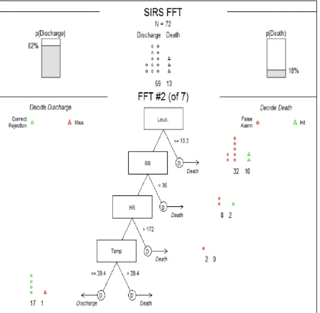
Figure 1: Example of Fast-and-frugal Tree for the response (R) element of PIRO…...29
Tables List:
Table 1: SIRS diagnosing criteria for cat and dog. (Sykes, 2014a)………....3
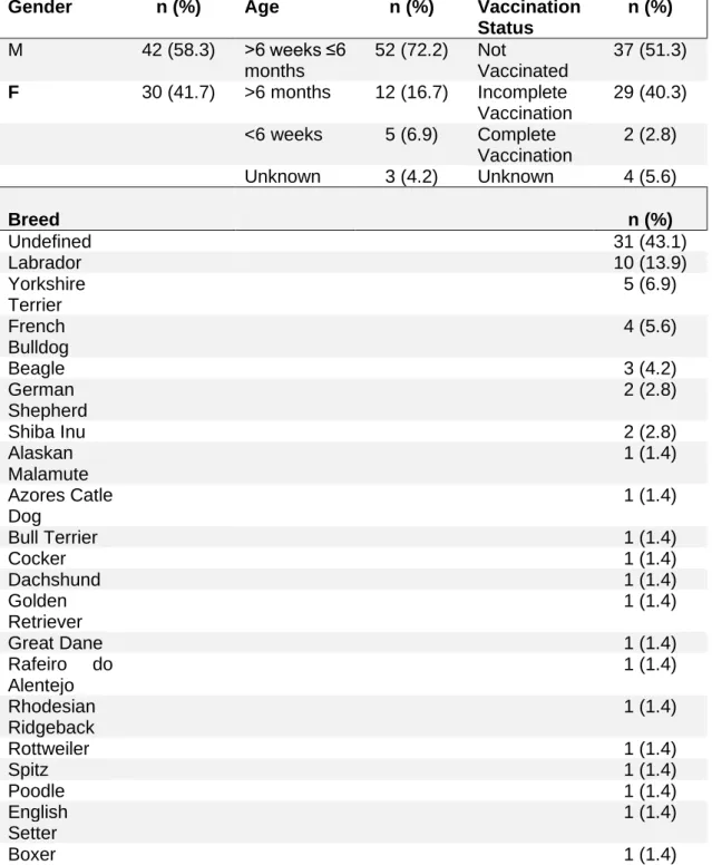
Table 2. Proposed predisposition (P) scoring criteria for parvovirus infected dogs,
considering age, breed and vaccination status. (Glickman et al., 1985; Houston et al., 1996; Goddard & Leisewitz, 2010; Greene & Decaro, 2012; Prata, 2017)………..……24
Table 3. Proposed response (R) scoring criteria for parvovirus infected dogs,
considering temperature, heart rate, respiratory rate and leucocytes count.
(Rijnberk & Stokhof, 2009; Boag, 2011; Sykes, 2014a; Marshall & Sweeney, 1990; Prata, 2017)……….25
Table 4. Proposed organ dysfunction (O) scoring criteria, based on clinical and
analytical parameters. Each evidence of organ system disfunction equals to 1 point...25
Table 5. Sample characterization of the animals subjected to SIRS criteria and to the
PIRO classification………..…27
Table 6. Fisher's exact test results for the evaluation of the
correlation between the different SIRS criteria and the outcome……….28
Table 7. Fisher's exact test results for the evaluation of the
X
Abbreviation List:
AKI – Acute Kidney Injury ALP – Alkaline Phosphatase ALT – Alanine Transaminase AP-1 – Activator Protein 1
APACHE – Acute Physiology and Chronic Health Evaluation
APPs – Acute Phase Proteins
ARDS – Acute Respiratory Distress Syndrome
ARF – Acute Renal Failure
AUC-ROC – Area Under the Receiver Operating Characteristics Curve
bpm – beats per minute ºC – degrees Celsius
CIRCI – Critical Illness Related Corticosteroid Insufficiency
CPV – Canine Parvovirus COX-2 – Cyclo-oxygenase CRP – C-Reactive Protein CRT – Capillary Refill Time
DAMPs – Damage Associated Molecular Patterns
DD – D-dimer
DIC – Disseminated Intravascular Coagulation
FDP – Fibrin degradation Products FFT – Fast-and-Frugal Tree FNA – Fine Needle Aspiration GI – Gastrointestinal
HEV-FMV-UTL – Veterinary Teaching Hospital of the Faculty of Veterinary Medicine of the University of Lisbon
HK – High Molecular Weight Kininogen HR – Heart Rate
ICU – Intensive Care Unit
IDIU – Infectious Disease Isolation Unit
IL – Interleukin
IRAP-1 – Interleukin 1 Receptor Antagonist Protein
iNOS – Inducible Nitric Oxide Synthase IV – Intravenous
LPS – Lipopolysaccharide
MDA – Maternally Derived Antibodies MEDS – Mortality in Emergency Department Sepsis
mmHg – millimetre of mercury
MODS – Multiple Organ Dysfunction Syndrome
NF-κB – Nuclear Factor Kappa B NO – Nitric Oxide
OR – Odds Ratio
PAI-1 – Plasminogen Activator Inhibitor Type 1
PAMPs – Pathogen Associated Molecular Patterns
PCO2 – Partial Pressure of Carbon Dioxide PIRO – Predisposition, Insult, Response, Organ Dysfunction
PK – Prekallikrein
PRRs – Pattern-Recognition Receptors PT – Prothrombin Time
PTT – Partial Thromboplastin Time qSOFA – quick SOFA
RR – Respiratory Rate
SIRS – Systemic Inflammatory Response Syndrome
SOFA – Sequential/ Sepsis-related Organ Failure Assessment
sTREM – soluble Triggering Receptor Expressed on Myeloid Cells
T – Temperature TF – Tissue Factor
XI TNF- α – Tumour Necrosis Factor-α
TNFR – Tumour Necrosis Factor Receptor
UIDI – Unidade de Isolamento de Doenças Infeciosas WBC – White Blood Cells
1
I - Activities developed during the curricular internship
This report concerns the curricular internship that occurred between 10th September 2018 and 11th January 2019 at VetOeiras - Central Cascais Veterinary Hospital and between 14th January 2019 and 15th March 2019 at the Isolation Unit for Infectious Disease, University of Lisbon's Faculty of Veterinary Medicine. The activities developed during the internship encompassed different clinical areas including surgery, internal medicine, and inpatient care with a rotating rota trough them with a total number of hours of 1264 (320h internal medicine, 300h surgery, 300h inpatient care and 344h in the Isolation Unit for Infectious Disease). The rotation included different working periods with day, night and weekend shifts that varied from 8 to 15 working hours a day. All the activities took place under the supervision of the Veterinary Surgeon or Veterinary Nurse on duty.
1. Internal Medicine
During the time spent in the internal medicine service there was opportunity to attend first time, revaluation and reference appointments during which it was possible to participate in the collection of prior clinical history and anamnesis of the patients as well as a thorough physical exam to each one of them. Under the supervision of the Veterinary Surgeon there was also the opportunity to participate in some medical procedures such as peripheral intravenous catheterization, blood samples analyses (hemogram and biochemical analyses), imagiological complementary exam (ultrasonography, radiography and endoscopy), to perform cytological evaluation (obtained via FNA or swab) and preparation and administration of medication. There was also the chance to discuss, with the responsible clinician, the different possible approaches, differential diagnosis and treatment options to each of the clinical cases.
2. Surgery
The scheduled surgeries took place from Monday to Friday morning, with emergency surgery occurring whenever needed. The activities developed in the surgery department of VetOeiras included reception of the animals and preoperative hemogram and biochemical screening, peripheral venous catheterization, preanesthetic drugs preparation and administration, trichotomy and surgical asepsis, anaesthetic induction and monitoring, assistance in the surgical procedures, and post-surgical monitorization. The surgeries witnessed fell mainly under the Orthopaedic and Soft Tissue surgery field with the opportunity to observe and participate in some Ophthalmological surgery and Neurosurgery.
2
3. Inpatient Care
During the hours spent in the inpatient care unit of VetOeiras there was the chance to follow the progress of the different clinical cases, participating on the animals clinical monitoring, feeding plan, and drugs preparation and administration according to the indications of the Veterinary Surgeon responsible for the case. There was also the opportunity to discuss not only the evolution of the animals’ condition with the clinician responsible, as well as thedischarge plan for each one. It was also possible to participate in some medical procedures including wound cleaning and dressing, blood sample collection and analyses, peripheral venous catheterization and urethral catheterization. Some of the cases offered the opportunity to participate in critical care treatments and monitoring and to assist in some emergency procedures such as cardiorespiratory resuscitation.
4. Infectious Diseases Isolation Unit
The activities developed in the Infectious Diseases Isolation Unit (IDIU) of the Faculty of Veterinary Medicine were fundamental in allowing the accomplishment of the present work since it gave the opportunity to accompany some Canine Parvovirus Infections, allowing the characterization of its´ clinical presentation. The activities that took place included clinical monitoring, medication preparation and administration and the correct hygienization of contaminated infectious material. All the activities took place under the supervision of Professor Solange Gil. It was possible to assist in some infectious disease consultations and to participate in the clinical discussion concerning the differential diagnosis and treatment options. The possibility to access the Unit´s clinical record allowed to obtain the data needed to accomplish the present Master Thesis.
II – Literature Review
1. Sepsis
1.1. General notions and SIRS:
The concept of sepsis has undergone a series of alterations since it has first been associated with bacterial infection approximately 100 years ago (Vincent et al. 2011). As it stands, according to the most recent scientific consensus, the term sepsis should be used to describe the organ dysfunction triggered by a deleterious inflammatory host response to infection (Singer et al. 2016). Despite being the most common cause of sepsis, bacteraemia is not a mandatory condition for sepsis to occur (Greiner et al. 2008). It is also important to note that bacteraemia can be present in non-septic animals. These
3
animals present positive blood cultures, and therefore viable bacteria in their bloodstream, but do not show any signs of inflammatory response. After the release of bacteria into the bloodstream as a result of an infection (abdominal and respiratory tract infections being the most common sepsis inducing for dogs, and septic peritonitis, pyothorax, and hepatic abscessation for cats (Ettinger et al. 2017)), sepsis only takes place if the host immune system is overpowered resulting in clinically significant bacteraemia. The most common organisms found in the bloodstream of dogs with bacteraemia are Staphylococcus spp. (including S. pseudintermedius, S. aureus, and coagulase-negative staphylococci), Streptococcus spp. (particularly S. canis, but also S. bovis complex organisms and seldom other streptococci) and Escherichia coli (Sykes, 2014a).
A related concept is endotoxemia which refers to the presence of lipopolysaccharide (LPS) of the Gram-negative bacterial cell wall in the bloodstream, without necessarily being associated with the presence of live bacteria (Silverstein & Otto, 2012).
To clarify the correlation between the systemic inflammatory response and sepsis a conference was held in 1991, from which arose the criteria to assess if a systemic inflammatory response syndrome is taking place in humans. Since the first adaptation of these criteria for animals (Purvis and Kirby 1994) they have been subjected to a series of alterations and the cut off values slightly vary among authors. On behalf of augmenting the sensitivity and specificity of the diagnosing capability of these parameters, it is important to use them in association with the clinical judgement when screening animals for sepsis, as they are not sufficiently accurate to establish a definitive diagnosis (Hauptman et al. 1997).
Table 1: SIRS diagnosing criteria for cat and dog. (Sykes, 2014a)
Criteria Dog Cat
Temperature <37.8 or >39.4 ºC <37.8 or >39.7 ºC HR >140 beats/min <140 or >225 beats/min RR >30 breaths/min or PCO2 < 32 mmHg >40 breaths/min Leukocyte count <6000 or >16,000 cells/μL, or >3% band neutrophils 19,500 cells/μL, or >5% band neutrophils
Legend: ºC, degrees Celsius; HR, heart rate; RR, respiratory rate; PCO2, partial pressure of carbon dioxide; mmHg, millimetre of mercury; cells/μL, cells per microliter
4
For a dog to be diagnosed with SIRS at least 2 of the 4 criteria need to be met: body temperature <37.8 ºC or >39.4 ºC, HR >140 bpm, RR >30 breaths/min or PCO2 < 32 mm Hg (venous or arterial), WBC <6000 or > 16,000 cells/µL, or >3% band neutrophils. Cats need to meet 3 of the 4 criteria for SIRS to be diagnosed: body temperature <37.8 ºC or >39.7 ºC, HR < 140 or >225 bpm, RR > 40 breaths/min, WBC > 19,500 or <5000 cells/µL, or >5% band neutrophils (Sykes 2014a).
As stated at the most recent human medicine consensus the term severe sepsis should no longer be used to describe sepsis-induced organ dysfunction or tissue hypoperfusion, as this description falls under the definition of sepsis. When the circulatory and cellular/metabolic abnormalities are severe enough to increase mortality, septic shock should be considered (Singer et al., 2016). This translates to a sepsis associated hypotension non-responsive to intravascular volume expansion, i.e., dogs with systolic blood pressure < 90 mm Hg or mean arterial pressure < 70 mm Hg that only respond to vasopressor therapy (Sykes, 2014a).
1.2. Pathogenesis
1.2.1. Innate immunity and inflammatory mediators
The instigation of the systemic inflammatory response begins with the activation of the innate immune system cells (mainly macrophages, monocytes, neutrophils, and natural killer cells), either by pathogen associated molecular patterns (PAMPs) or damage associated molecular patterns (DAMPs).
PAMPs are molecules originated from the pathogen invading the animal and can vary from endotoxin from Gram-negative bacteria to exotoxins, peptidoglycans, or superantigens from Gram-positive bacteria or even fungal cell wall material (Gyawali et al. 2019). On the other hand, DAMPs are endogenous molecules or material released by damaged cells as consequence of trauma, ischemia, malignancy, inflammatory diseases or other events capable of causing cellular stress (King et al. 2014).
Both PAMPs and DAMPs can be recognized by the innate immune system by binding to specific receptors denominated pattern-recognition receptors (PRRs). PRRs can be found in various different cells, including dendritic cells, macrophages, B cells, natural killer cells, endothelial cells, epithelial cells, and fibroblasts.(Lewis et al. 2012).
After the recognition of the molecular patterns, an intracellular signalment cascade pathway arises culminating in the activation of nuclear factor kappa B (NF-κB) and activator protein 1 (AP-1), whose function is to enter cell nucleus and activate the transcription sites of multiple genes that codify, among other, acute phase proteins,
5
coagulation factors and pro-inflammatory cytokines such as tumour necrosis factor (TNF)- α and Interleukins (ILs) 1, 6, 8, and 12 (Lewis et al. 2012).
Macrophages and T cells are the main producers of TNF-α. This molecule is responsible for many of the manifestations of the inflammatory status, as it promotes vasodilation and blood stasis as a result of inducible nitric oxide synthase (iNOS) and cyclo-oxygenase 2 (COX-2) production (Lewis et al. 2012). Another consequence of TNF-α production is neutrophils activation and leukocytes chemotaxis to the endothelial wall, subsequently to the upregulation of endothelial adhesion molecules (King et al. 2014).
1.2.2. Inflammation and acute phase response
Pro-inflammatory cytokines are answerable for the inflammatory clinical manifestations recognised during sepsis with alterations including hyperthermia, tachycardia, leucocytosis, exudation, hypotension, acute phase response and neutrophils chemotaxis to the affected site (Giamarellos-Bourboulis 2010).
ILs 1 and 6 are the principal accountable for the inflammatory response associated with sepsis. IL-1, in its two proinflammatory forms IL-1α and IL-1β, besides being responsible for the endothelial cell adhesion molecules and cytokines production, is also the major endogenous pyrogenic agent and acute phase response stimulator (Silverstein and Otto 2012). IL-6 takes an important role in leukocyte activation and myeloid cell proliferation, despite also being responsible for fever induction and triggering the acute phase response (Lewis et al. 2012).
In addition, consequently to the activation of PRRs, hepatocytes are triggered to release acute phase proteins (APPs) contributing to the animal´s response to the inflammatory stimulus by promoting fever, neutrophilia and activation of coagulation and complement cascades. Type I APPs are induced by IL-1α and IL-1β and TNF-α and type II are induced by IL-6. Even though some APPs, particularly procalcitonin and C-reactive protein (CRP), can be of predictive value when it comes to mortality in sepsis in humans and animal trials, there is still the need to find a better method to predict the outcome for SIRS affected animals (Chan et al. 2009; Lewis et al. 2012).
1.2.3. Anti-inflammatory agents and immunosuppression
Anti-inflammatory mediators are present alongside inflammatory mediators since the onset of sepsis with the animals’ state fluctuating between hyperinflammation and hypo-inflammation. The principal cytokine implicated in the anti-inflammatory response is IL-10. This interleukin anti-inflammatory action is a result of TNF-α, IL-1β and IL-6 release inhibition, as well as IL-1 receptor antagonist protein (IRAP-1) production (Lewis
6
et al. 2012). Tumour necrosis factor receptor (TNFR) 1 and 2 and soluble triggering receptor expressed on myeloid cells (sTREM) 1 also play a role in the anti-inflammatory response (Giamarellos-Bourboulis 2010).
One of the consequences of the anti-inflammatory response is the impairment of the immune system, prompting the host to secondary infections via bacteria, viral or fungal exposure. Alterations during the anti-inflammatory state that contribute to the immunosuppression include: Th2 and Treg response prevalence over Th1 response, CD4+ lymphocytes depletion and overall lymphocytes disfunction and apoptosis, inflammatory cytokines (such as IL-6 and TNF) impaired production and neutrophils reduced phagocytic and chemotaxis aptitude (King et al. 2014; Gyawali et al. 2019).
1.2.4. Inflammation and dysregulated coagulation
Parallelly to the inflammatory response there is a stimulation of the haemostatic system with variable clinical presentation raging from non-symptomatic thrombocytopenia to severe disseminated intravascular coagulation (DIC). During sepsis haemostasis deviates to a hypercoagulability state, with consequent thromboembolic events that contribute to organ failure, this is followed by a hypocoagulability state triggered by fibrinolytic events and by coagulation factors and platelets consumption (King et al. 2014; Gyawali et al. 2019).
Tissue factor (TF) is the main instigator of the coagulation abnormalities that take place during SIRS. TF becomes exposed to the bloodstream after endothelial damage but it´s also produced by cytokine and PAMPs stimulated monocytes, macrophages, parenchymal cells and neutrophils, causing activation of the extrinsic coagulation pathway resulting in thrombin production, platelets activation and fibrin-clot formation (Lewis et al. 2012; King et al. 2014).
In healthy animals anticoagulants counter the hypercoagulability events triggered by TF, thus balancing the coagulation state. During sepsis these counterbalance mechanisms are impaired due to decreased levels of antithrombin and reduced protein C activation. Antithrombin is diminished in sepsis as a result of a decreased hepatic synthesis, consumption subsequently to the formation of thrombin-antithrombin complexes, and deterioration by activated neutrophils elastase release.
Thrombin is responsible for thrombomodulin activation, which is accountable for converting protein C into activated protein C that promotes fibrinolysis and anticoagulation via cleavage of factors Va and VIIIa. In the septic process downregulation of thrombomodulin combined with overconsumption and reduced production of protein C tilts the animal toward hypercoagulability.
7
Increasing levels of inflammatory cytokines promotes the release of tissue plasminogen activators from endothelial cells with consequential increase of plasmin activation. Counterbalancing mechanisms result in plasminogen activator inhibitor type 1 (PAI-1) increment resulting in decreased fibrin clots degradation that contribute to the hypercoagulability state (King et al. 2014; Gyawali et al. 2019).
It is also relevant to mention the role of the contact phase system. When triggered by polyphosphate, collagen, protein aggregates, LPS, glycosaminoglycans, or nucleic acids, this system consisting of coagulation factors XII and XI, plasma prekallikrein (PK), and nonenzymatic cofactor high molecular weight kininogen (HK), contributes to the altered coagulation. Surface contact promotes factor XII activation into to factor XIIa which is responsible for PK activation and consequent kallikrein release. Kallikrein is then responsible for further activation of factor XII resulting in positive feedback cycle of coagulation stimulation (Lewis et al. 2012; Wu 2015).
1.2.5. Organ dysfunction
The definition of multiple organ dysfunction syndrome (MODS) has remained unchanged since it was defined as “the presence of altered organ function in an acutely ill patient such that homeostasis cannot be maintained without intervention” in the first sepsis consensus conference in 1991 (Bone et al. 1992). During sepsis MODS can be considered when at least two organ systems, distant to the infection site, became dysfunctional (Sykes 2014a).
The intricate pathophysiology of MODS makes it hard to come to an agreement, with different models being proposed. Different mechanisms act together contributing to the induction of organ failure: hypoxic cellular and tissue damage, apoptosis induction, gastrointestinal microbiota translocation, immune system dysregulation and mitochondrial dysfunction.
Even though all the above contribute to organ failure, immune system dysregulation and mitochondrial dysfunction are the ones thought to have a key role in MODS pathogenesis. Neutrophils stimulated by inflammatory cytokines chemotaxis to different tissues where they produce superoxides responsible not only for tissue damage, but also for the perpetuation of the inflammatory response. Mitochondrial respiration is impaired by the action of superoxide released from neutrophils and nitric oxide (NO) originated from vascular endothelium, this process is denominated cytopathic hypoxia and causes decreased cellular energy production with consequential cellular dysfunction or death (Mizock 2009; Osterbur et al. 2014).
The decrease of oxygen delivery and utilization can have an impact in several organ systems with different clinical presentations. Hypoperfusion secondary to septic
8
shock is the primary responsible for the hepatic injury verified in sepsis. This first stage of hepatic dysfunction is characterized by cellular damage with aminotransferases increments and diminished gluconeogenesis and glycogenolysis and consequent hypoglycemia. The later stage of hepatic dysfunction results from Kupffer cell activation and release of further inflammatory and oxidative agents, leading to added cellular damage. Parameters that can be used to assess hepatic dysfunction include hyperbilirubinemia, increased alanine transaminase (ALT) or alkaline phosphatase (ALP) and the presence of hepatic encephalopathy. It is important to monitor the hepatic markers as the liver is considered to be the most affected organ in septic dogs (Osterbur et al. 2014; Gyawali et al. 2019).
Respiratory capacity is also affected during sepsis. Animals subjected to a septic insult can develop an acute respiratory distress syndrome (ARDS) characterized by pulmonary edema, structural cellular damage and atelectasis resulting from loss of alveolar-capillary barrier, increased vascular permeability, neutrophil infiltration of the pulmonary tissue and action of proinflammatory cytokines (Wilkins et al. 2007; Osterbur et al. 2014).
The cardiovascular function is affected by both decreased cardiac output and loss of vascular resistance. Inflammatory cytokines promote these cardiovascular changes by increasing vascular permeability and peripheral vasodilation, which contribute to hypovolemia and blood flow variations. Cytokines, endotoxins and calcium changes act together causing downregulation of cardiac contractility. When vasopressor therapy is needed to reverse the resulting hypotension, the animal can be considered to be in septic shock.
The renal impact of sepsis usually leads to acute renal failure (ARF). More commonly, the sepsis associated ARF is triggered by the circulating cytokines, that promote apoptosis with no macroscopic tissue damage. ARF can also come as a consequence of renal hypoperfusion and ischemia, tubular necrosis takes place and glomerular function is impaired.
Gastrointestinal signs of sepsis include vomiting and diarrhoea, ileus and signs of mucosal ulceration like melena and hematochezia. Blood flow alterations, mitochondrial dysfunction and cell apoptosis promoted by cytokines all contribute for the loss of integrity of the intestinal wall, the consequent bacterial translocation can act as a perpetuating factor for sepsis. Dehydration and electrolytical changes can aggravate other organs hypoperfusion, predisposing the animal to organ failure.
Other organ systems like the central nervous system and the adrenal glands can also be affected by sepsis (Silverstein and Otto 2012; Osterbur et al. 2014).
9
1.3. Diagnostic approach
1.3.1. Physical exam
The clinical findings in animals suffering from sepsis reflect a systemic inflammatory state, with possible alteration of the vital parameters like temperature, heart rate and respiratory rate. These parameters should be measured upon admission and compared to see if they meet the SIRS criteria, as sepsis is diagnosed when the SIRS criteria are fulfilled and an infection is confirmed. The clinical signs tend to be unspecific as they correlate, not only to the organ system originally affected by the infectious agent, as well as any secondary organ dysfunctions mentioned above. Vomit, diarrhoea, dehydration, icterus and lethargy are just some of the common clinical signs observed.
Some dogs may already be in septic shock upon hospital admission. These will present either in the initial phase of shock, with pale mucous membranes, prolonged capillary refill time (CRT), and weak pulses, or in the hyperdynamic phase of shock. In this later stage, vasodilation subsists with resulting hyperemic mucous membranes, a decreased CRT (<1 sec), and strong or bounding pulses. Blood pressure should always bee part of physical examination of a suspected SIRS as hypotension may be present. (Hauptman et al. 1997; Drobatz et al. 2019).
1.3.2. Clinical laboratory findings
The diagnostic approach to a septic animal should include a complete blood count, biochemistry profile and coagulation tests. The hemogram may reveal abnormalities in different cellular lineages. Complete blood count most frequently reveal anaemia secondary to blood loss, haemolysis, oxidative damage and reduced erythrocyte production. Polycythaemia can also be present in hypovolemic animals due to haemoconcentration and splenic contraction. The majority of animals present with leucocytosis and band neutrophils and the blood smear reveal toxic changes to the neutrophils. Due to the immunosuppression and lymphocyte apoptosis it is also possible for lymphopenia and leukopenia to occur. Platelet consumption and DIC mean thrombocytopenia is also a usual finding (Girardot et al. 2016; Drobatz et al. 2019).
Biochemical abnormalities vary and reflect the organ dysfunctions taking place, either primarily affected by the infection or secondary to the inflammatory state. Common findings include hypoalbuminemia, glycemia alterations, hypocalcemia and hyperbilirubinemia. Hypoalbuminemia is a result of combined decreased hepatic synthesis and protein loss trough the gastrointestinal and urinary tract. Glycemia changes over the course of SIRS, with hyperglycaemia being more common due to cortisol and catecholamines release in response to stress and hepatic insulin resistance.
10
Hypoglycaemia ensues from increased consumption and decreased hepatic synthesis and reflects a more terminal stage of sepsis, in both dogs and cats. Haemolysis and cholestasis are responsible for the hyperbilirubinemia some animals present. Animals can develop azotaemia if chronic or acute kidney injury is present. In these patients urinalysis and urine culture should be carried out to assess any urinary tract infections, as kidneys can be the primary organ affected by the infectious agent (Sykes 2014a; Drobatz et al. 2019).
Coagulation tests should also be a part of the septic animal´s screening. In the initial hypercoagulable phase of DIC coagulation tests may show increased D-dimers and decreased concentration of activated protein C and antithrombin. With the development of the hypocoagulable state changes might include prolongated prothrombin time and activated partial thromboplastin time (Silverstein and Otto 2012; Drobatz et al. 2019).
1.3.3. Diagnostic imaging
Radiographs and ultrasound can help determining the source of infection causing the SIRS but can also elucidate the overall organ function and any secondary alteration. Different projections of thoracic radiographs (lateral and ventral dorsal) should be taken, especially in animals showing tachypnea or dyspnea. Images might show pulmonary patterns consistent with pneumonia or ARDS. The intervertebral disc space should also be evaluated since discospondylitis is also a possible cause of sepsis.
Abdominal ultrasound is a valuable tool, especially in animals with biochemistry values compatible with abdominal organ dysfunction. Ultrasound images can reveal primary lesions, like abscesses or septic effusions, and secondary lesion, like renal injuries and gastrointestinal ulceration. Any abnormal free fluid should be sampled for cytology, culture and susceptibility as it can help determine the origin of the infection and redirect the antibiotherapy (Silverstein and Otto 2012; Sykes 2014a).
1.3.4. Microbiologic test
The definitive diagnosis of sepsis implies the fulfilment of the SIRS criteria and the confirmation of infection, which require pathogen isolation and identification. First approach diagnosis must include imaging guided tissue and fluid sampling as well as cystocentesis urine sampling. Clinical history, presenting signs and lesion distribution must always direct the sampling, as it helps interpret the validity of the results. Cytology, histopathology, and both aerobic and anaerobic culture and sensitivity test, should all be performed in order to identify the infectious agent and help in the treatment decision
11
making. Ideally the samples should be taken before antibiotherapy is started but this is not an exclusion criteria (Hauptman et al. 1997; Drobatz et al. 2019).
Blood samples can also be useful if bacteraemia is suspected. Blood should be withdrawn from two different sites with at least 30 minutes apart and the skin should be prepared in a surgical fashion to prevent contamination (Sykes 2014a; Drobatz et al. 2019).
Polymerase chain reaction (PCR) is also a useful tool to identify infectious agents, especially when used in conjugation with culture. PCR assays are particularly useful in the detection of fastidious growing organisms. Antibody quantification can also be used to determine an acute response to infection (Miller et al. 2011; Sykes 2014a).
1.4. Treatment
1.4.1 Initial stabilization
The initial approach to sepsis must consider the early goal-directed therapy and medical management should focus on resolving the hemodynamic changes in the first hours upon presentation, especially in the high-risk patients (Gyawali et al. 2019).
One of the first steps toward the stabilization of a septic patient is to ensure tissues perfusion and oxygen delivery, preventing the development of MODS. Hypovolemic and distributive shock are the two most common phenomena contributing for the hemodynamic changes verified during SIRS. Therefore, intravenous fluid therapy should be initiated as soon as possible with the administration of crystalloids boluses, of up to 90mL/kg for dogs and 60mL/kg for cats. Isotonic crystalloids or hypertonic crystalloid solutions can both be used for initial resuscitation and it should be accompanied by clinical monitorization of heart and respiratory rate, blood pressure and urinary output, so the minimal amount of fluids needed to maintain adequate blood pressure is administered, thus avoiding overloading the animal. The use of colloids should be avoided as they have been proven to augment mortality in critical ill patients. Colloidal administration is associated with acute kidney injury (AKI) and coagulation dysregulation (Drobatz et al. 2019). Other transfusion therapies may be required depending on the clinical presentation for example severely anaemic animals may need packed red blood cells or fresh all blood transfusion. (Silverstein and Otto 2012; Dellinger et al. 2013; Drobatz et al. 2019).
1.4.2. Cardiovascular support
When animals present with septic shock, hypotension may be unresponsive to intravascular volume expansion alone and there is the need to resort to vasopressor
12
drugs to control the vasodilation. Vasodilation may present clinically as mucosal hyperaemia, decreased CRT and bounding pulses (Silverstein and Otto 2012).
Vasoconstrictor drugs usually used include vasopressin, dopamine, norepinephrine and epinephrine. Epinephrine has been associated with increased mortality when compared to norepinephrine and vasopressin and should, therefore, be avoided if other options are available (Minneci et al. 2004). Vasoconstriction might worsen the organ dysfunction by cutting the blood supply to some organ systems, vasopressin low-dose therapy is the safest drug choice as it spares cerebral, renal, pulmonary and mesenteric vessels constriction (Silverstein and Otto 2012; Drobatz et al. 2019).
Positive inotropic drugs, like dobutamine or pimobendan, may also be administered to help ensure tissue perfusion reduced by myocardial depression (Drobatz et al. 2019). Considering that critical illness-related corticosteroid insufficiency (CIRCI) causes vascular hyporeactivity, glucocorticoids may also be indicated in some cases of sepsis.
1.4.3 Antimicrobial therapy
Upon recognition of sepsis, antimicrobial therapy should be initiated as soon as possible, with intravenous antibiotic administration within the first hour being associated with higher survival rates. When possible, antibiotics should be chosen regarding possible pathogens involved, infection site and antibiotic resistance (Dellinger et al. 2013).
Initial approach, before culture and susceptibility results, includes the utilisation of a broad-spectrum antibiotics that should cover as many quadrants as possible. This empiric antibiotic coverage should only be maintained for up to 5 days, after which the antibiotherapy should be narrowed and adjusted according to the lab results (Drobatz et al. 2019; Gyawali et al. 2019).
Examples of first line antibiotics used in sepsis include: ampicillin and enrofloxacin, ampicillin and amikacin, ampicillin and cefoxitin, clindamycin and enrofloxacin, amoxicillin clavulanic acid and enrofloxacin. These options ensure coverage against Gram-positive and Gram-negative aerobes and anaerobes (Silverstein and Otto 2012).
Another step that helps with infection control is source control. This includes any procedure that physically contributes to infection control, either by removing the infectious site or by reducing the propagation of infection to adjacent tissues. Procedures involved in this step include surgical removal of infection focus and drainage of septic fluids (Gyawali et al. 2019).
13
1.4.4. Supporting care
Additional therapeutic measures after patient stabilization, should be directed to the organ dysfunctions taking place and adding good supportive care.
One organ system that requires attention in septic animals is the gastrointestinal (GI) tract, as it may contribute to the further release of bacteria into the bloodstream, contributing to the worsening of the infectious status. Gastrointestinal bleeding and stasis should be addressed with antiemetics (as maropitant, ondasetron), prokinetics (as metoclopramide), antiacids and sulcralfate. Enteral nutrition may also be helpful in restoring health to the intestinal villus (Silverstein and Otto 2012; Drobatz et al. 2019).
1.5. Prognosis
Sepsis is a serious condition with mortality rates higher than 50%, worse if shock is present (Silverstein and Otto 2012). Organs dysfunctions contribute to the mortality of animals, with prognosis being worse with the increase of organ systems affected (Kenney et al. 2010). Timely diagnosis and early treatment are key factors to maximize sepsis survival.
2. Canine Parvovirus
2.1. Etiology and epidemiology
Canine parvovirus enteritis is one of the most common infectious diseases affecting dogs. Canine parvovirus (CPV), are small nonenveloped DNA viruses. Two types, type 1 and type 2 are recognized, with type 2 being the most virulent form that underwent a series of mutations evolving into new strains: type 2a (CPV-2a), type 2b (CPV-2b) and type 2c (CPV-2c) spreading around the globe (Sykes 2014b).
The extremely resistant nature of the virus allows it to stay infective in the environment for long periods of time. CPV is expelled in the faeces of infected dogs and spreads via oronasal exposure. Even though the infection may occur in animals from any breed, age or sex with different individual response to infection, most of the acutely affected animals are unvaccinated puppies between the age of 6 weeks and 6 month, and mortality rates are high. Protection against the virus is long lived after infection, with maternally derived antibodies (MDA) being an effective protection for the first weeks of a neonate`s life. The most susceptible period is between the loss of maternal defences and the completion of the vaccination protocol. MDA protection declines with time and susceptibility to infection arise (Pollock & Carmichael, 1982). Rottweilers, Doberman pinschers, Labrador retrievers, American Staffordshire terriers, German shepherds, and
14
Alaskan sled dogs appear to be the breeds more susceptible to infection (Glickman et al., 1985; Houston et al., 1996; Silverstein and Otto 2012).
Multiple factors contribute for the severity of the infection. Clinical signs differ depending on the virus strain, host immunity and the presence of concurrent infections such as other enteric viral and parasitic infections (Sykes 2014b).
2.2. Pathogenesis
The CPV requires a mitotically active cell population to replicate, which makes young dogs (under 12 weeks) the target population. After oronasal exposure to CPV-contaminated feces, follows a 7 to 14 days incubation period. The virus first replicate in the oropharynx lymphoid tissue, thymus and mesenteric lymph nodes but, within 1 to 5 days, viremia occurs and hematogenous spread to the intestinal crypts of the small intestine takes place. Virus replication causes destruction of the germinal epithelium impairing the normal cell regeneration and causing villi collapse, this results in haemorrhagic diarrhoea and malabsorption. Shedding of the virus in the faeces starts as soon as 3 days post infection and can last for up to 4 weeks (Goddard and Leisewitz 2010; Sykes 2014b; Drobatz et al. 2019).
Viral infection within the uterus may result in reabsorption or abortion. When the virus affects neonates up to 2 weeks old, it can also target the mitotically active myocardial cells, causing viral myocarditis with signs of congestive heart failure and sudden death (Sykes 2014b).
2.3. Bacterial translocation and sepsis
Multiple factors contribute for the development of sepsis in CPV infections. Cellular destruction, intestinal hypomotility, dysbiosis, gut inflammation and tissue necrosis all contribute to the disruption of the gastrointestinal mucosal barrier, allowing Gram-negative and anaerobic bacteria translocation from the intestinal lumen to the bloodstream and bacteraemia to develop (Greene & Decaro, 2012; Kalli, 2016; Krentz & Allen, 2017).
Along with the mucosal barrier disruption, impaired immunity develops increasing the susceptibility to secondary infections. Marked leukopenia (mostly neutropenia and lymphopenia) is often observed in CPV infected dogs, as the virus also targets the mitotically active precursors of leukocytes and lymphoid cells of the bone marrow and lymphoid tissue like the thymus. Increased tissue demand and sequestration of neutrophils in the damaged GI mucosa also contributes for the neutropenia. Neutropenia
15
and bacteria overload impair the elimination of luminal bacteria from the bloodstream that occurs in healthy animals (Goddard and Leisewitz 2010; Kalli 2016)
The passage of viable bacteria or their products to extraintestinal sites can potentiate the development of sepsis, which increases the mortality rate of CPV infected animals since it predisposes to coagulation alterations, thrombotic events and MODS. With SIRS progression and the release of inflammatory mediators, the gastrointestinal barrier is compromised again, contributing to the cycle of bacterial translocation (Krentz & Allen, 2017).
2.4. Diagnostic Approach
2.4.1. Physical examination
The clinical signs of CPV-2 infection are unspecific and corelate to enteritis. In the initial phase of infection the dogs may present anorexia, lethargy and elevated rectal temperature, that later develop into gastrointestinal manifestations, including vomiting and diarrhoea. The faeces characteristics vary and can range from mucoid to haemorrhagic, with large volumes and liquid consistence. Abdominal pain is also a common finding and may be related to the severe enteritis or secondarily intestinal intussusception (Kalli 2016; Drobatz et al. 2019).
In consequence of the profuse vomiting and diarrhoea, and the resulting fluid loss, dehydration and hypovolemia may ensue. In these cases animals will present with impaired perfusion signs, including altered mucous membrane colour, tachycardia, prolonged CRT, weak pulses and altered mentation. Dogs that develop sepsis associated to bacterial translocation may present with clinical signs of SIRS or septic shock, which indicates a worst prognosis (Goddard and Leisewitz 2010; Drobatz et al. 2019).
2.4.2. Clinical laboratory findings
Leukocyte count on admission is of predictive value for 24h survival, with nonsignificant leukopenia being associated with 100% survival rate (Goddard et al., 2008).Leukopenia is the typical haematological change found in CPV-2 infected dogs. Destruction of hematopoietic linage precursors in the bone barrow, depletion of lymphoid organs and massive intestinal recruitment, all contribute for the low leukocyte count. Leucocytosis may also be present in a smaller number of animals and may be due to a bone marrow hyperplasia, compatible with a later stage of infection (Sykes 2014b; Kalli 2016).
16
Other haematological chances may include blood loss anaemia and alteration of the platelet count, with both thrombocytopenia and thrombocytosis being a possible finding.
Serum biochemical tests might reveal some unspecific alterations often including hypoalbuminemia and hypoproteinaemia, secondary to malnutrition and profuse vomiting and diarrhoea. Hypoglycaemia or moderate hyperglycaemia can also be present. Possible electrolyte abnormalities include hypokalaemia, hypomagnesemia, hyponatremia and hypochloraemia. Pre-renal azotaemia may ensue as dehydration progresses, animals that develop sepsis may also show other enzymatic changes reflecting organ systems dysfunction (Sykes 2014b; Kalli 2016).
Acid-base tests more commonly reveal acidosis, but alkalosis may also be present depending on the severity of the vomiting and diarrhoea (Goddard and Leisewitz 2010).
2.4.3. Microbiological test
The faecal parvovirus antigen ELISA is a test performed on the faeces obtained via rectal swab. Specificity higher than 90% and a practical use, makes it the most widely used test for CPV-2 enteritis. The test disadvantage is the low sensitivity that can vary between 18% and 60%. False negatives are associated with intermittent viral shedding, antigen neutralization by circulating antibodies and dilution of antigen on the stools. False positive results are rare but can happen 4 to 10 days after vaccination with modified live CPV vaccine. A strong clinical suspicion allied with a negative ELISA, should be followed by a more sensitive faecal antigen detection test including electron microscopy, viral isolation, faecal hemagglutination, immunochromatography, and PCR (Goddard & Leisewitz, 2010; Sykes, 2014b; Zoetis 2017).
Serology can also help in the diagnosis of a dog with typical clinical signs, if it reveals recently produced antibodies (immunoglobulins M). Care should be taken interpreting serological results, as positive results may be due to vaccination, maternally derived antibodies, or previous subclinical contact with the virus (Goddard and Leisewitz 2010; Kalli 2016).
2.5 Treatment
2.5.1. Fluid therapy
Early supportive care implementation is associated with higher survival rates. The first goal of the therapy is to correct dehydration and improve hemodynamic, as well as acid-base and electrolyte abnormalities correction. Intravenous (IV) fluid therapy is the
17
most effective way to restore the fluids lost through vomiting and diarrhoea, as subcutaneous fluid absorption is impaired in dehydrated animals. Intraosseous catheterization may also be considered. Aseptic measures should be taken when placing the catheter as the immune system is impaired in CPV-2 infected dogs (Goddard and Leisewitz 2010; Kalli 2016).
The initial fluid choice should be an isotonic crystalloid solution. Oncotic pressure and fluid deficits must be corrected according to the patient presentation and accompanied by monitorization of perfusion indicators including CRT, pulse and blood pressure. In puppies presenting hypovolemic, fluids rate must be enough to restore circulating volume in the first few hours upon admission. If shock is suspected 15-20ml/kg boluses can be given 15 minutes apart, until a shock dose of 80-90 ml/kg is reached. Once perfusion is restored IV fluid rate should be adjusted to cover maintenance requirements and fluid losses. Colloidal administration can be considered if perfusion restauration is refractory to crystalloids alone and if hypoproteinaemic peripheral edema occurs (Kalli 2016; Drobatz et al. 2019).
Since hypokalaemia and hypoglycaemia are commonly found in CPV-2 infected dogs, potassium chloride and 2,5%-5% dextrose supplementation can be required. Daily electrolyte and acid-base status monitoring is recommended, and abnormalities should be corrected accordingly. Fresh frozen plasma has also been indicated as part of the treatment protocol and, even though there is little prof of its efficacy, it is associated with higher survival rates (Goddard and Leisewitz 2010; Kalli 2016).
2.5.2 Antimicrobial therapy
Parvovirus enteritis predisposes for the development of sepsis and broad-spectrum antibiotic coverage should be warranted. The antimicrobial agents’ choice must cover Gram-negative and anaerobic bacteria, as these are the most commonly found in parvovirus associated bacteraemia. Treatment protocols based on a penicillin with a beta-lactamase inhibitor, like amoxicillin clavulanate, or second-generation cephalosporin, can be effective single-agent therapies. Usually additional antibiotic coverage is warranted by the addition of aminoglycosides or metronidazole.Even though quinolones ensure a good anaerobic coverage, they should be used with precaution in growing dogs, in order to avoid cartilaginous damage (Goddard and Leisewitz 2010; Sykes 2014b; Kalli 2016).
18
2.5.3 Antiemetic treatment
Vomiting control helps preventing further fluid losses, increases patient comfort and allows oral nutrition and medication administration. Among the antiemetic options the more commonly used include metoclopramide, maropitant and ondansetron. Metoclopramide (a dopaminergic antagonist), used as boluses or as a constant rate infusion, is an efficient antiemetic drug but requires monitoring for increasing risk of intussusception. Serotonin antagonists like ondansetron, can be used as second line antiemetic treatment for uncontrollable emesis. Maropitant (an antagonist of neurokinin 1 receptors) is also a very effective antiemetic option, as it warrants both peripheral and central emetic pathways regulation. A multiple drug therapy might be necessary (Goddard and Leisewitz 2010; Kalli 2016).
2.5.4. Nutritional support
Being a GI disease, nil per os until clinical sings remission for 12 hours has been recommended in the past. Recent studies have shown that early administration of nutritional support via tube feeding is associated with faster clinical improvement, weight gain and gut barrier improvement, which could help prevent parvovirus associated sepsis. Small amounts of easily digestive food should be given early in the treatment of this animals (Goddard and Leisewitz 2010; Kalli 2016).
2.5.5. Pain management
Abdominal pain is usually present due to enteritis or secondary intussusception, pain relief should be administered as pain negatively impacts the appetite and delays recovery. Commonly used drugs include buprenorphine (a partial agonist of μ-opioid receptors) and butorphanol (an antagonist of Κ-opioid receptors). Maropitant, apart from the antiemetic effect may also have a pain relief effect, since is also a P substance blocker, which is involved in visceral nociception. Non-steroidal anti-inflammatory drugs should be avoided, as they can further the GI mucosal damage via COX-1 inhibition (Kalli 2016).
2.6. Prognosis
Survival rates of dogs with CPV-2 enteritis vary depending on individual characteristics and immune competence, and the treatment offered. Considering this, survival rates can range from 9% to 90%. (Sykes 2014b).
Factors that have been associated with higher mortality include initial leukopenia, lymphopenia and neutropenia, and meeting SIRS criteria. Hospitalization time also
19
varies, with vomiting, lethargy, lymphopenia and hypoalbuminemia all being associated with prolonged hospitalization (Kalli et al. 2010). Survival has been associated with early positive shifts in leukocyte count. Being mostly an acute process, animals that survive the first 3 to 4 days of treatment tend to make full recoveries.
Prognosis is worse if complications arise. Complications include bacteraemia and sepsis, aspiration pneumonia, oesophageal stenosis and intussusception (Sykes 2014b).
2.7. Prevention
Proper immunization is the most effective method of CPV-2 infection prevention. Initial passive immunity is warranted by maternally derived antibodies, most of which are obtained from the colostrum. It is also important to note that MDA interfere with proper immunization and should be considered when planning the vaccination of a puppy. Maternal CPV antibodies have a half-life of approximately 10 days and will have waned enough to allow effective vaccine protection after 8 to 12 weeks (Goddard and Leisewitz 2010; Kalli 2016).
The most recent vaccination guidelines classify the type 2 parvovirus vaccine as a core vaccine, meaning all dogs, regardless of the circumstances, should receive it. Recommendation is to start the vaccination protocol with a modified live vaccine at 6 to 8 weeks of age and then every 2 to 4 weeks until, at least, 16 weeks of age. If the initial dose is due after 16 weeks of age, two doses of the vaccine should be administered 2 to 4 weeks apart, even though one dose is considered protective. Revaccination should be given at 6 months or 1 year of age, and then once every 3 years (Day et al., 2016). Post infection immunity is effective and long lived.
Despite the efficacy of the vaccination, other health care measures should be taken to prevent infection, including disinfection of faeces exposed surfaces and objects, quarantine of newly introduced animals, isolation of infected dogs and reduction of stress factors (Goddard & Leisewitz, 2010; Kalli, 2016).
3. PIRO: staging of sepsis
3.1.
PIRO
(Predisposition,
Insult,
Response,
Organ
dysfunction/failure)
Even with the application of consensual treatment guidelines, sepsis is still one of the leading causes of death, with some patients not responding to the proposed treatment. The heterogeneity of the infective process and of the individual immune
20
response to it, makes prognosis prediction and treatment choice difficult in these animals (Rathour et al. 2015).
In order to try and stage septic patients by both their risk of mortality/adverse outcome and their potential to respond to treatment, in 2001 (the international sepsis definitions conference) a new stratification system was introduced, the PIRO. This system allows to stratify patients based on their predisposing conditions, the nature and characteristics of the insult, the extent of the host immune response to it, and the associated organ dysfunction (Levy et al. 2003). The purpose of this system is to help in the enrolment of individual in clinical studies and prognosis of septic patients, allowing to adapt the therapy offered and improve survival.
3.2 Predisposition
When considering predisposition, all factors present previous to the onset of the acute illness and that might influence its incidence or outcome, should be considered. This include genetic predisposition, acquired comorbidities and concomitant diseases (Levy et al. 2003; Rathour et al. 2015). Apart from genetic factors, that include sex and breed predisposition, advanced age and concomitant disease including liver disease, neoplasia, heart conditions and chronic renal failure were all associated with higher mortality and should hence be considered as predisposing factor for sepsis prognosis (Rubulotta et al. 2009; Howell et al. 2011; Granja et al. 2013).
3.3. Insult / Infection
When considering septic animals, the insult is the infection that triggers the SIRS. Infection characteristics like extent, location and associated pathogen, can all have an impact on the outcome. Human medicine studies have linked higher mortality to sepsis originated from pulmonary, gastrointestinal and central nervous system infections, when compared to soft tissue or skin infections. The virulence of the infecting organism can also impact the outcome, with more virulent agents being linked to higher mortality. (Levy et al. 2003; Cohen et al. 2004; Opal 2005)
3.4. Response
The host response to the infectious insult is what defines sepsis. Since it´s SIRS that triggers the organ systems dysfunction found in septic patients, mortality is closely related to the individual response to an infection. Many of the factors mentioned above, including genetic factors and concomitant disease, can influence the host immune status and therefore the immune response mounted (Opal 2005).