UNIVERSIDADE DA BEIRA INTERIOR
Ciências
Toxicological effects of TiO
2
nanoparticles in two
freshwater species:
Carassius auratus and Corbicula fluminea
Joana Filipa Cardoso Lourenço
Dissertação para obtenção do Grau de Mestre em
Bioquímica
(2º ciclo de estudos)
Orientador: Prof. Doutor Mário Emanuel Campos de Sousa Diniz
Co-orientadora: Prof. Doutora Maria Eugénia Gallardo Alba
Acknowledgements
To my advisors, who gave me the opportunity to become a part of such an interesting work, Dra Eugénia Gallardo and especially to Dr. Mário Diniz, without whom would not be possible to develop and conclude this work.
To FCT/MCTES for funding project “Avaliação Integrada de Nanomateriais: Caracterização e
Avaliação da Toxicidade Ambiental – NanoTox”, FCT (PTDC/CTM/099446/2008), in which the
present work takes part.
To all my family, friends and colleagues.
A special thanks to Célia, Adérito and Ana Carolina.
“Why cannot we write the entire 24 volumes of the Encyclopeadia Brittanica on the head of a pin?”
“No matter how sophisticated knowledge is, it will always be subject to some degree of ignorance. To be alert to — and humble about — the potential gaps in those bodies of knowledge that are included in our decision-making is fundamental. Surprise is inevitable. Just as one basis for scientific research is the anticipation of positive surprises — ‘discoveries’ — so it will always yield the corresponding prospect of negative surprises. By their nature, complex, cumulative, synergistic or indirect effects in particular have traditionally been inadequately addressed in regulatory appraisal.”
Resumo
As nanopartículas (NPs) de dióxido de titânio (TiO2) têm uma vasta utilização, desde aplicações industriais a produtos para os consumidores. O uso crescente de nanomateriais pode levar à entrada de quantidades significativas de NPs no meio ambiente, sendo o meio aquático muitas vezes o seu destino final. Porém, o impacto e possíveis efeitos nocivos das NPs para o biota aquático ainda não estão totalmente clarificados. Neste contexto, este trabalho pretende avaliar a toxicidade de NPs de TiO2 em duas espécies de organismos de água doce (Carassius auratus e Corbicula fluminae).
Os organismos foram expostos a suspensões de NPs de TiO2 (±21 nm) com concentrações desde 0.01 a 800 mg TiO2/L e processados para a realização de análises enzimáticas e histológicas após períodos de 7, 14 e 21 dias. Foi determinada a actividade enzimática da superóxido dismutase, catalase e glutationa-s-tranferase e o grau de peroxidação lipídica, a fim de avaliar a resposta ao stress oxidativo. Os tecidos de órgãos alvo previamente seleccionados de acordo com a sua importância fisiológica foram observados através de microscopia óptica e electrónica e a presença de TiO2 foi determinada através de análise elementar de raio x.
A exposição dos organismos a suspensões de NPs de TiO2 não foi letal para os peixes da espécie C. auratus, mas níveis de mortalidade significativos ocorreram nos bivalves da espécie C. fluminae. Os resultados demonstram que as NPs de TiO2 podem causar toxicidade sub-letal envolvendo stress oxidativo, aumentando a peroxidação lipídica e induzindo variações significativas da actividade antioxidante comparativamente aos controlos e ao longo dos períodos de exposição. Foram observadas alterações histológicas nas branquias, fígado e intestino dos peixes e na glândula digestiva dos bivalves. Verificou-se que as NPs presentes em suspensão foram ingeridas pelos organismos, o que resulta na acumulação de aglomerados de NPs dentro do lúmen intestinal dos peixes. Confirmou-se ainda a ocorrência de internalização celular de NPs de TiO2, especificamente nas células do tecido branquial dos peixes.
Os resultados obtidos sugerem que existe um potencial risco para o biota aquático, relacionado com a entrada das NPs de TiO2 no ambiente aquático. O impacto ambiental das NPs é uma questão de enorme relevância e apesar de se observar um aumento dos estudos sobre os efeitos das NPs de TiO2, os mecanismos de toxicidade destas e de outras NPs metálicas permanecem por esclarecer.
Palavras-chave
Nanoparticulas de TiO2; Carassius auratus; Corbicula fluminea; stress oxidativo; actividade antioxidante; nanotoxicologia; ecotoxicologia
Abstract
Titanium dioxide (TiO2) nanoparticles (NPs) have a widespread use, from industrial applications to consumer products. The increasing use of nanomaterials can lead to significant releases of NPs into environment and the aquatic system is commonly the ultimate recipient for NPs. However, the impact and potential detrimental effects of NPs to aquatic biota remains unclear. In this context, the aim of the present work is to evaluate the toxicity of the TiO2 NPs exposure in two freshwater species (Carassius auratus and Corbicula
fluminae).
Organisms were exposed to suspensions of TiO2 NPs (±21 nm) within a range of concentrations from 0.01 to 800 mg TiO2/L and sampled for enzymatic and histological analysis after periods of 7, 14 and 21 days. Lipid peroxidation, superoxide dismutase, catalase and glutathione-s-transferase activity were determined in order to evaluate the response to oxidative stress. Tissues from target organs were analyzed by optical and electron microscopy and x-ray elemental analyses allowed detecting the presence of TiO2.
The exposure to TiO2 NPs in aquatic suspensions was not lethal for C. auratus, but significant mortality rates were found for C. fluminea. Results show that TiO2 NPs causes toxicity involving oxidative stress, increasing lipid peroxidation and inducing significant variations of the antioxidant activity in the exposed organisms compared to controls and over exposure time. Histological pathologies were observed in C. auratus gills, liver and intestine and in C.
fluminea digestive gland. NPs in suspension are ingested by organisms, resulting in the
accumulation of TiO2 NPs agglomerates inside C. auratus intestinal lumen. Cellular internalization of TiO2 NPs was confirmed in cells from fish gills.
The results suggest that, a potential risk to the aquatic biota exist related to the TiO2 NPs release to the aquatic environment. The environmental impact of the NPs is a matter of concern and despite an increase of studies of nanosized-TiO2 effects, the precise mechanisms of toxicity of this and other metal NPs remain unclear.
Keywords
TiO2 nanoparticles; Carassius auratus; Corbicula fluminea; oxidative stress; antioxidante activity; nanotoxicology; ecotoxicology
Table of Contents
Acknowledgements iii
Resumo + Palavras-chave vii
Abstract + Keywords ix
Table of Contents xi
Index of Figures xiii
Index of Tables xv
Abbreviations xii
1 Introduction 1
1.1 Background 1
1.3.1 Characterization of Nanoparticles 2
1.4.2.1 Nanoparticles in aquatic environments 6
1.4.2.2 Effects in aquatic organisms 7
1.5 Titanium dioxide nanoparticles 7
1.5.1 Effects and toxicity of TiO2 NPs 8
1.5.2 Toxicity of TiO2 NPs to aquatic organisms 9
1.6 Biological models 12
1.7 Oxidative stress enzymes 13
1.7.1 Superoxide Dismutase 14
1.7.2 Catalase 14
1.7.3 Glutathione S-Transferase 15
1.8 Lipid Peroxidation 18
1.9 Microscopy Techniques and histopathology 19
1.9.1 Light and electron microscopy 19
1.9.2 Histological examination 19
2 Objectives 21
3 Materials and methods 22
3.1 Nanoparticles characterization 22
3.2 Experimental procedure 23
3.2.1 Test organisms 23
3.2.2 Acclimation 24
3.2.3 Preparation of Test Nanoparticles 24
3.2.4 Exposure Assays 24
3.3 Sampling the tested organisms 26
3.4.1 Optical Microscopy 26
3.4.2 Transmission Electron Microscopy (TEM) 27
3.4.3 Scanning Electron Microscopy (SEM) 27
3.5 Determination of Total Protein 28
3.6 Determination of Enzymatic Activity 28
3.6.1 Catalase (CAT) 28
3.6.2 Glutathione-S-Transferase (GST) 29
3.6.3 Superoxide Dismutase (SOD) 29
3.7 Lipid Peroxidation 30
3.8 Statistical analysis 31
4 Results 32
4.1 Nanoparticles characterization in aquatic suspensions 32
4.2 Mortality and macroscopic observations 34
4.2.1 C. auratus characterization and macroscopic observations 34 4.2.2 C. fluminea characterization and macroscopic observations 35
4.3 Histological observations 37
4.3.1 C. auratus histopathological analysis 37
4.3.1.1 Liver 37
4.3.1.2 Gills 39
4.3.1.3 Intestine 41
4.3.2 C. fluminea histopathological analysis 43
4.4 Antioxidant activity and Lipid Peroxidation analysis 45
4.4.1 SOD activity in C. auratus 45
4.4.2 CAT activity in C. auratus 46
4.4.3 GST activity in C. auratus 48
4.4.4 Lipid peroxidation in C. auratus 50
4.4.5 C. fluminea antioxidant activity and lipo peroxidation analysis 52
5 Discussion 54
6 Conclusions 61
7 References 63
Annex I 75 AnnexII 76
Index of Figures
Figure 1 Potential cellular interactions of nanoparticles. 4
Figure 2 Possible routes of environmental exposure to engineered nps after release to the
aquatic environment and trough food chains. 6
Figure 3 Catalase heme group. 15
Figure 4 Enzymatic antioxidant defense lines against ROS oxidative damage. 17
Figure 5 Lipid peroxidation chain reactions. 18
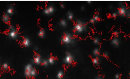
Figure 6 Typical image produced by NTA showing particle tracks. 23
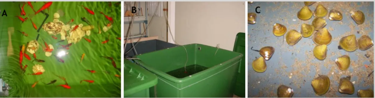
Figure 7 Sampling of C. fluminea at tagus river´s margin. 23
Figure 8 Organisms in acclimation tanks. 24
Figure 9 C. auratus in test tanks. 25
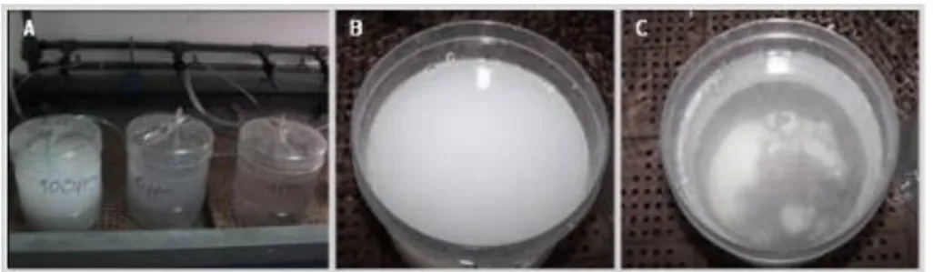
Figure 10 Tanks for C. fluminea exposure to TiO2 NPs suspensions. 25 Figure 11 Tanks for C. fluminea exposure to TiO2 NPs suspensions. 32
Figure 12 Scanning electron microscopy of TiO2 NPs. 32
Figure 13 Nanoparticle tracking analysis. 33
Figure 14 C. auratus macroscopic observations. 34
Figure 15 Macroscopic observations of NPs presence in C. auratus. 34
Figure 16 Representation of C. fluminea mortality. 35
Figure 17 Macroscopic observation of C. fluminea shell. 36
Figure 18 Observation of C. auratus liver sections by optical microscopy. 37 Figure 19 Electron microscopy observation of C. auratus liver tissue after exposure TiO2 NPs 38 Figure 20 Observation of C. auratus gill sections by optical microscopy. 39 Figure 21 Electron microscopy observation and elemental analysis of C. auratus gill tissue
after exposure to TiO2 NPs. 40
Figure 22 Observation of C. auratus intestine sections by optical microscopy. 41 Figure 23 Electron microscopy observation and elemental analysis of C. auratus intestinal
tissues after exposure to TiO2 NPs. 42
Figure 24 Observation of C. fluminea digestive gland sections trough optical microscopy. 43 Figure 25 Electron microscopy observation and elemental analysis of C. fluminea after
exposure to TiO2 NPs. 44
Figure 26 SOD activity in C. auratus. 45
Figure 27 CAT activity in C. auratus. 46
Figure 28 GST activity in C. auratus. 48
Figure 29 LPO in C. auratus. 50
Index of Tables
Table 1 Modeled concentrations of TiO2 nanoparticles released into environmental
compartments in Europe and United States. 5
Table 2 TiO2 nanoparticles physico-chemical proprieties, according to the manufacturer. 22 Table 3 Mortality rate, length and weight (mean±sd), after 7, 14 and 21 days of TiO2 NPs
exposure for C. auratus. 75
Table 4 Mortality rate, length and weight (mean±sd), after 7, 14 and 21 days of TiO2 NPs
Abbreviations
BSA Bovine Serum Albumine
CAT Catalase
CDNB 1-chloro-2,4-dinitrobenzene
DLS Dynamic Light Scattering
DNA Deoxyribonucleic Acid
EDS Energy Dispersive X-ray Spectrometry EDTA Ethylenediamine Tetraacetic Acid
EGF Epidermal Growth Factor
LC50 Lethal Concentration, 50%
LPO Lipid Peroxidation
GPX Glutathione Peroxidase
GR Glutathione Reductase
GSH Glutathione
GST Glutathione-S-Transferase
H&E Hematoxyline & Eosine
NADPH Nicotinamide Adenine Dinucleotide Phosphate-oxidase
NBT Nitroblue Tetrazolium
NF-kB Nuclear Factor kappa B
MDA Malondialdehyde
nm Nanometer
NPs Nanoparticles
NTA Nanoparticle Tracking Analysis
ROS Reactive Oxygen Species
SD Standard Deviation
SDS Sodium Dodecyl Dulfate
SEM Scanning Electron Microscopy
SOD Superoxide dismutase
TBA Thiobarbituric Acid
TBARS Thiobarbituric Acid Reactive Substances
TBT Tributylin
TCA Trichloroacetic Acid
TEM Transmission Electron Microscopy
TMP Trimethylolpropane
UV Ultraviolet
XOD Xanthine-Oxidase
1 Introduction
1.1 Background
The concept of nanotechnology was first introduced about 50 years ago, when the Nobel Prize-winner Richard Feynman presented a talk called “There’s Plenty of Room at the Bottom”, at the annual meeting of the American Physical Society at the California Institute of Technology. The audience was puzzled and intrigued with Feynman’s futuristic vision of how it could be possible to put a huge amount of information written in an exceedingly small space, while he was exploring the possibility of manipulating materials at the scale of individual atoms and molecules (Feynman, 1960).
Over the last 20 years, nanotechnology has emerged and is already a multidisciplinary reality, present in a wide range of fields including chemistry, physics, biology, medicine, engineering and electronics. Nowadays, nanotechnology can be defined as the research and development of structures, devices and systems by controlling shape and size at nanometric scale1 (RS, 2004), to create materials with new behaviors and chemical properties.
1.2 Nanomaterials
Engineered nanomaterials such as nanoparticles (NPs) are increasingly being used for commercial purposes in products within medicine, electronics, sporting goods, tires, textiles and cosmetics. In the past decade, the Project of Emerging Nanotechnology launched a Nanotechnology Consumer Product Inventory, available at http://www.nanotechproject.org/. From an initial number of 212 products identified in the year of 2006, over 1300 manufacturer-identified nanotechnology-enabled consumer products have entered the marketplace to date, according to this inventory. The nanomaterial potential applications seem endless, promising great benefits for society and bringing high economic expectations. This is considered to be one of the major technology sectors of the 21st century (Delgado, 2010), which is reflected on an increasing global investment of multi-billion euros/dollars. However, the same special properties that make nanomaterials so distinctive and useful also may represent and be the cause of potential risks and unpredictable effects to living beings and environment. Unfortunately, there is a great knowledge lack between nanotechnology
and its potential toxicity. Although the nanotoxicology is a very young research field, there is an increasing of studies that demonstrate hazards associated with nanoparticles, bringing the awareness of their potential adverse effects.
1.3 Nanoparticles
Nanoparticles (NPs) comprise diverse types of materials from metals, polymers, ceramic to biomaterials and have been defined as particles with at least one dimension in the order up to 100 nm (RS, 2004). However, without reliable methods for characterization and determination of the physicochemical properties of NPs it is also difficult to assess human or animal exposure to NPs (FSAI, 2008).
1.3.1 Characterization of Nanoparticles
Although NPs characterization is usually performed by using diverse techniques for estimating their physicochemical properties (e.g SEM, TEM, DLS, XRD) there is an urgent need to establish calibration standards and procedures for the characterization of NPs since the reliability, precision and accuracy of these techniques on the nanoscale are however often questioned. In this sense, the ISO TC229 Technical Committee on Nanotechnologies was established in 2005 to address these issues (FSAI, 2008). Accordingly, NPs characterization is a major challenge since it requires considerable care and there are many difficulties and uncertainties, especially regarding NPs aggregation, size, purity, and batch variations. Additionally, characterization is further complicated by the incorporation of NPs into biological matrices which may change their properties and requires further characterization beyond that of the pristine nanoparticle (FSAI, 2008).
Regarding NPs properties despite having the same chemical composition as bulk materials, they may exhibit new or enhanced size-dependent properties compared with larger particles of the same material (Hodes, 2007). According to Nel at al. (2006) the properties of nanomaterials are related to their size, structure and a large surface area-to-volume ratio relative to larger-sized chemicals and materials. In addition, in terms of size they are included in a transitional zone between individual atoms or molecules and the corresponding bulk material. Moreover, as the size of a particle decreases, more molecules are present at the surface giving rise to a larger surface area for chemical interaction. Thus, the higher surface area to the volume ratio plays a major role in the increasing of chemical reactivity and the change of the magnetic, conductive, optical and diffusion properties (Nel et al., 2006). Hence, materials that are inert in larger size can become reactive at the nanoscale. The size at which materials start changing their properties can vary from less than one nanometer to the micrometer range (Hodes, 2007).
Another important feature of NPs is their tendency to aggregate, often as a result of the drying stage during the synthesis process, and with considerable implications for the determination of the size and surface area of NPs but also for assessment of exposure to living organisms. The aggregation is often overlooked or even ignored when characterizing NPs size and surface area because they can exist both in very large and small particle size with a heterogeneous distribution. This is an important fact because sizes recorded are often only a small fraction of the sample, rather than a true representation of the sample composition. In this way, many studies use several techniques to disperse nanoparticles (e.g. ultrasound sonication, dispersing and milling). However, even following these procedures, the dispersions are often very polydisperse, and the surface may have changed, due to exposure of “fresh” surface due to the breaking up of clusters. Therefore, an important issue is to distinguish between the primary particle size, which is typically on the nanoscale, and the cluster size due to aggregation, which may be either nanoscale or micron scale (FSAI, 2008). In a toxicological point of view size and surface area are extremely important since the small size and a large surface area allows a great proportion of its atoms or molecules to be displayed on its surface rather than within the material´s interior. As a consequence, these nanomaterial´s atoms or molecules may be chemically and biological reactive and have potential negative effects on living organisms. Other factors such as shape, surface coating, aggregation potential and solubility also affects physicochemical and transport properties of nanomaterials but also its toxicity potential. As an example, some types of NPs are associated to DNA damage, production of reactive oxygen species oxidative stress and neurologic problems, among others effects. Moreover, the ever-increasing use of these materials, soon can lead to the release and accumulation of heightened levels of these materials into environment. Still, there’s a considerable gap in the regulation of the commercialization of products containing nanotechnologies (Falkner and Jaspers, 2012).
1.4 Environmental and Human Risks
1.4.1 Human and animal exposure to Nanoparticles
Human beings always have been exposed to airborne nanosized particles. However, such exposure has increased dramatically over the last decades due to the development of NPs from anthropogenic sources (Oberdörster et al., 2005).
The same properties that make NPs unique and so wide useful may also become a trap to our health. The higher toxicological potential of NPs is mostly due to their small size, wide surface, increase of their chemical reactivity and biological activity and the capacity to generate free radicals (Nel et al., 2006). NPs also can have the ability to penetrate trough the
biological barriers and to move easily through the biological systems (Oberdörster et al., 2005; Nel et al., 2006).
Figure 1 Potential cellular interactions of Nanoparticles.
The diagram shows the potential effects of NP with emphasis on potential oxidative stress induced effects and their consequences. (A) Particle-associated characteristics induce lipid peroxidation, intracellular oxidative stress and increased cytosolic calcium ion concentration; (B) NP may be actively endocytosed. In phagocytic cells phagocytosis triggers activation of NADPH oxidase and generation of ROS; (C) Particles and their associated metals, as well as oxidative stress, can activate the EGF receptor; (D) Oxidative stress, receptor activation and increased calcium ions activate transcription of pro-inflammatory genes via transcription factors such as NF-kB; (E) NP may enter the cell by passive diffusion and remain non-membrane bound from where they may enter mitochondria; (F) and disrupt normal electron transport leading to oxidative stress. (G) Free particles may also enter the nucleus via the nuclear pore complex and interact with the genetic material. (H) Lipid peroxide-derived products such as 4-hydroxynonenal form DNA adducts that may lead to genotoxicity and mutagenesis (in Oberdörster et al., 2005).
Nanotoxicological research has already associated some NPs to several toxicological effects as damage to DNA (Donaldson et al., 1996; Dunford et al., 1997), disruption of cellular function (Sayes et al., 2006), production of reactive oxygen species (Long et al., 2006), asbestos-like pathogencity (Poland et al., 2008), neurologic problems (NIH and NCCAM, 2010), organ
damage including significant lesions on the liver and kidneys (Wang et al., 2007), gill damage, respiratory problems and oxidative stress in fish (Federici et al., 2007).
1.4.2 Environmental Impacts of Nanoparticles
Nanoparticles (NPs) are not only artificial, but they also always existed in environment from natural sources. Carbon NPs have been found in 10 000 years ice cores (Murr et al., 2004). Other natural NPs nanoparticles can also be found in soil, water sources, atmospheric dust or volcanic ash (Handy et al., 2008).
Engineered nanomaterials can enter the environment through deliberated releases, which includes their use to remediate contaminated soils and groundwater, unintentional releases such as atmospheric emissions and also from the use of consumer’s products with NPs, as sunscreens and cosmetics (Klaine et al., 2008). NPs have the potential to contaminate soil, migrate into surface and groundwater and interact with biota (Klaine et al., 2008). Also, NPs in solid wastes, wastewater effluents, direct discharges, or accidental spillages can be transported to aquatic systems by wind or rainwater (Klaine et al., 2008).
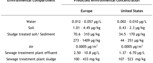
Table 1 Modeled concentrations of TiO2 nanoparticles released into environmental
compartments in Europe and United States (adapted from Menard et al., 2011).
Environmental Compartment Predicted environmental concentration
Europe United States
Water 0.012 – 0.057 µg/L 0.002 – 0.010 µg/L
Soil 1.01 – 4.45 µg/kg 0.43 – 2.3 µg/kg
Sludge treated soil/ Sediment 70.6 – 310 µg/kg 273 – 1409 µg/kg
34.5 – 170 µg/kg 44 – 251 µg/kg
Air 0.0005 µg/m3 0.0005 µg/m3
Sewage treatment plant effluent 2.50 – 10.8 µg/L 1.37 – 6.70 µg/L Sewage treatment plant sludge 100 – 433 mg/kg 107 – 523 mg/kg
1.4.2.1 Nanoparticles in aquatic environments
The aquatic environment receives daily substantial amounts of environmental pollutants that can be up taken by aquatic organisms from sediments, suspended particulate matter with toxic properties and food sources, depending on the particular dietary and ecological lifestyles of the organisms (Valavanidis et al., 2006).
Aquatic systems contain natural complex colloid2 materials. These include inorganic minerals, typically hydrous iron and manganese oxides, and as well organic matter, such as humic substances, proteins and peptides (Klaine et al., 2008; Lead and Wilkinson, 2006). Their small size and large surface area per unit mass make them important binding phases for both organic and inorganic contaminants. This way, NPs can be accumulated and transported by the colloid fraction (Lead and Wilkinson, 2006). Once within an aquatic environment, NPs can also enter a process of aggregation which is closely related to the deposition and sedimentation of particles (Wiesner et al., 2009). This process is determined by the NPs surface properties, which are mainly dependent on parameters such as temperature, ionic strength, pH, particle concentration and size, among others (Navarro et al., 2008).
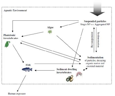
Figure 2 Possible routes of environmental exposure to engineered NPs after release to the aquatic environment and trough food chains. (in Baun et al., 2008)
2 In aquatic systems, colloid is the generic term applied to particles in a size range between 1 nm to 1
1.4.2.2 Effects in aquatic organisms
Although several studies and reports have been recently published there is still a lack of knowledge on the ecotoxicity of NPs in aquatic biota. To date most of the data available are on freshwater species, particularly those used as standard species in regulatory toxicology such as Daphnia magna or Danio rerio and using several types of NPs (e.g. TiO2, nanodiamonds, C60 fullereens) (Handy et al., 2008). It is also evident that more studies are needed on marine and terrestrial organisms and other vertebrates such as amphibians, reptiles and birds.
In aquatic invertebrates, the exposition to CeO2 and SiO2 NPs caused an increase of the mortality of D. magna and CeO2 NPs also induced DNA damage and potential reproduction reduction for these organisms (Lee et al., 2009). Moreover, nanosized copper and silver had a 48 h LC50 of less than 1 mg/L for daphnids and algae (Griffitt et al., 2008) and also ZnO NPs showed toxicity to these organisms (Adams et al., 2006). For fish organisms, studies showed that Ag NPs provoked a 48 h LC50 value of 1.03 mg/L in Japanese medaka (Oryzias latipes), with additional developmental, morphological and histopathological changes, including edema production, abnormalities in the spine, fins, heart, brain, and eyes (Wu et al., 2010). In zebrafish, Ag and Cu NPs had a 48 h LC50 of less than 10 mg/L (Griffitt et al., 2008). Aggregation of single walled carbon nanotubes has been visible on the gill mucus of trout (Smith et al., 2007) and also TiO2 NPs induced histological changes and oxidative stress in the rainbow trout (Federici et al., 2007).
1.5 Titanium dioxide nanoparticles
Conventional titanium dioxide (TiO2) is a naturally harmless occurring mineral, which has been used since the beginning of the 20th century for numerous industrial applications and consumer products, particularly for coatings and pigments (Chen et al., 2007).
In addition, as the size of TiO2 particles decreases into the nanoscale, higher is the potential for photocatalytic properties and UV absorption (Shao and Schlossman, 1999). These properties led TiO2 nanoparticles (NPs) to a wide range of industrial applications and consumer products such as water treatment agents, self-cleaning surface coatings, light-emitting diodes, solar cells, disinfectant sprays, sporting equipment, sunscreens and other cosmetics (Chen et al., 2007).
Consequently, the commercial production of nanosized TiO2 is increasing at a very high rate every year. For example, the estimated production between 2006 and 2010 was about 5000 tons/year, while the predicted production for between 2011 and 2014 is about 10 000 tons/year (UNEP, 2007). The high production of TiO2 NPs due to its widespread use, soon can
lead to significant release of these NPs into the environment (Hall et al., 2009). However, little is known about the NPs fate, behavior and toxicity once released into the environment and surface waters.
1.5.1 Effects and toxicity of TiO
2NPs
Conventional TiO2 has been considered to be biologically inert and harmless for living beings, and fine TiO2 particles were used as controls in toxicological studies of various particles, for example in numerous pulmonary toxicity studies (Sager et al., 2008).
TiO2 reflects and scatters UVB and UVA in sunlight, thus has been applied for e.g. as a safe physical sunscreen. However, TiO2 absorbs about 70% of incident UV and especially in aqueous environments this leads to generation of hydroxyl radicals which can initiate oxidations (Dunford et al., 1997). Also TiO2 NPs are photo-inducible, redox active and thus generators of potential reactive oxygen species (ROS) at its surface (Menard et al., 2011). Dunford et al. (1997) showed that TiO2 present in sunscreen samples was able to catalyze oxidative damage to DNA both in vitro and in human cells. Moreover TiO2 in nanoparticle size showed to be able to produce ROS not only in the presence of UV irradiation (Armelao et al., 2007), but also in the absence of photoactivation (Gurr et al., 2005; Reeves et al., 2008). ROS and free radicals are oxidative stress inductors and may play a major role in the NPs potential toxicity to organisms.
Concerning mammalians, some pro-inflammatory effects resulting from TiO2 NPs exposure were observed both in vitro and in vivo in pulmonary cells. Studies reported that TiO2 NPs can induce respiratory toxicity, epithelial inflammation and cytotoxicity within the lung of rodents (Ferin et al., 1991; Oberdörster et al., 1992; Bermudez et al., 2004; Warheit et al., 2006) but also in human lung cells (e.g. Gurr et al., 2005; Lai et al., 2008).
Exposure of rats by intracheal instillation to a suspension of TiO2 NPs caused dose-dependent pulmonary damage and inflammation, which persisted 42 days post-exposure (Sager et al., 2008).
The administration of TiO2 NPs into an air pouch in mice, provoked an acute inflammatory response, inducing a rapidly leukocyte infiltration with predominance of neutrophils and an increase expression of pro-inflammatory mediators as chemokines (Gonçalves and Girard, 2011).
Rabbit erythrocytes treated in vitro with TiO2 NPs underwent hemagglutination and dose dependent hemolysis (Li et al., 2008). Moreover intragastric administration of TiO2 NPs in mice provoked the damage of blood system haemostasis, reduction of the immunity in association with a seriously damaged liver function (Duan et al., 2010).
After an intraperitoneal injection in mice, TiO2 NPs were retained in multiple organs and tissues, mainly in spleen but also in liver, kidney and lung, inducing significant pathological changes and various degrees of organ lesions, severely in spleen (Chen et al., 2009). An accumulation of NPs in mice spleen due to a TiO2 NPs exposition by intraperitoneal injection for consecutive 45 days, was also reported by Li et al. (2010), leading to congestion and lymph nodule proliferation of spleen tissue, spleenocytes apoptosis, ROS accumulation, resulting in a decrease of immune capacity.
TiO2 NPs induced formation of micronuclei and apoptosis in hamster embryo fibroblasts (Rahman et al., 2002). In addition, several studies showed that TiO2 NPs caused hepatocyte necrosis in mice livers and changes in some enzymes levels (Wang et al., 2007; Liu et al., 2009) and hepatocytes apoptosis and inflammation related to alterations of both mRNA and protein expression levels of diverse inflammatory cytokines (Ma et al., 2009). Moreover, according to the same author the increase of lipid peroxides in brain and liver mouse tissues caused by TiO2 NPs was associated to an oxidative attack activated by a decrease of the antioxidative defense mechanisms.
TiO2 NPs were also associated with changes in gene expression, including alterations concerning brain development in mouse (Shimizu et al., 2009) and expression of apoptosis-related genes (Carinci et al., 2003).
With respect to skin, several studies report that TiO2 NPs are able to penetrate animal and human skin (Wu et al., 2009, Monteiro-Riviere et al., 2011). Dermal exposure to TiO2 in vivo, using mice as biological model, revealed that these NPs are able to penetrate skin, reach different tissues and induce lesions in different organs mostly at skin and liver (Wu et al., 2009). Results of this study also showed that prolonged exposure to TiO2 NPs can cause oxidative stress by increasing lipid peroxidation products and cause collagen depletion leading to skin aging, concluding that TiO2 NPs may pose a risk to human health after dermal exposure over a relative long time period (Wu et al., 2009).
1.5.2 Toxicity of TiO
2NPs to aquatic organisms
Regarding the ecotoxicity of TiO2 NPs to aquatic biota, the most studied group of aquatic organisms are freshwater invertebrates, followed by algae and for last freshwater fish (Menard et al., 2011).
Algae play an important role in the equilibrium of aquatic ecosystems, being the first level of the trophic chain to produce organics and oxygen (Sadiq et al., 2011).
The exposition of TiO2 NPs to freshwater green micro algae produced a growth inhibition (Hartmann et al., 2010; Metzlera et al., 2011; Sadiq et al., 2011) and also a concentration dependent decrease in chlorophyll content (Sadiq et al., 2011).
Hartmann et al (2010) observed that in addition to the generation of reactive oxygen species, possible mechanisms of toxicity to algae included the adhesion of TiO2 to algal cells and physical disruption of the cell membranes. Microscopy techniques confirmed that TiO2 NPs have a strong affinity toward the cell surface, demonstrating probable interactions between the particles and the surface active sites of the cell membrane (Metzlera et al., 2011; Sadiq et al., 2011). The adhesion/adsorption of NPs to the cell surface may interrupt the nutrient transfer; enhance the ROS reaction rates and membrane lipid peroxidation (Metzelera et al., 2011).
The observation of TiO2 NPs aggregates entrapping algal cells, suggested that it may play the major role in the toxicity of TiO2 NPs to algae species (Aruoja et al., 2009; Sharma, 2009; Sadiq et al., 2011).
Cherchi et al. (2011) showed that the internalization of TiO2 NPs through multilayered membranes in algal cells can occur, generating observable alteration in various intracellular structures and inducing a series of recognized stress responses. Therefore, NPs may be transported along the ecological food web and ultimately impact important biogeochemical processes, such as the carbon and nitrogen cycle (Cherchi et al., 2011).
Daphnia magna (a cladoceran freshwater water flea) is widely used as a biological model for
testing ecotoxicity. D. magna is a vital connection in the aquatic food chain between the algae that they consume and the ecologically and economically important fish that consume those freshwaters crustaceans (Lovern and Klaper, 2006).
The exposure of D. magna to TiO2 NPs induced significantly the activity of several antioxidant enzymes as CAT and GST, with a concentration-dependent increase (Kim et al., 2010). This suggested that the toxicity was mediated by ROS, generated by TiO2 NPs, via oxidative stress in D. magna (Kim et al., 2010).
Filtered TiO2 NPs were reported to cause an increase of the mortality of D. magna with the increase of TiO2 concentration (Lovern and Klaper, 2006). In a chronic bioassay, Kim et al. (2010) observed an increase in mortality, probably due to the accumulation of TiO2 NPs in the intestine of D. magna, which might induce effects such as oxidative stress relating to the induction of antioxidant enzymes.
Other studies also showed that D. magna ingest TiO2 NPs from aqueous suspension and their deposition is visible inside the gastrointestinal tract (Baun et al., 2008; Kim et al., 2010; Zhu et al., 2010a). D. magna displayed difficulty in eliminating TiO2 NPs from their body, resulting in a high level of bioaccumulation, which may interfere with food intake, growth and reproduction (Zhu et al., 2010a and 2010b).
The chronic exposure of D. magna to TiO2 NPs resulted in severe growth retardation, reproductive defects and increasingly mortality (Zhu et al., 2010a). The toxicity of NPs was shown to increase with the exposure duration, demonstrating that it may also be an important factor in the toxicity mediated by NPs (Zhu et al., 2010a).
Zhu et al. (2010b) showed evidence for TiO2 transfer from D. magna to zebrafish (Danio rerio) through a simplified freshwater food chain.
In larval zebrafish, TiO2 NPs affected significantly swimming parameters, as average and maximum velocity and activity level (Chen et al., 2011).
In vitro studies reported intrinsic genotoxic and cytotoxic potential of TiO2 NPs on fish cell lines derived from rainbow trout gonadal tissue (Vevers and Jha, 2008) and from gold fish (Carassius auratus) skin cells (Reeves et al., 2008). Also Reeves et al. (2008) indicated that ·OH radicals are the predominant radical species generated both in aqueous solution as in the fish cells, thus playing the major role in producing the genotoxic effects in terms of oxidative DNA damage.
Griffitt et al. (2009) reported that TiO2 NPs exposure to zebrafish altered the expression of genes involved in ribosomal function, which may be related to inhibition of protein synthesis by cellular stress. Moreover the microinjection of TiO2 NPs in zebrafish embryos caused significant changes in the expression of genes related to circadian rhythm, cell kinase activity, intracellular trafficking and immune response, detected by microarray analysis (Jovanovic et al., 2011).
In vivo studies using as a biological model different species of fish, also showed changes in
the activity of antioxidant enzymes, in lipid peroxidation levels and histopathological changes, as a result to the exposition to TiO2 NPs.
The exposure of rainbow trout (Oncorhynchus mykiss) to TiO2 NPs caused respiratory distress and sub-lethal toxicity involving oxidative stress, induction of antioxidant defense system, increase in lipid peroxidation and organ pathologies in gills, liver, intestine and brain (Federici et al., 2007). Also Frederici et al. (2007) suggested that the observation of a severe erosion of the trout gut epithelium can be a consequence of drinking contaminated water with NPs.
Xiong et al. (2011) exposed zebrafish to TiO2 NPs, concluding that these NPs were able to cause toxicity effects without entering the cells, despite the formation of aggregates in suspensions. They observed that extracellular hydroxyl radical (·OH) generated by TiO2 NPs could induce oxidative damage directly on the cell membranes of gill tissue.
In juvenile carp (Cyprinus carpio), TiO2 NPs modified the antioxidant enzymatic activity (SOD, CAT, POD) and elevated the lipid peroxidation levels most evidently in liver, inducing liver
disorders (as necrotic and apoptosis hepatocytes) and also gills pathologies (as edema and thickening of gill lamellae and filaments) (Hao et al., 2009).
The effects of dietary exposure to TiO2 NPs in rainbow trout were studied by Ramsden et al. (2009), showing the occurrence of Ti accumulation in gills, gut, liver, brain and spleen, with Ti not clearing in some organs following recovery, especially the brain. They also observed disturbances of Cu and Zn levels, a 50% inhibition of Na+/K+-ATPase activity in the brain and a 50% reduction of thiobarbituric acid reactive substances in the gill and intestine during exposure. Comparing their results for TiO2 NPs against the know hazard from other metals, Ramsden et al. (2009) concluded that the dietary hazard from TiO2 NPs might be considered more toxic than dietary Cu and Zn, and similar to Hg at equivalent oral doses.
Recent studies are revealing that the presence of TiO2 NPs can exacerbate the toxicity of other contaminants, having an indirect impact on aquatic organisms by varying the toxicity of coexisting pollutants. The presence of TiO2 NPs greatly enhanced the accumulation of cadmium (Cd) and arsenic (As) in carp (Cyprinus carpio), especially in viscera and gills (Zhang et al., 2007; Sun et al., 2009), acting as a carrier of these metals into fish. Hartmann et al. (2010) observed that the algal toxicity of Cd was enhanced in the presence of TiO2 NPs, indicating either a combined effect of Cd and TiO2 NPs or an increase of the bioavailability of Cd for the algae caused by TiO2 NPs. Also Hu et al. (2011) showed that TiO2 NPs in humic acid solutions can act as a carrier to facilitate the Cd bioaccumulation in zebrafish and potentially other heavy metals. The toxicity of tributyltin (TBT, a highly toxic marine antifouling compound) to abalone (Haliotis diversicolor supertexta) embryos increased with the presence of TiO2 NPs, as a result of the combined effects of TBT adsorption onto TiO2 NPs aggregates and the internalization of these aggregates by abalone embryos (Zhu et al., 20111). Thus, not only the direct impacts of NPs should be a matter of concern but also their interactions with other environmental pollutants.
1.6 Biological models
Aquatic organisms are widely chosen as test species, as they are more sensitive to exposure and toxicity compared to terrestrial organisms including mammals. Besides being easily to cage and having a filtration capacity, they can provide model systems for evaluation of oxidative damage concerning to chronic exposure or sublethal concentrations, mutagenicity and other adverse effects of pollutants (Valavanidis et al., 2006).
1.7 Oxidative stress enzymes
Oxidative stress is an important subject for terrestrial and aquatic toxicology, where its molecular biomarkers found widespread applications in mechanisms of environmental toxicity in aquatic organisms exposed to different chemical pollutants (Livingstone, 2001).
Reactive oxygen species (ROS), such as the superoxide anion radical (O2-), hydrogen peroxide (H2O2) and hydroxyl radical (·OH) are continually produced in biological systems as toxic byproducts of normal oxidative metabolism, but can be increased by interactions with pollutants by various mechanisms (Livingstone, 2003).
In living aerobic organisms, the neutralization, detoxication and removal of ROS is effected by intracellular antioxidant defense systems, whose roles are to intercept and inactivate reactive radicals (Valavanidis et al., 2006). The antioxidant defense includes specific antioxidant enzymes, such as superoxide dismutase (SOD; converts O2- to H
2O2; EC 1.15.1.1), catalase (CAT; converts H2O2 to H2O and O2; EC 1.1.1.6), glutathione-s-transferase (GST; conjugates and detoxifies products of lipid peroxidation; EC 2.5.1.18), but also non enzymatic cellular defenses, such as reduced glutathione (GSH), vitamins A and E, ascorbate and urate (de Zwart et al., 1999; Livingstone, 2003). The between the generation and the neutralization of ROS by antioxidant mechanisms within an organism is called oxidative stress (Davies, 1995).
Antioxidant enzyme activities are found widely distributed in tissues of aquatic organisms, with a mostly higher activity in liver of fish and in the digestive gland or equivalent in invertebrate organisms (Livingstone, 2001). Assaying antioxidant enzymes can indicate the antioxidant status of the organisms, working as a potential biomarker for contaminant-mediated oxidative stress (Valavanidis et al., 2006).
Cellular antioxidant defense systems may be increased or inhibited under chemical stress, depending on the intensity, duration of the stress applied and on the susceptibility of the exposed organisms (Cossu et al., 2000). The exposure to organic pollutants and metals may induce significant increases in antioxidant enzymes in response to ameliorate oxidative stress, but these are transient and variable for different aquatic species. Studies with fish observed that, in response to toxicant-induced inflammation by ROS, the concentrations of certain antioxidant enzymes are increased, but under high levels of pollution the antioxidant defenses can be reduced (Valavanidisa et al., 2006). An induction of the antioxidant defense can be considered an adaptation of the organisms to prevent toxicity, while a decrease suggests a precarious state characterized by a higher susceptibility to environmental stress, resulting in adverse effects (Cossu et al., 2000). The knowledge of the regulation of antioxidant systems in aquatic organisms in relation to sources of ROS is limited (Livingstone, 2001). The complexity of pollution in aquatic ecosystems provides a non-specific response to a kind of contaminants, but antioxidants constitute useful biomarkers reflecting not only an
exposure to pollutants but also their toxicity. A multiple marker approach can be more relevant than a single antioxidant parameter to the evaluation of the total antioxidant status.
1.7.1 Superoxide Dismutase
Superoxide dismutase (EC 1.15.1.1) is the antioxidant enzyme that catalyzes the dismutation of the highly reactive superoxide anion (O2-) to O2 and to the less reactive species H2O2 (reaction (1)), which can be further destroyed by catalase or GPX reactions.
-
→
(1)
Another function of superoxide dismutase is to protect dehydratases (dihydroxy acid dehydratase, aconitase, 6-phosphogluconate dehydratase and fumarases A and B) against inactivation by the free radical superoxide (Matés and Sánchez-Jiménez, 1999). Four classes of SOD have been identified, containing either a di-nuclear Cu, Zn or mononuclear Fe, Mn or Ni cofactors (Matés and Sánchez-Jiménez, 1999).
At physiological pH, the rate of the non-enzymatic dismutation of superoxide is significant, but it is considerably increased in the presence of SOD. The turnover numbers of SODs are indeed very high over a wide range of pH. In animal cells, the fact that intracellular SOD concentrations range from 10-6 to 10-5 M supports the concept that superoxide is strongly toxic (Chaudière and Ferrari-Iliou, 1999).
1.7.2 Catalase
Catalase (CAT; EC 1.11.1.6) is included in the subclass of oxidorredutases, being present in practically all aerobic organisms and in many anaerobic organisms. It can also be named hydroperoxidase as it catalyzes the conversion of hydrogen peroxide (H2O2), a powerful and potentially harmful oxidizing agent, to water and molecular oxygen (reaction (2)).
→
(2)
Much of the H2O2 that is produced during oxidative cellular metabolism comes from the breakdown of one of the most damaging ROS, namely the superoxide anion radical (O2-), by superoxide dismutases into hydrogen peroxide and oxygen.
Each catalase molecule can decompose millions of H2O2 molecules every second, thus this is one of the most efficient enzymes found in cells. It is so efficient that it cannot be saturated by H2O2 at any concentration.
CAT also acts as peroxidase, at low hydrogen peroxide concentration, using reducing co-substrates, as a variety of metabolites and toxins donors of hydrogen such as alcohols, formic acid, formaldehyde or phenols.
Structurally, most catalases exist as tetramers, each subunit containing an active site heme group deep within its structure, but accessible from the surface through hydrophobic channels. This very rigid and stable CAT structure makes it more resistant to unfolding, to pH, thermal denaturation and resistant to proteolysis than most of other enzymes (Chelikani et al., 2004).
Figure 3 Catalase Heme Group. In the middle of the heme group sits an iron atom. The
catalase enzyme uses this iron atom to help it break the bonds in the two molecules of H2O2, shifting the atoms around to release two molecules of H2O and a molecule of O2.
H2O2 is broken down by CAT within a two-stage mechanism in which H2O2 alternately oxidises and reduces the heme iron at the active site. In the first step, one H2O2 molecule oxidises the heme to an oxyferryl species. In the second step, a second hydrogen peroxide molecule is used as a reductant to regenerate the resting state enzyme, producing water and oxygen (Chelikani et al., 2004).
The ubiquity of the enzyme, its ease of assay, involving a cheap, readily available substrate, H2O2, and the outstanding display of oxygen evolution have combined to make it an attractive target for biochemists and molecular biologists alike.
1.7.3 Glutathione S-Transferase
Glutathione-S-transferases (GSTs; EC 2.5.1.18) are large family (cytosolic, mitochondrial, and microsomal) of phase II biotransformation enzymes, with substrates that include products of oxidative stress and electrophilic xenobiotics. Besides their enzymatic activity, GSTs can
protect cells from toxicants through chemical removal of the agents by noncatalytic binding. GSTs and ligandin (a protein binding to physiologic and exogenous ligands) bind toxic xenobiotics and their metabolites and this noncatalytic activity of GSTs can provide protection to the cellular environment by acting as a shield to protect DNA, proteins, and lipids from the deleterious effects of xenobiotics. GSTs are also involved in the biosynthesis of leukotrienes, prostaglandins, testosterone, progesterone, as well as in the degradation of tyrosine. GSTs are among the most abundant proteins in some tissues, including kidneys and especially in the liver, which plays a key role in detoxification.
Oxidative stress usually leads to enhanced generation of endogenous electrophiles and electrophilic toxins generated from lipid peroxidation or that are converted to genotoxic electrophilic intermediates by the catalytic action of cytochromes P450. GSTs catalyze the nucleophilic attack of the sulphur atom of the reduced glutathione (GSH) on electrophilic groups of a range of hydrophobic substrates, thereby neutralizing their electrophilic sites and rendering the products more water-soluble. Glutathione conjugates are metabolized further by cleavage of the glutamate and glycine residues, followed by acetylation of the resultant free amino group of the cysteinyl residue, to produce the final product, a mercapturic acid. The mercapturic acids, i.e. S-alkylated derivatives of N-acetylcysteine, are then excreted. Most substrates are inactivated by conjugation with GSH, however some are converted to more reactive compounds increasing toxicity (bioactivation of a toxin).
Although GSTs do not decompose the ROS per se, these enzymes constitute an all-important second line of defense and provide protection against oxidative stress by attenuating lipid peroxidation and by detoxifying the toxic end-products of lipid peroxidation. As antioxidant enzymes, GSTs complement the role of primary defense enzymes in protecting organisms from the deleterious effects of ROS. While superoxide anion and H2O2 are effectively disposed of by the cells through highly efficient enzymes (superoxide dismutase, catalase, and GPx), the ROS escaping this line of defense can initiate the autocatalytic chain of lipid peroxidation through the generation of free radicals capable of abstracting a single hydrogen atom from unsaturated fatty acids. GSTs prevent propagation of lipid peroxidation by reducing lipid hydroperoxides, and GSTs also detoxify the toxic electrophilic end-products of lipid peroxidation. Thus constitute the all-important second and third lines of defense against oxidant toxicity (Sharma et al., 2006).
Figure 4 Enzymatic Antioxidant defense lines against ROS oxidative damage. (in Sharma et
1.8 Lipid Peroxidation
Lipid peroxidation is probably the most extensively investigated process induced by free radicals. Membrane phospholipids of aerobic organisms are continually exposed to oxidant challenges, being a target rapidly affected by free radicals. Thus, peroxidized membranes and lipid peroxidation products represent constant threats to aerobic cells. The group of polyunsaturated fatty acids is especially highly susceptible to oxidative reactions by ROS, because of their double bonds.
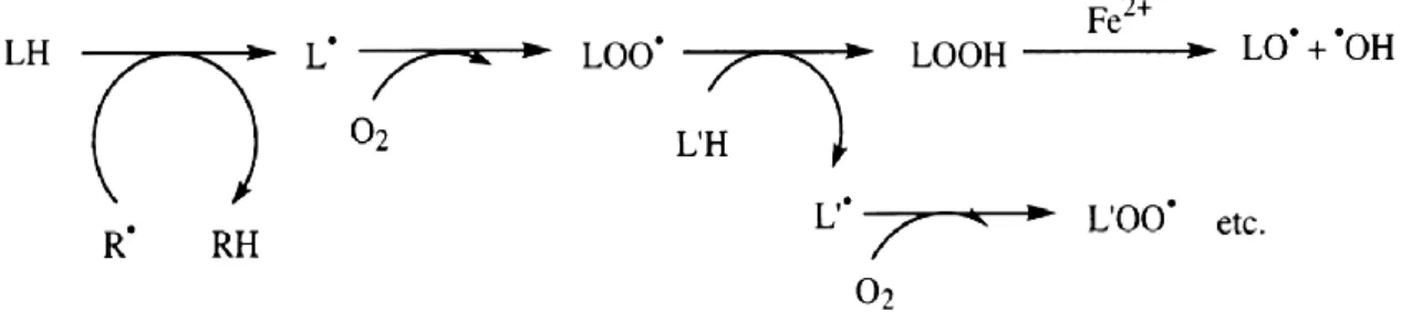
The process of lipid peroxidation is composed of a set of radical chain reactions, initiated mainly by hydroxyl radicals, especially in transition metal-catalyzed reactions, resulting in the formation of many equivalents of lipid peroxides (LOOH).
Figure 5 Lipid peroxidation chain reactions. Schematic proceed of lipid peroxidation chain
reactions, resulting in the formation of many lipid peroxide radicals.
These degenerative propagation reactions in lipid membranes are usually accompanied by the formation of a wide variety of products, as the resulting LOOH can easily decompose into several reactive species including lipid alkoxyl radicals, aldehydes, alkanes, lipid epoxides, and alcohols. Most of these products are toxic by themselves, especially hydroxyalkenals, and active mutagens, acting as second messengers for radical damage (Valavanidis et al., 2006). These products may form DNA adducts giving rise to mutations and altered patterns of gene expression and the peroxidized membranes become rigid, losing permeability and integrity. Thus, products resulting from lipid peroxidation may be important parameters to monitor radical damage and several of the most important products (de Zwart et al., 1999).
The most widely used assay for lipid peroxidation is malondialdehyde (MDA) formation as a secondary lipid peroxidation product, often assayed with the thiobarbituric acid reactive substances (TBARS) test. MDA levels in hepatic homogenates can be used for metal-induced oxidative stress in fish.
1.9 Microscopy Techniques and histopathology
1.9.1 Light and electron microscopy
The alterations in cell structure resulting from chemical exposure can be evaluated by different types of microscopy, depending on the purpose of the study. Changes which alter the cells and tissues of an organism and do not result in death can be observed under the light microscope or electron microscope. Therefore, light microscopy is commonly used for examination of histopathological alterations caused by exposure to a chemically altered environment while electron microscopy is used for the observation of alterations in the cell ultra-structure and is commonly used to assess tissue and cell changes. Both the scanning electron microscope (SEM) and the transmission electron microscope (TEM) are useful in such evaluations. SEM images provides a tri-dimensional view of tissues and cell surface while TEM provides a bi-dimensional image of cell´s inside and allows for the observation of structural changes which cannot been assessed by light microscopy.
1.9.2 Histological examination
Histology has been widely used for assessment of negative effects on living organism´s tissues and cells. Regarding fish, histopathological biomarkers or cellular changes in tissues such as gill, liver, kidney and spleen have received much attention in assessing the effects of exposure to pollutants and other substances. Therefore, histology can be a powerful tool, especially when used in conjunction with measurements of other biomarkers and other morphological studies. Additionally, the diagnostic power of histopathology may be further enhanced by employment of additional histological techniques such as immunohistochemistry. An added value of histopathology is in its capability to analyse the mechanistic effects of exogenous substances or materials and to characterize effects more specifically. For instance, histopathological analysis of target organs can reflect fish health more realistically than biochemical biomarkers. By comparing the impact of known or unknown samples on indicative histological parameters with that of specific reference compounds, the nature and magnitude of the evoked effects can be then determined (Wester et al., 2002). Thus, it can be used as a useful tool to assess the level of sub-lethal and chronic effects of toxicity, as indicator of the exposure to pollutants and may provide a better extrapolation to community and ecosystem-level (Au, 2004; Bernet et al., 1999).
Histopathological biomarkers are also useful as they can specify the target organs, tissues, cells and organelles of a single or group of toxin(s). Understanding the specificity of a contaminant to damage a particular organ system gives an insight into the mechanism of action of the toxicant which would not be available from doing classical 96h-LC50 testing. Chronic exposure to low levels of a toxicant can be studied at a broader scale on a light structure basis rather than conducting LC50 test which only study acute exposure. LC50 values
may be misleading because they reflect total metal concentration while particular attention should be given to the chemical processes that control chemical speciation. With respect to histopathological indicators they are beneficial since they show the net effect of biochemical and molecular changes in the organism resulting from exposure to a contaminant. Light structure of tissues and organs is altered when levels of the contaminant are still at low levels, therefore histopathological evaluation provide a valuable screening method of an ecosystem before severe ecological damage occurs.
2 Objectives
Within the present context, the main objective of this study is to provide a toxicological assessment of the TiO2 nanoparticles exposure (which is already present in consumer products) to two different freshwater species, goldfish (Carassius auratus) and freshwater clam (Corbicula fluminea).
For this propose, the methodology included the evaluation of the activity of some antioxidant enzymes (SOD, CAT, GST), lipid peroxidation and histopathological observations of different potential target organs.
3 Materials and methods
3.1 Nanoparticles characterization
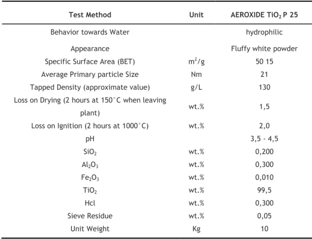
Commercial TiO2 nanoparticles Aeroxide TiO2 P25 (Aerosil, USA), with an average primary
particle size of 21 nm in the powder form (Tab. 2), were used for the exposure assays.
Table 2 TiO2 nanoparticles physico-chemical proprieties, according to the manufacturer.
Test Method Unit AEROXIDE TiO2 P 25
Behavior towards Water hydrophilic
Appearance Fluffy white powder
Specific Surface Area (BET) m2/g 50 15
Average Primary particle Size Nm 21
Tapped Density (approximate value) g/L 130
Loss on Drying (2 hours at 150°C when leaving
plant) wt.% 1,5
Loss on Ignition (2 hours at 1000°C) wt.% 2,0
pH 3,5 - 4,5 SiO2 wt.% 0,200 Al2O3 wt.% 0,300 Fe2O3 wt.% 0,010 TiO2 wt.% 99,5 Hcl wt.% 0,300 Sieve Residue wt.% 0,05 Unit Weight Kg 10
Nanoparticle Tracking Analysis (NTA) (NanoSight LM10-HS, United Kingdom) was used to characterize the behavior of nanoparticles in liquid suspensions. NTA is a method that uses Brownian motion to locate and follow individual particles (10-1000 nm) in solution (Fig. 6), from which size-distribution profiles can be obtained (Carr, 2009).
Figure 6 Typical image produced by NTA showing particle tracks. Nanoscale particles can be directly and individually visualized and counted in liquid in real-time (in Carr, 2009).
3.2 Experimental procedure
3.2.1 Test organisms
The biological materials used to carry out the experiments were a freshwater fish (Carassius
auratus) and a freshwater bivalve (Corbicula fluminea). C. aurautus has been widely used as
a model species in several ecotoxicological studies, since they are commercially available, easy to maintain and handle in laboratory (Ostrander, 2000). C. fluminea is also an important tool currently used as a biomarker for monitoring water contamination (Legeay et al., 2005). The fish were obtained from commercial suppliers (Koi Park, Portugal) and transported to the laboratory facilities for a period of acclimation before the exposure assays.
The bivalves were collected manually on Tagus River, near the locality of Escaroupim and imediatley transported to laboratory for a period of acclimation before being used in exposure tests.
3.2.2 Acclimation
Previously to the beginning of the assay, both species were acclimated for two weeks in a closed circuit system with filtered de-chlorinated tap water, at a pH of 7.4 ± 0.2, temperature of 19 ± 1ºC, photoperiod: 12hL:12hD and with continuous aeration enough for keeping the dissolved oxygen always higher than 6 mg/L.
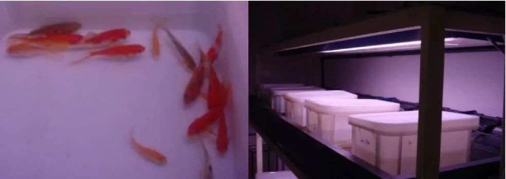
Figure 8 Organisms in acclimation tanks. C. auratus (A) acclimated in a 400 L polystyrene
tank (B). C. fluminea in acclimation tank (C).
3.2.3 Preparation of Test Nanoparticles
Stock solutions of TiO2 nanoparticles were prepared using distilled water and then ultrasonicated (10 min, 35 KHz, 100/400W) using an ultrasound bath (Elma, Germany) for dissolution. Then, solutions were added to 10 L or 2 L of dechlorinated tap water in exposure tanks, in order to obtain nominal concentrations of 0.01, 0.1, 1, 10, 100, 400 and 800 mg TiO2/L.
3.2.4 Exposure Assays
Carassius auratus
After the acclimation period, Carassius auratus (N=105; 5.9 ± 0.4 g; 4.4 ± 0.7 cm standard length), of both sexes, with less than one year of age, were randomly distributed into 15 L capacity polystyrene tests tanks, in groups of 15 fish per tank (Fig. 8). Fish were exposed to different concentrations of TiO2 nanoparticles, from 0.01 to 800 mg TiO2/L. An additional tank with clean tap was used for a group of control fish. The fish were tested under a constant temperature of 19 ± 1ºC, pH of 7.4 ± 0.2, photoperiod: 12hL:12hD and continuous aeration. The experimental conditions in each tank were renewed every 48 hours and the assay had duration of 21 days. During the experiments fish were daily fed ad libitum with commercial flakes of dry food (Tetra brand). Tanks were monitored constantly for the counting of dead fish.
Figure 9 C. auratus in test tanks.
Corbicula fluminea
After the acclimation period, C. fluminea (N=60; 1.5 ± 0.3 g ; 2.8 ± 0.2 cm standard length), were randomly distributed into 2 L capacity tests tanks, in groups of 10 individual per tank (Fig. 10). The individuals were exposed to the previously prepared solutions of different concentrations of TiO2 nanoparticles from 0.01 to 100 mg TiO2/L. An additional tank with clean tap was used for a group of control individuals. The bivalves were tested under a constant temperature of 19 ± 1ºC, pH of 7.4 ± 0.2, photoperiod: 12hL:12hD and continuous aeration. The experimental conditions in each tank were renewed every 48 hours and the assay had duration of 14 days. Tanks were monitored constantly for the counting of dead individuals.
3.3 Sampling the tested organisms
Carassius auratus
Fish were collected for sampling after 7, 14, and 21 days of exposure. At the beginning of the experiment, one additional fish group (n=5) was also collected from the acclimation tank for sampling.
Fish were sacrificed by decapitation and dissected to remove the liver, intestine and gills. Tissue samples from liver, intestine, gills were collected and fixed in a solution of
Bouin-Hollande´s fixative for histological processing.
For enzymatic and biochemical analyses, tissue samples from the target organs were homogenized on-ice in cold buffer 100 mM potassium phosphate (Sigma-Aldrich, Germany) pH 7.0 containing 2 mM of EDTA (Riedel-Haën, Germany). Tissue homogenates were centrifuged at 10,000x g for 15 minutes at 4 ºC. Supernatant was removed and freeze at -80 ºC for further analysis.
Corbicula fluminea
Organisms were collected for sampling after 7 and 14 days of exposure.
Bivalves were dissected to remove and separate the digestive glandule and gills.. Then, samples of the removed digestive glands were fixed in Bouin-Hollande´s fixative for 48 hours. For enzymatic and biochemical analyses, sub-samples of the tissues were homogenized on-ice in cold buffer 100 mM potassium phosphate (Sigma-Aldrich, Germany) pH 7.0 containing 2 mM of EDTA (Riedel-Haën, Germany). Tissue homogenates were centrifuged at 10,000x g for 15 minutes at 4 ºC. Supernatant was removed and freeze at -80 ºC for further analysis.
3.4 Histology
3.4.1 Optical Microscopy
Histological procedures for light microscopy followed essentially Martoja and Martoja (1967). Briefly, after a fixation period of 48 h in Bouin's fluid, the samples were washed in tap water and passed through a series of alcohols (70º, 95º and 100º) for dehydration, followed by a bath of xylene (Lab-Scan, Belgium) for intermediate impregnation.