UNIVERSIDADE DO ALGARVE
FACULDADE DE ENGENHERIA DE RECURSOS NATURAIS
UP-REGULA TED CENES DURING IN VITRO MINERALIZATION
OF Sparus aurata OSTEOBLAST-LIKE CELLS
(dissertação para a obtenção do grau de mestre em Biotecnologia)
Vera Alexandra Garcia da Fonseca
NOME: Vera Alexandra Garcia da Fonseca
DEPARTAMENTO: Faculdade de Ciências do Mar e do Ambiente ORIENTADOR: Dr. M. Leonor Cancela
CO-ORIENTADOR: Dr. Vincent Laizé DATA: 30 de Janeiro de 2005
TÍTULO DA DISSERTAÇÃO: Up-regulated genes during in vitro mineralization of Spams aurata osteoblast-like cells.
Júri:
Presidente: Doutora Deborah Mary Power, Professora Associada da Faculdade de Engenharia de Recursos Naturais da Universidade do Algarve.
Vogais: Doutora Maria Leonor Quintães Cancela da Fonseca, Professora Associada da
Faculdade de Ciências do Mar e do Ambiente da Universidade do Algarve.
Doutor Carlos José Fialho da Costa Faro, Professor Auxiliar da faculdade de Ciências e Tecnologia da Universidade de Coimbra.
Acknowledgments
I am grateful to Professor Leonor Cancela for supervising and supporting this thesis. I also would like to thank her for critically reading the manuscript and for helpful discussions.
I would like to thank my co-supervisor Dr. Vincent Laizé that supelvised directly this thesis. Thank you for critically reading the manuscript, for helpful discussions and above all for helping me with lab work and with long sequence analysis.
Thank you so much Asuncion Lago for always being there to help me with everything, including of course for the clucial help on the subtractive libraries.
Thank you Sandrinha for the long scientific discussions, for helping in the lab and most of all for being really my friend.
I would like to thank my colleague António Pombinho for helping me in the lab and for always being cool in the weirdest situations.
Thank you Carla Viegas for helping in the lab and for helpful discussions.
Thanks Natércia Conceição for all the articles and the imaginary “cappuccino mit zhãne” talks.
Thank you Paulo Gavaia and Nuno Henriques for lending me some space on your bench. . .it was not easy!!!
I Would also like to thank the rest of the colleagues for all the attention and friendship shown: Dina Simes, Juan Bosco, Pedro Rodrigues, Rita Ascenso, Jorge Pinto, Ricardo Afonso, João Fidalgo, Marta Rafael, Marta Valente, Ricardo Leite, Sofia Cavaco, Susana Domingos, Brigitte Simões e Daniel Tiago.
Special thanks to my husband Fred for being an amazing person thanks for making me
laugh and push me up when things look a bit grey.
Abstract
Vertebrate bone formation involves numerous and complex mechanisms and despite many studies, essentially performed in mammals, most of them remain largely unclear. Few in vitro studies, all in mammalian systems, have identified various genes differentially expressed during mineralization but no data is available yet from lower vertebrates. In the present study, it was used the recently developed Sparus aurata osteoblast-like cell line VSa16 to constmct a cDNA subtractive library (control vs. mineralizing conditions) aimed at the identification of genes associated with fish in vitro mineralization. Suppression Subtractive Hybridization (SSH) combined with Mirror Otientation Selection (MOS) identified 194 cDNA clones representing 20 different up-regulated osteoblast-related genes. Among them, four were related to calcium mechanisms: osteopontin-like (SaOP-like), S100-like (SaS100-like), mucin-like (SaMUC) and transgelin2 (SaTGL2). The full length cDNA of SaOP-like (2138 bp) and SaS100-like (8648 bp) was obtained by RACE-PCR. SaOP-like and SaS100-like relative gene expression was analysed in 1) larvae and juvenile fish representing different developmental stages, 2) a broad selection of adult tissues, and 3) cell lines derived from bone and cartilage recently developed in our laboratory. Op-like Was shown to be expressed essentially in calcified tissues and to be important for late stages of mineralization. In contrary, SIOO-like was ubiquitously expressed and found to be important at early stage of larval development. The role of the proteins encoded by these two genes in the process of mineralization is likely to be related to their capacity of binding calcium and hydroxyapatite cxystals.
Key words: subtractive libraries, gene differential expression during mineralization, osteoblast and chondrocyte-like cells, S. aurata osteopontin-like, S. aurata calcium-binding like S100
Identificação de genes expressos em condições de mineralização em células derivadas de osso de Sparus aurata e estudo da sua expressão em culturas in vitro
Resumo
A formação do osso em vertebrados envolve mecanismos numerosos e complexos e embora se tenham desenvolvido muitos estudos essencialmente em mamíferos, a maioria deles permanece ainda por esclarecer. Poucos estudos in vitro, todos em mamíferos, identificaram vários genes diferencialmente expressos durante a mineralização contudo não existem dados disponíveis em vertebrados inferiores. No presente estudo, utilizou-se um linha celular desenvolvida recentemente com células de Sparus aurata semelhantes a osteoblastos VSa16 de forma a construir um biblioteca subtractiva de cDNA (condições control vs. mineralizantes), com o objectivo de identificar genes associados à mineralização in vitro de peixes. A hibridação subtractiva de supressão (SSH) combinada com a selecção orientada tipo espelho (MOS) permitiu a identificação de 194 clones de cDNA que representava 20 genes differencialmente expressos relacionados com osteoblastos. Quatro candidatos desta biblioteca subtractiva estão relacionados com mecanismos de regulação de cálcio, osteopontina-like (SaOP-like), S100-like (SaS100-like), proteína mucin-like (SaMUC) e transgelin2 (SaTGL2). Clonou-se 2138-pb e 648-pb correspondentes ao comprimento total da SaOP-like e SaS100-like, respectivamente. Analisou-se a expressão relativa da SaOP-like e SaS100-like em 1) diferentes estádios de desenvolvimento de larvas e juvenis de peixe, 2) vários tecidos de dourada adulta e 3) linhas celulares de origem óssea e cartilagínea. Estes dois genes podem estar envolvidos em mecanismos de regulação de cálcio e deposição de minerais durante o processo de mineralização, devido à sua expressão especialmente em tecidos ósseos e estádios de desenvolvimento específicos na dourada.
Palavras-chave: bibliotecas subtractivas, expressão diferencial de genes durante a
mineralização, osteoblastos e condrócitos, S. aurata osteopontin-like, S. aurata calcium-binding like S100.
Abbreviations
1,25-(OI-I)zD3 1o‹.,25-dihydroxyvitamin D3“P
aabp
cDNA cds dCTP ddHzO DEPC DMEM DMSO DMF DNA DNase ECM EDTA FB S Gla HEPES IPTG kb MCS MOPS mRNA phosphorus isotope 32 amino acid base pairs(s)complementary DNA to RNA complete coding sequence deoxycytidine 5' tnphosphate double-distilled water
diethylpyrocarbonate
Dulbecco s modified Eagle medium
dimethyl sulfoxide dimethyl formamide deoxyribonucleic acid deoxyribonuclease extracellular matrix ethylenediaminotetraacetate Fetal bovine seium
Y- carboxyglutamic acid
N-2-hydroxyethylpiperazine N -2 ethanesulfonic acid isopropyl-B D-thiogalactopyranoside
kilobase
Multiple cloning site
3-[N-morpholino] propanesulfonic acid messenger RNA
ORF Open reading frame
PBS phosphate-buffered saline
Rpm PCR
rotations per minute polymerase chain reaction
RNA ribonucleic acid
RT-PCR reverse transcriptase PCR SDS SSC TAE UTR U U.V. vol
X-GAL 5-bromo-4-chloro-3 -indolyl-B-Dgalactopyranoside
sodium n-dodecyl sulfate sodium-chloride sodium-citrate Tris-acetate-EDTA untranslated region enzyme unit(s) ultra-violet volume(s)
ERRATUM
Page Where is... Should be. __
iv iv 8 1 2 13 8648 bp 648 bp
found to be important could be important
cell differentiation. cell differentiation (Chang et al., 2000).
using recently using a recently
(Pombinho et al., 2004) (Pombinho et al., 2004)
13, 14 (see Appendix I for composition) (Appendix I)
15 1 |.il cDNA 1 ul of cDNA
16, 22 (see Appendix III) (Appendix III)
21 22 25 27 27 30 30 30 31 31 34 34
3-4 min under UV light (Fonseca et al., in 3-4 min under UV light. press).
(accession number not yet attributed) (accession number AY787209)
PolyA+ RNA were PolyA+ RNA was
SHH S SH
All positive clones Were selected, giving a total number of 672 clones from single SSH and 960 clones from SSH+MOS. The search for up-regulated genes. _.
The search for up-regulated genes. ._
by 83 clones by 123 clones
1 15 1 14
encodes a 86-aa encondes an 86-aa
accession number has not been attributed yet.
did not identified did not identify
with the accession number AY787209
aligned sequences from aligned sequences
38 in.brain, gall bladder in brain, gall bladder
40 efficient subtraction subtraction eficiency
41 Altogether, results Altogether, the results
42 mechanisms of related mechanisms related
50 given not its only to bone specificity given not only its bone specificity
(Adapted from (Beck et al., 2000; (Adapted from Beck et al., 2000; Maeda et
51
Maeda ei at. _, 2004)).
al., 2004).
52 calcified tissues and in mineralized calcified tissues, in mineralized osteoblasts
_ Bsteoblasts (OP-likeiand to be _ (O_P-like) and during larval _
5 References cited_in_t_'ext but iiiissiiig iii Bibliograpliic Ilefereiice 1
Agüero, F., Campo, V., Cremona, L., Jãger, A., Noia, J.D_, Overath, P., Sánchez, D. and Frasch, A. (2002). Gene Discovery in the Freshwater Fish Parasite Trypanosoma carassiiildentification of trans-Sialidase-Like and Mucin-Like Genes. infection and Irnmunity. 70: 7140-7144.
Bellows, C., Heersche, J. and Aubin, J. (1992). Inorganic phosphate added exogenously or released from betaglycerophosphate initiates mineralization of osteoid nodules in vitro.
Bone Miner 17: 15-29.
Fonseca, V.G., A. Lago-Lestón, V. Laizé and M.L. Cancela. Rapid identification of differentially expressed genes by in situ screening of bacteria. Mol Biotech (in press). Kawasaki, K., Suzuki, T. and Weiss, K. (2004). Genetic basis for the evolution of vertebrate
mineralized tissue. Proc Nati Acad Sci U S A 101: 11356-61.
Kerr, J., Fisher, L., Termine, J. and Young, M. (1991). The cDNA cloning and RNA distribution of bovine osteopontin. Gene 108: 23 7-42.
Nomura, S., Wills, A., Edwards, D., Heath, J. and Hogan, B. (1988). Developmental Expression of 2ar (Osteopontin) and SPARC (Osteonectin) RNA as Revealed by In Situ Hybridization. J Celi Biot 106: 441-450.
Young, M., Kerr, J., Termine, J., Wewer, U., Wang, M., McBride, O. and Fisher, L. (1990). cDNA cloning, mRNA distribution and heterogeneity, chromosomal location, and RFLP analysis of human osteopontin (OPN). Genomics. 7: 491-502.
INDEX Acknowledgments._. _ Abstract... _ Resumo... Abbreviations Erratum_.. I-INTRODUCTION...
1.1- Bone function and structure. _. ._
1.2- Bone cells...
1.3- Bone niineralization. _ _ _ _ _ _ 1.4- Bone matrix proteins. _.
1.4.1- Collagen._.
1.4.2- Adhesion inolecules. ._ _. 1.4.3- Acid and alkaline phosphatases. ._ 1.4.4- Glycoproteins...
1.4.5- Vitamin K-dependent proteins. _. _. 1.5- Bone cell cultures...
1.6- Gene expression and detection... _ 1.7- Objectives._. _ 11- MATERIALS AND METHODS _ ._ _.
11.1- Cell culture...
11.l.l- Cell lines and culture conditions... _. 11_1.2- Extracellular matrix mineralization and nodule detection. _ _ _ ._ _ 11.2- Total RNA extraction and polyA+ RNA preparation...
11.3- Suppression subtractive hybridization (SSH). ._ ._ 1I.3.l- cDNA synthesis and Rsal digestion. ._ _ 11_3_2- SSH: adaptor ligation and hybridization __
11_3.3- SSH: PCR amplification of subtracted products... ._ 11.4- Mirror orientation selection (MOS). ._ _ _. _ 11.5- Cloning DNA fragments... _
11.5 _ 1- Cloning of subtracted cDNA templates. ._ _. 11.6- Preparation of subtracted probes_ ._ _ 11.7- Differential screening... _
11.8-Northeni blot_..
11.9- Cloning of S. aurata osteopontin-like full leng1;h cDNA. ._
11.10- Protein sequence analysis... _
|.||¡¡¡ +¡*I*I¡'I Ii fri--|-I nnnnn-1 _...iii _. .._..iv .vi vii __l __l 1|:1:-1-1-I _.6 _7 .|.n.¿n.|.|.¡¡ .7 .8 “Fl .U +.,.¡.¡|.¡|.E: 10 13 .[3 l3 ._ __._..I3 I4 15 lri 16 .lfi ___...l8 ______l8 19 211 .E0 El .22 22 23
111.1- Preparation of total RNA from control and mineralized VSa16 cell cultures... 11I_1_1- Invitro mineralization ofVSa16
111.l_2- Total RNApreparation._..___..___....__._...__.._..__._._..___.._..._... 111.2- Suppression subtractive hybridization (SSH) and differential screening...
111_2.2- Differential screening_..__.._.
111.3- Identification of genes up-regulated during mineralization. __ _ ._ _ ._ ._ _
111.4- Cloning and characterization of S. aurata S100-like and OP-like full length cDNAs..__ 111_4.1- Characterization ofSl00-like full length cDNA... _
111.4. l_l- Characterization of S 100-like putative domains... _._ ._ _.
111_4_2- Cloning and characterization of osteopontin-like full length cDNA. _. _. _ _ 111_4.2_2- Characterization of SaOP-like protein domains... __ 111.5 Analysis of S100-like and OP-like gene expression...
111.5.1- Expression pattenis in S. aurata bone-derived cell lines... _ 11I_5_2- Gene expression during larval development. ._ _ 111_5.3- Gene expression in adult tissues. _.
IV-IV. 1- Suitability/efficiency of the subtractive method used...
1V.2- Differentially expressedgenes____.._______..__....______._.._.___...._____..____.___.___..._____ 1V.3- S100-like cloning and expression in S. aurata cells, stages of development and tissues 1V.4- OP-like cloning and expression in S. aurata cells, stages of development and tissues... 1V.5- Genes temporal pattern ofexpression_._
1V_6- Final considerations and perspectives. __ _. V- BIBLIOGRAPHIC REFERENCES _ __ _ APPENDIX 1: solutions and protocols
APPENDIX 11: cloning Appendix 111: primers
Appendix IV: PCR-select cDNA subtraction teclmique Appendix V: Electrophoresis gel ladder
Appendix V1: Northem blot procedure
1-INTRODUCTION
I- INTRODUCTION
The Vertebrate skeleton has specific developmental and functional characteristics that define its identity in biologic and pathologic terms. Skeleton is composed of multiple elements of various shapes and origins spread throughout the body (Karsenty, 1999; Karsenty, 2003). Two different tissues form most of these skeletal elements: cartilage and bone (Karsenty, 1999; Karsenty & Wagner, 2002). Bone tissue and mechanisms of bone formation will be the focus of this study.
I. I - Bone function and structure
Bone is a highly specialized form of connective tissue that is nature”s provision for an internal support system in all higher vertebrates. It is a complex living tissue in which the extracellular matrix is mineralized, conferring marked rigidity and strength to the skeleton while still maintaining some degree of elasticity (Marks & Odgren, 2002).
Bone has mesenchymal and hematopoietic origin and provides mechanical protection for internal organs, allows direction of motion, and facilitates the locomotion process (Lian & Stein, 1999; Tate et al., 2004). In addition, bone provides a protective housing for blood-forming marrow and seives as a reservoir (70-90%) for mineral ions (Ca2+, Mg2+, PO43`) (Lian
& Stein, 1999; Young, 2003).
There are two histologically defined bone types: dense or compact bone and spongy or
cancellous bone (also known as trabecular bone) (Boskey, 1999; Loveridge, 1999;
Sommerfeldt & Rubin, 2001). The general features of both compact and trabecular bones are similar. Both are solid mineralized matrices With small canals (canaliculi), spaces (lacunae), and bone cells. In cancellous bone, the matrix lacunae and mineral-encased cells (osteocytes) are organized in the form of thin interconnecting spicules_
1-INTRODUCTION
In cortical bone, the tissue is organized in Harvesian systems, or osteons, consisting of blood vessels surrounded by concentric and interstitial lamellae (Fig. 1) (Boskey, 1999; Sommerfeldt & Rubin, 2001).
Lacunae containing nstancytes
Osteon: of lc-ompactbene
Lam B"ae`_` z-_, Trabec-uiae of spongy*
Cariafltuli -- bone -n.
Osteon
Haversian
'canal
Perinsteum Éf.olI<mar¬|nFs canal.-' _ _ 1 _ -M _Figure 1: Compact and s_pn›ngy bone featnrezsz.
{Jm.viv_lraining_se:r.cm1¢er gnv]
Bone may also be described based on its mechanism of formation during development. Bones that are developed by the replacement of a cartilage structure (endochondral ossification) are often distinguished from those that fonn directly (intramembranous ossification) (Boskey, 1999). In intramembranous ossification, the bone matrix mineralizes directly from mesenchymal condensations_ In contrast, during endochondral ossification, intermediate steps are involved in which cartilaginous templates prefigure future skeletal elements and play a major role in regulating the developing skeletal elements (Ducy et al., 2000; Mackie, 2003; Nakashima & Combrugghe, 2003).
I-INTRODUCTION
1.2- Bone cells
Bone has its own specific cell types: osteoblasts, osteocytes and osteoclasts (Karsenty, 1999; Karsenty & Wagner, 2002). Osteoblasts and osteocytes are of mesenchymal origin and share a common progenitor, whereas osteoclasts derive from the myelomonocytic lineage (Karsenty & Wagner, 2002).
Osteoblasts are bone-forming cells and osteoclasts are bone-resorbing cells. Osteocytes, support bone structure and are organized throughout the mineralized bone matrix (Fig. 2).
"5'mph°°i'te5 _i,,QM‹-.zsenzzhymzi wii
Gstenclast Moriocyte __ :~,;-'_;§; 1-"' ';f'T."'-¬._""- -:_ -" " ri F ,fg . `"¬;~. "¬~._ _J . L '__ Qstenblasiâ - -` ' L-_-ui'-'-'*'“"= - ¬.,,_:__,is_--' _ §_¿--'~4_____ ' _
;_›.~f_.=.'".-1§:=.-¬_; __ _ Q
- :__ I _-___:-:___ Mm _ _ - .ir___-_
f . .iu..~i_'f' ' _ ¬ ni* -'r:---_--* ."¬- ‹- .-.¬ - _ _ -'-" _ _ Í-,Ç '_ '- ¬ . - ._ . ' ' -' ú'-¬_,.{"J ' ' ' ' '_ ' I J' "l'i"'.€|p¡_'' J .r-_: ,, Ji 1 __.. .- _I' Íefvivi; ¡1'--=-¬~'_ -.--.~¬ .à TíƒF: _J __|'¡LÇ i.-Hr.. "|| ...Ç .if__Í. 'fi'_ I1.Í _r 1-. ' Ç'J;'I L' f""-:L. _ Í'.--I'...¡I- Ii-_ __:_I lu- - -.-.-- I. . - - r~ _ '- _ --_ _ '_ ;~_ _ _ i._ ._ _-, *-I...-~"" '-.""' ' I ' " ' _'=- r ¿_q-` -Iris.-J' _|I _ sl I 11-3 fl' ___- ._ , ___. HL. Fi -E--*-¬ J ' ~ ` ' _' _ -'- 'F-.'Ii'¬-¬¬_`_.__fd. _* -I u_u-1-,__ I 1.i.I--\_ I-¡_,¡Dstencytes _ _ -.. \I |l|..||¡_ II ¿z .-_' .-1. .ITÚP J Ir. -i-i*11:fi _Ã.f'l____ i F'-.I -' rt _
-I-F 'Il 'IF“'|'='__ r|%i_.ƒii
1.1. : l.I:'_'5l' 1:1'':.-r¬E__-_-¬d'I._-1.l' ui I
Figure 2; Bone cells.
(Iwwii-_mche_conv'pages¡3
The main function of osteoblasts is the secretion of a complex mixture of matrix proteins (known as osteoid); active osteoblasts have a prominent Golgi complex and abundant rough endoplasmic reticulum (Bowtell, 1999; Loveridge, 1999; Sommerfeldt & Rubin, 2001; Mackie, 2003).
Initially, pre-osteoblasts start to grow and proliferate while accuinulating glycogen and acid phosphatase_ This will decrease with the onset of calcification and the differentiation in mature osteoblasts (Lian & Stein, 1999). On completion of their bone-forining activity most mature osteoblasts become embedded within bone matrix to form the osteocytes.
1-INTRODUCTION
Some osteocytes will gradually stop secreting the osteoid while others undergo apoptosis (Marie, 2001; Mackie, 2003). Osteoclast degrades mineralized tissues (Karsenty & Wagner, 2002) by dissolving bone after isolating a region of the matrix and secreting HC1 and proteinases at that site. Synergy between osteoblast and osteoclast function leads to successive cycles of removal and replacement of bone matrix and allow bone growth, repair and remodeling (Lian & Stein, 1999; Lemer, 2000; Blair et al., 2002).
Proliferation and differentiation of all cells of the osteoblast lineage occur under the influence of a number of transcription factors, growth factors and hormones_ As osteoblasts differentiate from their precursors they begin to secrete bone matrix proteins (Lian & Stein, 1999; Ducy et al., 2000; Karsenty, 2000; Mundy et al., 2001). Type 1 collagen is the major protein in bone matrix, representing about 90% of the organic matrix. Osteoblasts also secrete non-collagenous proteins, including proteoglycans, glycoproteins and y-carboxylated (Gla) proteins, known to be involved primarily in regulation of bone cell differentiation, migration, proliferation and differentiation (von DER Mark, 1999; Yamaguchi et al., 2000). Osteopontin and bone sialoprotein are adhesive proteins, and appear to be important for cell adhesion of both osteoclasts and osteoblasts while osteocalcin plays an important role in preventing excessive mineralization (Yamaguchi et al., 2000; Marie, 2003; Nakashima & Combrugghe, 2003; Maeda et al. , 2004).
In teleost fish, two types of bone have been obseived: the cellular bone (osteocytic) and the acellular bone (anosteocytic)_ In osteocytic bone, osteoblasts secrete a collagenous pre-osseous matrix in which they become emneshed and become pre-osteocyte (Weiss & Watabe, 1979; Witten et al., 2001). VVhen the pre-osseous matrix mineralizes, pre-osteocytes differentiate in osteocytes that are then completely surrounded by bone. In anosteocytic bone, osteoblasts recede from the mineralizing front and never become trapped as osteocytes (Weiss & Watabe, 1979).
1-INTRODUCTION
I.3- Bone mineralization
The bulk of the mineral of bone is a crystalline substance, hydroxyapatite (HA: Cam [PO4]6 [OH]z), composed mainly of calcium, phosphate and hydroxyl ions (Boskey, 1999) that deposits norinally in the skeletal tissues (bone tissue, calcified cartilage, tooth) (Boivin & Meunier, 2003). It is thought that, during the initial phase of mineralization, calcium is deposited in the form of amorphous calcium phosphate precipitate_ It is then transforined to hydroxyapatite by the addition of hydroxyl ions to form a ciystal lattice into which, by substitution and inclusion, carbonate, citrate and fluoride ions as well as magnesium, potassium and strontium may be introduced (Boskey, 1999).
Physiological bone mineralization in mammals refers to the ordered deposition of hydroxyapatite on a type 1 collagen matrix (Boskey, 1999), and it is restricted to bones, teeth and hypertrophic growth plate cartilage (Schinke et al., 1999). Several molecules were found to be involved in this process (Schinke et al., 1999) and their sequential expression depends on the location and stage of differentiation of the producing cells (Sommer et al., 1996).
1-INTRODUCTION
1.4- Bone matrix proteins
An important component of the cellular enviromnent of bone is the extracellular matrix (ECM) (Heinegard et al., 1999), which is a complex of versatile proteins and polysaccharides that are secreted and assembled by cells residing on or in its interstices (Nakajima et al. , 2002). The composition and organization of the matrix, and spatial relationships provide specific environmental infonnation to cells (Bidwell et al., 1998; Nakajima et al., 2002). In most vertebrates, bone ECM is composed essentially of collagens, adhesion molecules, enzymes such as acid and alkaline phosphatases, glycoproteins and various noncollagenous proteins such as Vitamin K-dependent proteins (Bidwell et al., 1998; Lian & Stein, 1999; Gundberg, 2003)
1.4.1- Cflllúgen
Collagen provides the structural framework of bone and cartilage and corresponds to 90% of the dry mass in bone and 60% in cartilage. There are numerous types of collagen: the fibril-forining collagens are dominant in both tissues. Collagen types 1 and V can be found only in bone while collagen types 11, X and X1 are typical of cartilage (Hardingham, 1999; von DER Mark, 1999; Young, 2003).
Z 4. 2- Adhesion molecules
Cell-matrix and cell-cell interactions are mediated by adhesion molecules. These molecules not only play an important role in the function of fully differentiated cells in the skeleton, but are also increasingly implicated in their developmental pathways (Helfrich & Horton, 1999; Karsenty & Wagner, 2002). The main classes of adhesion molecules implicated in bone and cartilage turnover are integrins (matrix ligand receptors), cadherins (calcium dependent), syndecans (surface proteoglycans) and selectins (leukocyte extravation) (Helfrich & Horton, 1999).
1-INTRODUCTION
I. 4. 3- Aflialnnii niiieiíne phfl.rp_¡ii:rte:res_
Acid and alkaline phosphatases are enzymes expressed in a variety of tissues, but are also widely used as markers for bone cell types. Acid phosphatase is an iron-binding glycoprotein that appears to be secreted by osteoclasts and implicated in bone resorption while alkaline phosphatase is secreted by osteoblasts and involved in cell differentiation and mineralization (Henthom et al. , 1999).
I. 4,4- Givcopi-nreins
Glycoproteins play an important role in the regulation of both cartilage and bone development. They include: osteonectin (ON), growth factors, small integrin-binding ligands with N-linked glycosylation (SIBLINGS), osteopontin (OP), bone sialoproteins (BSPs), dentin sialoprotein (DSP) and matrix extracellular protein (MEPE) (Croucher & Russel, 1999; Gundberg, 2003). Growth factors are primarily responsible for the initial events involved iii bone remodeling and comprise various family including the insulin-like growth factor (IGF) family, transforining growth factor-beta (TGF-B) superfamily, fibroblast growth factor (FGF) family and members of the epiderinal growth factor family (EGF) (Miyazono, 2000; Mundy et aL,200l)
Bone morphogenetic proteins (BMPs) fonn a large family of proteins that share coimnon structural features with the TGF-[3 proteins, and are the most potent inducers of bone formation (Croucher & Russel, 1999; Ducy & Karsenty, 2000; Mundy et al., 2001). OP, MEPE and BSPs are members of the family of small integrin binding ligands with N-linked glycosylation (SIBLING) and together with osteonectin, represent acidic proteins with calcium and hydroxyapatite-binding properties (Hunter et al., 1996; Gundberg, 2003; Young, 2003; Murshed et al. , 2004).
I-INTRODUCTION 1.4.5- Vitamin K-depeiident p.rntein_i'
The members of Vitamin K-dependent proteins family play an important role in various tissues and cellular functions (Furie et al., 1999). These proteins contain a variable number of gamma-carboxyglutainic acid (Gla) residues that confer to the protein a high affinity for calciuin and hydrohyapatite crystals (Ferland, 1998; Furie et al., 1999; Seibel & Robins, 1999). Bone and cartilage were among the first tissues where Gla proteins were identified and characterized (Gundberg, 2003; Yoimg, 2003). These include: (i) osteocalcin (OC, also named bone Gla protein or BGP), that is mainly produced by osteoblasts and odontoblasts and accuinulated in bone and dentin tissues, and (ii) matrix Gla protein (MGP) that is produced mainly by chondrocytes and vascular smooth muscle cells and is accumulated in a variety of tissues. Both proteins are clearly related to the control of the mineralization process (Hunter et al., 1996; Burgoyne & Weiss, 2001; Gundberg, 2003; Murshed et al., 2004).
Members of the family of Vitamin K-dependent proteins, namely osteocalcin and matrix Gla protein (MGP), exhibit high affinity to mineral ions and have therefore the capacity to regulate and/or inhibit mineral deposition in normal conditions (Schinke et al., 1999; Gundberg, 2003). Additionally, osteopontin promotes osteoclast attachment to mineralized surfaces, and osteonectin binds collagen, hydroxyapatite, and growth factors (Gimdberg, 2003).
_I.5- Bone cell cultures
A common goal for cell biologists is the establishment of in vitro model systems that faithfully recapitulate a particular biological process that occurs in vivo. Cell cultures represent an important tool to understand many biological processes, as gene activity, cell division, protein expression and cell differentiation.
1-INTRODUCTION Numerous studies using bone-derived cell cultures to understand mechanisms of bone cell differentiation and function in matrix mineralization have been published. To provide useful information regarding the in vitro process of mineralization, a bone cell culture must be able to fonn an extracellular matrix that can mineralize (Chang et al., 2000) and for that several mineralizing agents are added like [3-glicerophosphate ([5-GP), vitainin C (Standford et al., 1995; Chang et al., 2000; Sugawara et al., 2002; Beck et al., 2003; Rochet et al., 2003) and calcium (Pombinho et al., 2004). Bone-derived cell lines have been developed successfully in mammals (Standford et al. , 1995; Kato et al. , 1997; Declercq et al. , 2004) and more recently in fish (Pombinho et al., 2004). These fish bone-derived cells lines (named VSa13 and VSa16) were obtained from Sparus aurata vertebra and are capable of mineralizing in vitro their ECM and express genes found in chondrocyte (VSa13) and osteoblast (VSa16) cell lineages (Pombinho et al., 2004).
[_ 6- Gene expression and detection
Despite having different bone morphology, fish and mammalian bone-specific cell types exhibit similar mechanisms of gene regulation (Wagner et al., 2003). These mechanisms are still not completely imderstood (Karsenty & Wagner, 2002; Marie, 2003; Safadi et al., 2003) but many genes (essentially in mannnals) have been identified (Kobori et al., 1998; Haudenschild et al. , 2001; Doi et al., 2002; Raouf & Seth, 2002; Jong et al. , 2004).
I-INTRODUCTION There are characteristic changes in gene expression at each stage of bone cells formation (Yainaguchi et al., 2000; Aubin, 2001) in response to systemic and local signaling factors, like fibroblast growth factors (Marie, 2003), bone morphogenetic proteins (Jong et al., 2004) and insulin growth factors (Maeda et al., 2004). Only few genes corresponding to proteins involved in fish bone and cartilage fonnation have been cloned in seabream, including osteocalcin and, matrix Gla protein (Cancela et al., 1995; Pinto et al., 2001; Pinto et al., 2003; Simes et al., 2003)
Identification of genes associated with nonnal growth and differentiation dining osteogenesis may help elucidating the molecular mechanisms underlying mineralization (Doi et al., 2002). Mineralization is a major characteristic of vertebrates and lower vertebrates represent biological simple study models. ln the case of bony fish, they are considered to be an evolutionaiy success since they represent the largest class of vertebrates with thousands of species with high improvements at the skeletal development. Ftutherinore, the identification of genes responsible for mineralization in fish can help to find and interpret differences at the evolutionary level of genes and proteins, when comparing to mammals_
The global investigation of changes in gene expression in a biological system can be carried out using several tools that lead to the identification of differentially expressed transcripts between two populations of mRNA (Munir et al. , 2004). Various techniques such as differential display, subtractive hybridization (Kobori et al., 1998; Haudenschild et al., 2001) and, more recently, gene array analysis (Doi et al., 2002; Raouf & Seth, 2002; Jong et al., 2004) have led to the identification of novel factors that regulate bone cell development and function in mammals (Safadi et al. , 2002).
Despite the fact that these methods have proven their suitability in isolating of differentially expressed genes, sometimes they are incapable of isolating rare transcripts (Munir et al., 2004).
I-INTRODUCTION A novel technique called suppression subtractive hybridization (SSH) generates an equalized representation of differentially expressed genes irrespective of their relative abundance (Diatchenko et al., 1996; Boengler et al., 2003). SSH provides high-fold enrichments of differentially expressed mRNAs_ This method has been successfully used in numerous works to identify differentially expressed genes in two transcriptomes (Rebrikov et al., 2000; Kiss et al. , 2003). Furtherinore, since it allows the isolation of differentially expressed cDNAs without a prior knowledge of their sequence, it is highly desirable for studying differential gene expression in systems where information on the genomic sequence is scarce (Munir et al., 2004)
Once isolated by SSH, libraries are enriched with both differentially low and high expressed transcripts through Mirror Orientation Selection (MOS) and selection of different transcripts takes place by differential screening. In differential screening, dot blot arrays of clones from the subtracted library are hybridized with labelled probes from both control and mineralized populations of cDNA.
1-INTRODUCTION
If: .OÔÍEÇIÍFEJÍ
The main objective of this study is to identify up-regulated genes during tissue mineralization using recently developed Sparus aurata osteoblast-like cell line and a combination of suppression subtractive hybridization and differential screening methods. A secondary objective is to clone the full-length cDNA of potentially important genes for tissue mineralization and characterize their expression pattern in various cell lines and tissues as well as during fish development.
11- MATERIALS AND METHODS
II- MATERIALS AND METHODS
II.l- Cell culture
II_1.1- Cell lines and culture conditions
The cell line used in this work, nained VSa16, has been recently developed and characterized in the laboratory (Pombinho et al., 2004) and is derived fi'om Sparus aurata vertebra. Cells were routinely grown in D-MEM (lnvitrogen) supplemented with 10% FBS (lnvitrogen), 1% penicillin-streptomycin, 1% fungizone and 2 mM L-glutamine (lnvitrogen), and incubated at 33°C in a 10% COz humidified atmosphere. When confluent, cells were trypsinized using solution T (see Appendix 1 for composition) and diluted 1:2.
II.1.2- Extracellular matrix mineralization and nodule detection
Confluent VSa16 cells were grown for 3 weeks in D-MEM medium under mineralizing or normal conditions. Mineralization of the extracellular matrix (ECM) was induced in confluent cultures by supplementing medium with 50 ug/ml of L-ascorbic acid (vitamin C), 10 mM of B-glycerophosphate and 4 mM of CaClz to give, according to medium composition supplied by the manufacturer, a final phosphate concentration of 10.9 mM, and a final calciuin concentration of 5.8 mM. Medium was changed and supplements were added twice a week until mineral deposits were revealed by von Kossa staining. For this, cells were washed 3 times with PBS (see Appendix I for composition) at room temperature, fixed with 10% formaldehyde (in PBS) for 1 h at 4°C, washed 3 times with distilled water and then stained with 5% silver nitrate for 30 min under UV light. Staining solution was discarded and cells were washed 3 times with distilled water.
II- MATERIALS AND METHODS
Formation of mineralized nodules in the ECM was observed under an Axiovert 25 inverted light microscope (Zeiss) equipped with phase contrast and linked to a C-3030 digital camera
(Olympus).
II.2- Total RNA extraction and polyA+ RNA preparation
Total RNA was extracted from confluent VSa16 cell cultures grown in D-MEM using the acid guanidiniuin thiocyanate-phenol-chloroforin method as described by Chomczynski & Sacchi (1987). Briefly, 5 ml of solution D (see Appendix I for composition) containing a strong denaturating agent (guanidinium thiocyanate) were added to cultures grown in 100-mm culture dishes then cells were detached using a cell scrapper_ At this step the genomic DNA was sheared by pipetting up and down the cell suspension 4-5 times with a 5 ml syringe fitted with a 19 gauge needle_ Then, 0.1 vol. of 2 M sodiuin acetate (pH 4.0), 1 vol. of acid phenol (pH 4.3) and 0.2 vol. of chloroformzisoamyl alcohol (49:1 v/v) were added and the mixture was homogenized for 10 s, incubated on ice for 15 min and centrifuged at 10,000×g for 15 min at 4°C_ The aqueous phase was transferred into a clean tube and 1 vol. of 100% isopropanol was added. RNA solution was inverted several times and placed at -30°C for at least 1 h. The mixture was centrifuged at l0,000×g for 15 min at 4°C and the pellet resuspended in 500 |.il of solution D. One vol. of 100% isopropanol was added and the mixture was placed at -3 0°C for at least 1 h. Total RNA was recovered at 10,000× g for 15 min at 4°C, washed with 75% ethanol, air-dried and re-suspended in 100 ul of DEPC-treated water. RNA quantity was estimated from absorbance at 260 mn and RNA quality from the 260 mn/280 nin ratio.
+
Starting with 1 mg of total RNA, polyA mRNAs were purified using oligo-d(T)-cellulose chromatography kit (QIAGEN), following manufacturer°s protocol.
11- MATERIALS AND METHODS
II.3- Suppression subtractive hybridization (SSH)
Suppression subtractive hybridization (SSH) was performed using the PCR-Select cDNA Subtraction Kit (Clontech, Palo Alto, CA), following manufacturer°s protocol.
II.3.1- cDNA synthesis and Rsal digestion
First strand cDNA was synthesized with 1 ul cDNA synthesis primer (10 uM) in a mixture containing 2 ug of mineralized (prepared from mineralized cultures) and nonnal (prepared from non-mineralized cultures) mRNA, 1 ul of dNTP mix (10 mM each nucleotide), 1 ul of AMV reverse transcriptase (20 U/ul), 2 ul of 5× first-strand buffer in a final volume of 10 ul at 42°C for 1.5 h. Then, second-strand cDNA synthesis was carried out by adding 10 ul of first-strand reaction with 4 ul of 20× second-strand enzyme cocktail, 48.4 ul of DEPC-treated HzO, 1.6 ul of dNTP mix (10 uM), 16 ul of 5× second-strand buffer and by incubating this mixture for 2 h at 16°C. Polyinerization reaction was perforined at 16°C for 0.5 h after addition of 12 U of T4 DNA polymerase and stopped with 4 |.il of 20× EDTA/glycogen mix. cDNA solution was cleaned from proteins by 2 rounds of phenol:chloroforin:isoamyl alcohol (25:24:1 v/v) and chloroformzisoamyl alcohol (24:1 v/v) extractions then precipitated in the presence of 4 M NH4OAc and 95% ethanol_ cDNAs were recovered by centrifugation at 14,000 ×g for 20 min, and washed with 80% ethanol, air-dried and dissolved in 50 ul of sterile HzO_ A 6-ul aliquot of this solution was kept for later analysis by agarose gel electrophoresis (Appendix I) and the remaining solution was digested with 15 U of Rsal in a final voluine of 50 ul at 37°C for 1.5 h. Digestion was stopped by adding 2.5 ul of 20× EDTA/glycogen mix.
11- MATERIALS AND METHODS
After standard extraction and precipitation, digested cDNA was recovered by centrifugation at 14,000×g for 10 min and washed with 80% ethanol, air-dried and dissolved in 5.5 ul of sterile HzO.
II.3.2- SSH: adaptor ligation and hybridization
Note: Forward and reverse subtractions will represent specific cDNAs up- and down-regulated during mineralization, respectively_
The forward subtracted cDNA was prepared as follows: 1 ul of the mineralized digested cDNA was diluted into 5 ul of sterile water and splitted into 2 aliquots (2 ul each). Each aliquot was ligated with 2 ul of different adaptor (NP1 or NP2, see appendix 111) in the presence of 400 U of T4 DNA ligase, 3 ul of DEPC-treated Hz0, 2 ul of 5× ligation buffer, in a final volume of 10 ul. In parallel, 2 ul of each adaptor-linked mineralized cDNA were mixed, and this mixture was used as an unsubtracted cDNA control sample. The two pools of cDNA-subtracted samples bearing the different adaptors and the unsubtracted cDNA sample with both adaptors were incubated at 16°C overnight for adaptor ligation. The reaction was stopped by adding 1 ul of EDTA/glycogen mix and heated for 5 min at 72°C to inactivate the enzyme.
Subtractive hybridization was performed in two rounds. First, 1.5 ul of each adaptor-linked cDNA population was separately mixed with an excess of nonnal cDNA in 1.0 ul of 4× hybridization buffer solution at 68°C for 8 h, after denaturation for 90 s at 98°C. This first round enriches cDNA population in cDNAs specifically up-regulated during mineralization. Then, the two reaction products were mixed in the presence of a 5-fold excess of denatured normal cDNA, placed at 68°C for 16 h and finally supplemented with 200 ul of dilution buffer. During this second round, single-stranded cDNAs specific for the mineralized
II- MATERIALS AND METHODS
conditions, bearing different adaptors, formed hybrids that will be subsequently amplified by two rounds of PCR.
The reverse subtracted cDNA was prepared by using the same protocol but switching the mineralized and nonnal cDNA samples.
II.3.3- SSH: PCR amplification of subtracted products
Hybridization step was followed by two rounds of PCR amplification: First PCR amplification was conducted for both forward and reverse subtractions using diluted subtracted products obtained from the second hybridization and the diluted unsubtracted cDNA as control. One microliter of sample was added to 24 pl of PCR master mix and PCR amplification was performed as follows: 75°C for 5 min to extend the adaptors; 94°C for 25 s to denature DNA; 25 cycles [94°C for 10 s, 66°C for 30 s, and 72°C for 1.5 min]. Amplified products were diluted 10-fold in sterile water and 1 pl of diluted primary PCR products were added to 24 ul of secondary PCR master mix containing nested primers, NP1 and NP2R (Appendix III) to ensure specific amplification of double-stranded templates containing both adaptors. Considering the amount of amplification of the first PCR, only 10 to 12 cycles for second PCR amplification were performed at 94°C for 10 s, 68°C for 30 s and 72°C for 1.5 min. Primary and secondary PCR products were analyzed on a 2% ethidium bromide agarose gel.
All PCRs were done on a GeneAmp PCR System 2400 thermocycler from Applied Biosystems.
II- MATERIALS AND METHODS
II.4- Mirror orientation selection
To decrease the number of false positive clones after the second PCR amplification, the method “mirror orientation selection” (MOS) described by Rebrikov et al. (2000) was used.
Briefly, secondary PCR products from SSH were purified using the DNA and Gel Band Purification kit (Amersham Biosciences) and dissolved in DEPC-treated water to give a cDNA concentration of 20-30 ng/ul. Adaptor l was removed by digesting 5 pl of cDNA sample at 37°C for 1 h with 10 units of Xmal (New England Biolabs) in a final volume of 20 ul. The enzyme was inactivated by adding 2 ul of 200 mM EDTA and incubated at 70°C for 10 min. One microliter of Xmal-digested cDNA was mixed with l ul of 4x hybridization buffer (2 M NaCl, 200 mM HEPES pH 8.3, 0.8 mM EDTA) and 2 ul of HzO and incubated at 98°C for 1.5 min then at 68°C for 4 h. Then, 200 ul of dilution buffer (50 mM NaCl, 20 mM HEPES pH 8.3, 0.2 mM EDTA) was added and the resulting solution was placed at 70°C for 7 min. One microliter of diluted cDNA was added to 20 ul of PCR mix (lx Advantage KlenTaq Polymerase Mix, 200 |.tM dNTPs, 0.6 |.tM of NP2Rs). The primer used for this amplification was NP2Rs (Appendix III), which is shorter than NP2R to reduce the strong suppression PCR effect that occurs for short DNA fragments. The PCR mixture Was incubated at 72°C for 2 min then submitted to a 25 cycle amplification at 95°C for 7 s, 62°C for 20 s and 72°C for 2 min).
II- MATERIALS AND METHODS
II.5- Cloning DNA fragments
Ligation reaction is performed at room temperature for at least 1 h, using 50 ng of pGEM-T Easy vector (Promega) (Appendix II), l× pGEM-pGEM-T easy buffer, l U of pGEM-T4 ligase enzyme and 3 ul of PCR product. A tube of DH5oL competent cells (ca. 100 ul) was thawed on ice, supplemented with 1-5 ul of either the plasmid solution or the ligation reaction, placed on ice for 30 min and heat-shocked at 42°C for 45 s. Bacteria were then placed on ice for 30 min and 500 ul of SOC (Appendix I) were added. Bacteria were incubated at 37°C at 200 rpm for 45 minutes then 5 ul of a 1 M IPTG were added. Bacteria were plated onto LB-agar plate supplemented with 50 ug/ml of ampicillin and 0.8 mg/plate of X-Gal (Appendix I). Plates were incubated overnight at 37°C and positive clones were identified according to the white/blue colour selection [intenuption of the B-galactosidade gene by DNA insert prevents its expression and the formation of the enzyme that would degrade the X-Gal present in the medium and give the colonies a blue colour; the positive (containing the insert) colonies should be, therefore, the white ones].
II.5.l- Cloning of subtracted cDNA templates
After the second step of PCR amplification, only forward-subtracted products with and without MOS (forward MOS/ forward) from each cDNA population were cloned into the pGEM-T Easy vector (Promega) and transformed into competent DH5ot Escherichia coli cells as referred previously. Colonies were grown ovemight at 37°C in Luria-Bertani (LB) agar plates containing ampicillin (100 ug/ml), X-gal (0.8 mg/plate) and IPTG (1 M) were added for blue/white colony screening. All positive clones were grown in 96 well plates (each well
II- MATERIALS AND METHODS
II.6- Preparation of subtracted probes
Four tubes of either forward or reverse subtracted secondary PCR were pooled and supplemented with 5 ul of 0.2 M EDTA. DNA was cleaned using one round of phenol-chloroform-isoamyl alcohol (25:24:1 v/v) and phenol-chloroform-isoamyl alcohol (24:l v/v) extractions were then precipitated in the presence of 4 M NI-I4OAc and 95% ethanol. The DNA pellet was washed in 80% ethanol, air-dried and dissolved in 24 pl of sterile HzO. Adaptors were finally removed from the ends of cDNAs to avoid hybridization with themselves during differential screening, by digestion with 10 U of Rsal and 10 U of Eael for 2 h at 37°C then with 10 U of Smal for 2 h at 25°C, using adequate restriction enzyme buffers supplied by the manufacturer.
Adaptors were separated from cDNAs by electrophoresis on a 2% low-melting point agarose/ethidium bromide gel. When well separated, the region containing the adaptors was cut off and discarded, and gel was run backwards until cDNAs were concentrated in a small region. DNA was purified from gel using DNA and Gel Band Purification kit. Adaptor-free cDNAs from forward and reverse subtraction were radiolabeled (Appendix III) with [oc-32P]dCTP (3000 Ci/ml; Amersham Biosciences) using the random priming Rediprime II kit (Amersham Biosciences) and purified by spin filtration (Microspin S-200 HK columns, Amersham Biosciences).
II- MATERIALS AND METHODS
II.7- Differential screening
To identify differentially expressed cDNAs in the forward-subtracted products, clones were subjected to differential screening. For that, 3 pl of each forward MOS and forward positive clones, grown at 37°C for 16 h in 96-well plates supplemented with LB and ampicillin, Were spotted onto nylon membranes in duplicates (Hybond-XL, Amersham Biosciences). Membranes were air-dried and cells were lyzed by placing the membrane on top of 2 ml of lysis solution (0.5 N NaOH) with the colony facing up and incubated at room temperature for 10 min. Afterwards, membranes were neutralized twice using 2 ml of Tris-Cl 1 M pH 5.4, air-dried and the DNA cross-linked for 3-4 min under UV light. Membranes were washed twice for 5 min in boiling 0.1% SDS (Appendix I) and pre-hybridized for 2 h at 42°C in 40 ml of UltraHyb hybridization solution (Ambion). Each duplicate membrane was hybridized with the forward or reverse radiolabeled DNA probes ovemight at 42°C. Membranes were washed twice in 2× SSC/ 0.1% SDS at 42°C for 5 min, twice in 0.l× SSC/ 0.1% SDS at 42 °C for 20 min (Appendix I), air-dried and exposed to a Kodak XAR film (Amersham Biosciences) overnight. Films were revealed using standard methods and Kodak developer and fixative.
Differentially expressed clones, identified manually, were sequenced (Macrogen, Korea) and identified by homology Searches in public databases using BLAST facilities at the National Center for Biotechnology Information (www.ncbi.n1m.nih.gov).
II- MATERIALS AND METHODS
II.8- Northern blot
Total RNA was extracted as described by Chomczynki & Sacchi (1987). Ten micrograms of total RNA were fractionated on 1.2% agarose formaldehyde gel and transferred to Hybond-XL nylon membrane (Amersham biosciences) by capillary blotting with l0× SSC (Appendix VI). Membranes were probed, as described in paragraph II.7, using either the 83 8-bp osteopontin-like cDNA fragment (accession number n°AY651247) or the 648-bp S100-like cDNA (accession number not yet attributed) obtained during this study. Relative levels of mRNA were determined by densitometry methods using the Quantity One (Bio-Rad) software and normalized using the house keeping cDNA of L27a ribosomal protein (accession number n°AY188520) of S. aurata.
II.9- Cloning of S. aurata osteopontin-like full length cDNA
Osteopontin-like full-length cDNA was cloned using MarathonTM cDNA
Amplification Kit (Clontech) following the manufacturer°s instructions. Briefly, purified mRNAs from VSa16 cells were used to construct an adaptor-ligated cDNA library. A complete osteopontin-like cDNA was obtained through 5°- and 3°-RACE PCRs using AP1 or AP2 oligonucleotides (Clontech, see appendix III), and specific primers designed based on the osteopontin-like partial cDNA previously obtained. PCR fragments were size fractionated by agarose gel electrophoresis, purified and subcloned into pGEM-T Easy vector.
II- MATERIALS AND METHODS
II.l0- Protein sequence analysis
Several Intemet tools were used to analyze OP-like and S100-like protein sequence.
These include: SignalP for the prediction of signal peptide
(www.cbs.dtu.dk/services/SignalP); NetPhos for the prediction of serine, threonine and tyrosine phosphorylation sites in eukaryotic proteins (www.cbs.dtu.dk/services/NetPhos); Sequence Manipulation Suite to analyse and format DNA and protein sequences
(http://Www.bioinformatics.org/sms/); Prosite for the prediction of protein domains
(au.expasy.org/prosite); Blastp to search for homologous protein sequences
III- RESULTS
III- RESULTS
III.1- Preparation of total RNA from control and mineralized VSa16 cell
cultures
III.1.1- In vitro mineralization of VSa16 cells
Confluent ,cultures grown in D-MEM (Fig. 3a and 3b) were supplemented for 3 weeks with ascorbic acid, calcium chloride and [5-glycerophosphate to induce extracellular matrix mineralization. Mineralization of VSa16 cells was then evaluated using von Kossa staining method (Fig. 3c and 3d). Mineral nodules of various sizes were found in treated cultures at sites where cells condensed more (Fig. 3d), while no staining was observed in control cultures (Fig. 3c)
Figure 3: Phase contrast micrographs of S. aurata VSa16 cells grown tmtil confluence in D-MEM (a and b) or silver-stained according to von Kossa technique after 3 weeks under control (c) or mineralizing conditions (d). Black arrow indicates silver stained mineral nodules. Bar represents 20 um in a and b, and 100 um in c and d.
III- RESULTS III.1.2- Total RNA preparation
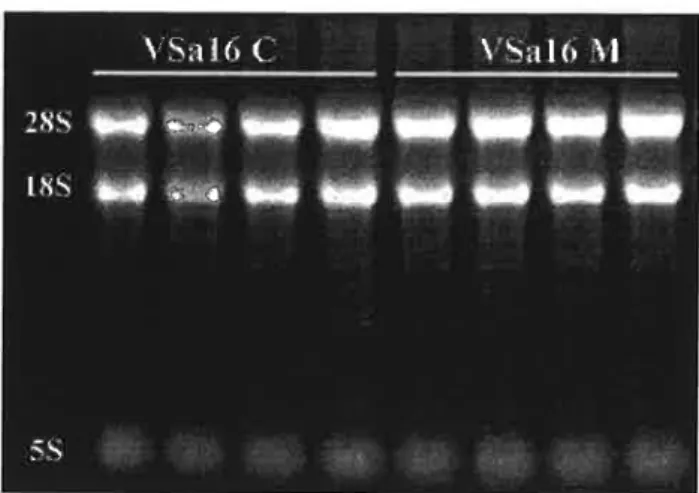
Total RNA from control and mineralized cells was extracted from 4×100-mm culture dishes and 10 ug of each preparation was size-separated in a 1% agarose formaldehyde gel (Fig. 4). Characteristic bands corresponding to 28S, l8S and 5 S ribosomal RNAS were clearly visible demonstrating the good quality of the RNA preparations.
Figure 4: Ten micrograms of total RNA prepared from VSa16 cells grown under control (C) and mineralizing (M) conditions (4 plates each) and separated on a formaldehyde gel.
III.2- Suppression subtractive hybridization (SSH) and differential screening
III.2.1- SSH
PolyA+ RNA Were purified from total RNA using a commercially available kit and 2 pg of either control or mineralized samples were reverse-transcribed to cDNA and subtracted according to the protocol provided by Clontech.
cDNAs up-regulated during mineralization (forward-subtraction) were then selectively amplified during the primary and secondary PCR reactions (Fig. 5). The result of the primary PCR was a smear of 0.2-2 kb, without distinct amplified bands whereas several bands were
III- RESULTS
observed after the secondary PCR. The pattem of amplification observed for the unsubtracted cDNA samples was clearly different from the subtracted one.
Êkh-
&2kh-Figure 5: Primary and secondary PCR amplifications of VSa16 forward-subtracted cDNAs. Lane M, 100-bp DNA ladder (Fermentas, Appendix V).
A second subtraction needed for the differential screening of forward-subtracted cDNAs (see section III.2.2- Differential Screening) was prepared in the reverse direction (reverse-subtraction). Differentially expressed cDNAs from control sample were selectively amplified during the primary and secondary PCR reactions and results similar to those obtained for the forward-subtraction were observed (Fig. 6).
2
kh-ÍL1
I-:l:-Figure 6: Primary and secondary PCR amplifications of VSa16 reverse-subtracted cDNAs. Lane M, 100-bp DNA ladder.
III- RESULTS
Forward-subtracted cDNAs were submitted to the Mirror Orientation Selection (MOS) technique, in order to reduce the background level as illustrated in Figure 7. Although, it was already possible to distinguish individual bands with SSH alone, the use of the MOS in addition to the SHH strongly increased their number and intensity.
3
kh-fi.2í
kb-Figure 7: PCR amplification of forward-subtracted cDNAs after SSH (lane 1) or after SSH + MOS (lane 2). Lane M, 100-bp DNA ladder.
III.2.2- Differential screening
The search for up-regulated genes during mineralization was accomplished by hybridizing forward library (blotted on 2 identical membranes) With forward or reverse probes. Clones exhibiting a stronger signal when hybridized with forward probes (by comparison with membranes hybridized with reverse probes) were considered as positive clones and further analyzed. A total number of 672 clones from single SSH and 960 clones from SSH+MOS were selected (Fig. 8).
When comparing both approaches (SSH versus SSH+MOS), SSH+MOS clearly allowed the identification of more clones.
III- RESULTS SSH SSH + MOS ` `` F '1-'n".¬ ~ Í *Ii Í.I› '”*"i iii Í* Q. .ii-._g zig.. *OI gn;
tn.
*'I~.
ú
Iljjj .-Iii..É
;1
li " Q Ú* e"I'`:I #1!' '_anti
¢ O ISnlIfi**' amiuliat
gxfiaoiin
.a;<‹'Í#'°=i*""Í`gucofiifii
._.|¡ _- '-Í |5 -I 1. ` *F ..+' g-_-'+9 :I i.
RV/
-| ' -ii p_.= I» if _ .ti lu _ . Q I. _- Q G '; tl-7 '-II ` .;_ E H. E .I *Í .. Ú'Figure 8: Screening of differentially expressed genes in a subtractive library prepared from VSa16 cells undergoing mineralization. DNA from 96 different bacterial clones obtained using SSH or SSH+MOS techniques were blotted onto nylon membranes and hybridized with the forward (FW) or reverse (RV) probes.
III.3- Identification of genes up-regulated during mineralization
More than 1600 positive bacterial clones were screened. From these, 194 were confirmed as differentially expressed (up-regulated) during mineralization. cDNA fragments corresponding to each clone were sequenced and identified by similarity search using Blast facilities at NCBI. A total number of 20 different genes representing different class of proteins were finally identified (Table II). These included proteins involved in cell metabolism and calcium binding, and proteins with no similarity with any' sequences present in public databases. Interestingly, 2 genes accounted for more than 84% of the positive differential clones. These genes encode proteins identified as S. aurata osteopontin-like protein (the most abundant with 68% of the positive differential clones) and S. aurata sniffer-like protein (16% of the positive differential clones). This observation suggests that these 2 genes are likely to be strongly up-regulated
Table II: Genes up-regulated during VSa16 cells mineralization
111-RESULTS
osteopontin-1i1<e protein (o1>-1i1<e)*
Ê
1331
2133
...-_. ._--- --.-..--- - ..-.--_-_--- _ - ---I» --- -_---Jr
--_ Cytochrome oxidase subunit I (COX I) 3 1437
''''''
OOiÊrànege1in'2°i¿ri{ë2)i
E É
1
O 2
;
P455
sniffer-1i1<e protein (sN1F)
'
312 g
515
L_....-....---__ _ .--..--- -_-_ _ ._ -.--- _
_.-_-_._---l--__---¬---i---í¬-¬.---Unknown 1 (Un1<1)
4
_
519
1
Moein-1i1<e protein (MUC)
2
Í
576 ,
Ubiquitin-conjugating enzyme E2 (UBQE2) _ 2 ¿ 168
Un1orown3(Un1<3)
1
2
f
235
Unlorowns (L'n1<5)
í
1
É
I
_- .. ... _. _ ---|¿ ¡
_.-_-_--- _____=--- -- _ __ ________--- 1 ___;-z ---- -1 ¬- l--- -_ ....---|.-___--n---
--_ Calciurn-binding protein (S100-like) 1 :_ 648 |
Cytochrome c oxidase subunit Vlb (COX Vlb) 1 f 303
_ Putative fish transposase (fishTRP) 1 Ê! 458
Unknown 4 (Un1<4)
1
y
389
606 263
15-eotin(13AcT)
1
2
444 _
Glucose 6 phosphate 1 dehydrogenase (G6PD) :E 1 g 399 '
I -.¡_&
Lnlorown 7 (Ln1<7)
y
1
1
_...í...-.---.--- - __ __---._--- _ ___...-...- -_'_____,,______-- _______-E915
Uniorown 8 (Unks)
1
1
266
|Ribosomal protein L23a (RPL23 a) 1 I' 245
1 Three different fragments corresponding to different regions of OP-like cDNA were identified (OP-likel, 2 clones; OP-like2, 8 clones; OP-like3, 123 clones).
2 Two different fragments corresponding to different regions of SNIF cDNA were ident1fied (SNIF 1, 20 clones; SNIF2, ll clones).
III- RESULTS Ten up-regulated genes were identified using the SSH technique alone (MUC, OP-likel, UBQE2, Unk4, G6PD, Unl<3, S100-like, COX Vlb, Unk6 and Unk7), 6 using the SSH/l\/IOS technique (Transg2, BACT, Unkl, Unk2, fishTRP and RPL23a) and 6 were common to both techniques (SNIF, COX I, like2, like3, Unk5 and Unk8). It should be noted that OP-like3 was represented by 83 clones (43% of total clones).
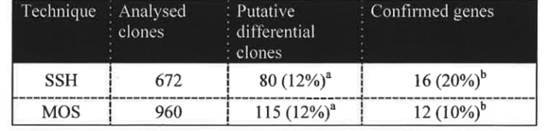
From a total of 672 clones from SSH and 960 clones from MOS forward libraries, the percentage of confirmed genes was twice as much in the SSH than in the MOS technique
(reble 1).
Table I: Comparison of enriched subtractive cDNA libraries generated either by SSH or SHH+ MOS techniques.
T
_
so (12%)fl
_
16 (2o%)*°
I IIE/J|¡cz:E1I II -1111'-Inllwuliillr I I I II xo;oxCD|l\J II'I I I ir-I-_'-_IIt_tÇi-3. 1' IIIIIII I I I III ml I I -L-_-rx-|Il-__-._-_ I III IIIII III I III tz;-II IIII I I I MI..i
Mos
115 (12%)
12 (10%)
3 Percentage of all analyserl clones b Percentage of putative genes
III.4- Cloning and characterization of S. aurata S100-like and OP-like full
length cDNAs
III.4.1- Characterization of S100-like full length cDNA
Analysis of the 648-bp S100-like clone obtained previously using bioinformatic tools identified a complete coding sequence (cds) with an ATG initiation codon at position 43 in frame with a stop codon at position 302 (Fig. 9). This cds encodes a 86-aa protein that exhibited sequence identity (from 35% to 40%) to numerous members of the S100 calciurn-binding protein family (including Ictacalcin, calbindin D9k, S100A 1-10).
É 45 šií 23€ šâí 226 273; BES 351 ëãifi 451 49€ ãêš. 535 631 *-112 ACA T Hi? H *Êâä E ACA T GAC Ê GSA FL TEÂE ã".1`€ TTP. CTG TT? ETE AÀG .FJl.|'-I. TTT GÃT tt 1".iP.'E ii ETE L TCC S GGG G GBC J-'L CPA. ãškflf CJLG AGA TGT TST ILRC ILFIJIL TTÊ? EXC šz GQ? A T5? 5 FERA ší Ê? «ETE-Í. CTE' RFLP. Tflš ZEAT TÀC ÊTG *Ski ãlírâ AAG fiíí' Ê -GGA E GAE E Hà-É H SST .Q JLC T T TÊT THE LET AAA TQT TRT FIAT 11.31.11. ECT AÊA Ê Prkä E CTG Ê.. IÍ.'¬1`-U5. ã QTT 'äàf GTE-Ê? 'IGT' ÊTT ÃCÀ .IICT TEE-ÃTE TAA GET ÊÂÊÊ EQR Pr äí* Ê Q TC ir TEE Y.. GTE' gv .PET É E CT? Tšâ Tišãt TTE ETT AQ? .FL'š.`fG TG? AGE ATG H SGA Q CGC E GAC E .ILGT S TEE C .FLC T AAC REA Té'-EA E-ELG GI-'LT TCA T?? GGJL G GAC D GQ? Ê. .ãäš K Ê?? F 1£iil`1.'.'ã` âšsã TEF; TTE' .F-LGA RGE Ê?? TFLT TCPI. em L raca r me E Tire 5° 431.4: E. rsrsrs c TG? ÊTQ 214321' ERC TG? *Etlâä T Tfi TEC- FIJLC CTC ELGG L E âšrfi IQ? K S 'ITI' CE? F' F ETE? ILFLE-É ãl Sã-E E'I"š B- F GFLII. TAL E sua ÊIÊ. CÊLR ÉÃT S?? GAP. G‹G'£“ 'PET ííäü i~'t..F-III. FIT? .F-LTG CTC 'ITI' ACA 'ETT ÀCA T TTG .L i.`z.I-'LG E TC42 E 51€ 1; ÊAT SIT GET TTA EÀT ÃAG Gflà QEÊC GTS V AAC ëë GE G A CÊ13' ÀCT T CGI: GET G«1I.".FL CAA TCÀ GTE-`› 'IGJL AAA Afãíâ TTÉ F ÊAG K É/IÊÊL É SAT "FIT ÀS? ããã GSE TA! *ÇÍÀG 'CÉTA TGP. RAP. ÂTG M .ILAG Eš Àäiš K TCC E ÊIAC H GIGA Ei GRE .REA GTB. GTI ATQ EIÊT IL TG ILFIA III- RESULTS
Figure 9: Nucleotide and deduced amino acid sequences of S. aurata S100-like cDNA. Nucleotides are numbered on the left and amino acid on the right. ATG start codon and TAA stop codon are indicated in bold. Sequence has been deposited in GenBank but an accession number has not been attributed yet.
III.4.1.1- Characterization of S100-like putative domains
In order to identify putative features of S100-like protein, sequence was analyzed using PROSITE, SignalP and NetPhos Intemet tools. No signal peptide was found indicating that S100-like protein is not a secreted protein but an EF-hand domain responsible for calcium binding and putative serine and threonine phosphorylation sites were identified (Fig. 10). Analysis of protein sequence did not identified any proteolytic cleavage sites therefore the sequence presented in Figure 10 is likely to correspond to mature protein.
III- RESULTS F
1 __¡;
“G
QR
°T°"
/fíí-í 643 bp
" .tt-_¡.\u uaQN _.-If /E 'Í É Í \ Í ._ ;. -_-,'.¡-'_-:_ II. ; ._ z z ¿-z' 35 ai' `."1'z IL' ' _ 1 _-.?`¬' - EF-Hand domain OQ Putative Ser, Thr phosphorylation sitesFigure 10: Structure of SaS100-like cDNA and protein.
III.4.2- Cloning and characterization of osteopontin-like full length cDNA
Non-overlapping clones OP-likel, OP-like2 and OP-like3, representing the same gene identified as S. aurata osteopontin-like protein, were used to design specific primers (Table III) in order to clone OP-like full length cDNA.
Table III- Primers used to clone OP-like full length cDNA.
SaOPl-F01 É 5°- CGCTCCAGCCGCTGAACTCCTGAAGC -3 ” SaOP2-F02 ¡ 5 '- CCACCCCTCAGCCCATCGACCCTACC -3°
""""sooP2;Ro4ii
3°- GGTAGGGTCGÃTGõäETõÂ`öëZšš§Të2šÍš`*```````T
A I So0P3¿F0IšÍMI"I05*ÍGÕÉÕëÕÃÕÕf¿§ÃëÃÍÊ`ÕÃÕÕÀCT‹§ÃÕÂ13*""""i
The complete OP-like cDNA (GenBank accession number AY651247) was amplified by PCR and RACE-PCR, cloned in pGEM-T Easy and subsequently sequenced. Full length cDNA is 2138-bp long with an ATG initiation codon at position 130 in frame with a stop codon at position 1253 (Fig. 11).
1 23€ 182 226 ZTE 31€ 363 áüõ fl51 äãã 542 535 fifil ãíã 722 756 êil üãfi 991 846 ÉQE 1636 ÍQEÊ 112€ ilíš 1215 íiëi 130€ 135â 1396 iáúi šáãš lãšä 15?ä Íá2Ê ÊÉGS 171ä 1156 lãüí iüüã iügí iflšã ÊQBÍ 262€ ZQÊÊ 2116 GTE V CC-G E* QTÊ IF GTI EF .FL É-CG H. .PLGII S AKG K =Gñ.€.~`$ E GGÉ 43 *STS V ÉCT F' GG-P1. G 3§.F¡.Ê'f. K GIT V' GHG EI 'FÉT É flñéíã Q CGÉ: E. -ECC A *..`.'-E-AG E .ILEC T ECC .FL *GAC li* .Pai T ACT .F|..I1..ñ. CCE. .ñ.'I".l' EAT .RAT .HJLT "Z`.Fâ'I' ACI' GTF1. CÍIZG .Fnñäíi =5.i|t.<3 zP¡.'1'T 'IGT GLT .?t°.`1'iS %`.1".l'.P|. 131.51 íññ GIST F1. Gfñ ã. .Fa.G-H. R QC! E' G-C1' FL 1131.5 *ãë frftza s GIILL' Bt ERG E Gãlt.-'Zi E .¡t."i.'Z.ñ. 1' ñfiãfií 1' EGEG IG ACC E GGC 'è äfšëi U %.`â‹'ÃI E 'G-GE BG CH.-G Q* .Fâäfi 1' ããktfr É A455. E R$151. 'š ã.fi'šI E ñfifã 'E GÍHÉ 551.55. ÊAI ir?? flëíl' ñ..i'Lñ. EEE FLSG *Eñ.i'.' ARE. ãfíñ "IGG ãfiñ ãfã. *HGB .P|.=«G.FL £'?‹.=Gz CÊT Ch? ñññ 511 E AGJ1. E ñññ E 'Chã Q fififl E EEF: 3:. E ñëší E Fl.>É5t`E S TÊÚ S ›€šiIt.&.“'.I-B ñífl Z TPLÉ3 Y TAC 3' ãffih. Í Ehfiš E III: I E375 E-r 458€ .Vi .FLQCÍ X QFLG E Chã? Ê QCG il. EEE F1. HÊZÍT Il' TÉ5 §*'Câ'š TUÊÍ LT? TIG =šÍ.°›*Cí.Fz ETE .F:T*5= CTG GG? TE? Rã? RGE Gñššl .'1`T.?s ãrfiif-.iá TEC HCL. .ÊTI .H..F|..i'|. =§EÍI`T 14" HIS K QCÊL LE* ECC .FL GGI: G GCI. .Fa ACF: T £Fu'-'L Ez tãšhñ. E É GCH. .F1 GW V ECC E* Tñš: 71' Gášii. Q HLEÊ E. CTG L-=IšF¡.áI E? `I*{P§Z` Ê FLGT E .i\i.i`73I É 'E.'‹ñ.¿3 E .PLUG T 32; GAC Cê GFJI. *Cñ'.=E .FLÚT °C'.I.'=C TTÊ. TTT TTE ATG É .Pei-Z,.F|. .?:..'I'fi ëãäiã. ET I EGCI' ATG .Pt.E.'..I|. €§'.I'ã. ëG`.l'ê`-'|. .H.i'Lñ. ITC' F GYT V 55,1%. ii. ÊICT 5.? ICE S ECT P GC T Fi. GFLC Tí .FLÇC 1' ?â.CC 'E' CCC P .FLEFE I .FLGT B F|.Â:¿G K .ä..Fs.:G K. Iílã. 35 'GFLG IE. R$593 É CIC E.: Shi E =°5>E.f.ñ 35. TCA. S Êí F? Plí-›T 5 .FLIG E .F1.Gr`T ACT GPÍZF1. ñ..Pt.'T Gññ ÃET 531.1: .Fâ.PLFt. FLCC .iâäiãí Cíã. EFLH. CCE .RIA TIG fišññ. >1.`.`.Fi.'l' €.'š.T EEE .ñ..Ff.Ft. G If \“f SCE Fr. >ÇCfG ii' Gíâ Pá. 531.5 É ='$.F1..ñ. E 1`£C S fi Êiñã É ãflš' Fi QTG V GRC-É Gñšjl E GTCC V .FLF|.=Gé K '51 'E5 L. Gla. G E TÇG S flflfi. P E4595' S .FL*G‹€Í Fr Gñã E G'-GG šè *SEC É 'i`.`§°+ZF:. š' flG.F1.@ SCE ~*G~G‹‹G-535 ñâíáfiâ íšfiã. *CEI CÍYG T?? Tñã-=-5% ACE TE I CTI TTQ .ñífi .Ff.=Jã. GT? TE E .F|..F|.h G-EFI. 1GC>G€ AAC ã¡.'§"ãÍ` 155.43 GGFL P..i°t=.F:. 415 .:`1ã.C Êflñ. íl' CCG T5? `.~I`€?€Í“ ÉCÉ *Qi CTT =§I-IE Chã ECT ›G€C.ä. .F:.F¡.*C› .ññfã ñtäñ
ET-E I.: íšãtl D WÕCPL É Eüñ 3:. ‹í3..¡:.㧠E ÍÊÉT É-'L 3'-'1.â".I‹§Z T 5116-°¿ã'›'‹.. E G-PHS E ë3`I'T 'V 횧í.'3'l` F2. Jsflñ. T THE Y ÚCT E* .ETI-Tt BS 131.5 Q GAC Cê Tüà Ê M E GFIÉS E RK T GIZHT Ú GC'-C Pt. Tflñ E ETE. 'U' TEE H eo 'CCZÊ ATT .BAT Gflñ. *SCA TEE *C5 IC .I-'t_F:.F|. IGE. `E.h{`.' .ñ..`.I"I' ELE CTA .ilãã -FLCJ1.. BG-T1' âL'-TH. .I|.F|..ñ. 41.21%? L ?t.›í'.'z‹"E` E 5-‹..'.Í=ã Ft. IZYIS E' É .IUGC 5 -ãflët. E Iflñ S EFLE I? GAE E. ÊC C. E ããh g Ghü E Aki; K fi`!`ET V ECC .E *EI5 1.1' PLÇCÃ É 456€ ü G.Ft..H. E ?1.GÇr S GCC .R fiñëé E. 43€ T A GñT É -«a'.5'I*§; 15 PLUG ñ.G5'EF š.ã.C ELE 'I ññü-M .F-JCÍ1' *CCC š.G›.`ñ. $Z.FI.€ E-'LÊ T Êhñ 31. 'IT GU? GGI URE T5! .PLPLFL HH.
Figure 11: Nucleotide and deduced amino acid
TTI" F TE?? E 5 ICÍ ãt W-GÉ V Tí?-'.Í¡ E =š§"š`.F¡ 'EF EÊC E) .FREI 5' '%.'šFL`§ ä ñššífi 5 ñëišl-'E ñlflñ E Tfiã E ÉEPCJ ä' TRC Y Tñfl Y Úãiíí S Ft Tãf. E <í'zš.i'-lâãš E Tëflfl S ñfiãfl S ããñã E GRC E GÀG E .FLTÊE I G T5 Irã 'lí 3.43 G =C.?|.Et. TÍ1' äšz TG I üññ. G CE. TT É ACA 'FG E GT É .i'-'1=.'1'ñ. ñ.F1. 1? ññ. 'E ñšíñ TC C CE C, .i|u?'|. 1' GCCZ 3.. GAE E ñšâáš K 'EÍFLG Q ELG E š.`=ã§.F1. E .?â.€.'t'z T GIILT [ii 31.5'-É E FLC' E T .RCC T GÊÍÍ G ATC I. TAE Y GRC Ú LHE K ãflü T TCÉT É Fu€.'=EC 5 ÊLGC É Gññ. E .Pzãš-É Si EEE S {'.P‹.'G *ü nar T GTE G.I|:C .HIÍT TII HAS A545 Qãfãlí LÀG .ETT TIT *CIT 'ÊZCÁÊ ¬?|.T'CÍ Gi TPLT .Fi.'I`5 Tãñ. ñ.*§=§§â Z' E. GC E .FL A3: K üñü Q F1. ET se *ETC V “GCC Fi. š.3.ñ.T zé' šãfi E. ÉÍI Cí L EQT E' ízfizü H. EEE I? ãñã Ef. FL `!`šš M GC-'I' H. ÉIÃZTI P Ch-'3 Q .Fuã 1' É FLCC 'E GAC H CILG Q Gñ.C H G-EG ñ G E-L' Í-'L 3`€~'I' ARG F1. IC €-'EG IEC EIT 1% 'FGCÊ TC T ñtäñ Eãi ÊIFG Siãšíñ. I.ñ.C Lfiñ 'ETC TCP; TÍG Zlññ .err 1.: Tex" s me Q HTE' I Tfiñ E CA. G É TE 'ã' 'S E1 FUIÍZGÍ E .kfií 'E *CBG Q. ÁEGÉ-5 Tñfi Y "1"v°.'.'.`3=CÍ 3 .hflñ T ÊTÉ 'ie =ã`3.Ã.'í.'š. E %€í EÍI ñíšü S CC-E ? fiëlfl .El .ã.á?fi $ 'l`=CPr. -5 Êšíläš ã. RFLP; ZÉ. ?`G'!¬I *CCÍZ «UAE TG?-f. =G=Ê.Í‹€§ *S11 'IQ Êâãfl Cíízfi Cãã TCA E E E A Eä ATI Cí-I .ÃFLÍE T?? T?? ñäñ *€Í$'°-C, ãrãfíš AGE. `.I.'ñ.Ft. ÊLÊT Cññ CCE fifüiff GCI fl.-šññ CTE E-TE füüñ. ?t.l1.É `1'@.'5`§ 'Eli A423. 11% *ETC L GRE E ECF.. Ê. =i°5.'1'ñ. 'EF 'ÍS.F|.T É tcg 3 G'TC V GRI G* TÊC 3 ÉZÍÍIT E. Úílfi P' GPU: É *GTE V ".`l'?¡.íÍ Y >§%€z`. E *€Í.Pf.G Q GTÊ 1! 'C-ILG Q 5-'LQÊ T Eli E .IILG-É 55 ñiãfl E =š`_š‹ÀT G GIII.. G THH. _-ti se 1» EEE. CÊT 435€ 1'.P1.C 'ÊTF'-1. =šZ*£Í×$ ñ'.I.'*~`¬3 ñãäfiš 'CZ TC .F¡.››.z›*`"Íl' I TG .Ft.<C;F1. ñ.`I"1' Ífifl GET fiñfl' GÍÍFL ÊCT TÉT M 'IGE 5. ELE E GCI; F; GEL. .P-L GJLFI. E TÊT 3 .F1.ã.4'=I`-lã *G.Fi.`Fe E iãñit. Ez Gfëš 'U' R EQ. Z H. GC E €v.F.\.G E ELG E G-GC G *EEE V GLHG EL 'GL'-'Z ü ñfifl E 55.3: E. ã.*E1z".';‹ 'I =\G.P|.\G Ez -E-.HE E ÊC T P .FLCC irc rafa 165. err IA? -err; nu. .HG-E car can nxr âââ zrà IG: ›i:»Gz"r ara. cita GTI; 21'1-€=`u'-'sí .1ã.G~ ññã at: -Q 54:. R ara 1; G rs v ez 45:. 1.: Q esa.: Q .2ri:;4:. 5 mts. E Tita 5 Arc 1: taste n IAÇ Y Gm. E -crer. 5. FLF¡.¬-.T N *flñfiílš II-E ñfiëfi E STÇ ir D GPLG E .ñ.=šÊ§Í E ÊAE E .Pt.'»¿'51"š S Gñü El ÊTQ AÍCR RT 'E GTÉ 'FIA GEL *CTA T fã GGI? GTE TF; É T5-LF1. ÂILGC CTG GER TELE 'ITQ PLT 5 IÉXS III- RESULTS É 1? 32 ã? fiã T? QE IQT 1â2 13? 152 16? lâã 1?? 212 22? 242 25? 272 23? EGZ 31? 332 34? 352 3?á
sequences of Spams aurata osteopontin-like cDNA. Nucleotides are numbered on the left and amino acid on the right. ATG initiation codon and TAA stop codon are indicated in bold. Sequence has been deposited in GenBank with the accession number AY651247).
III- RESULTS S. aurata OP-like cDNA encodes a 374-aa protein that exhibits ca. 40% identity with other OP-like obtained from Oncorhynchus mykiss (rainbow trout), Salvelínus fontinalis (brook trout), Danio rerío (zebrafish), and Ictalurus punctantus (channel catfish) (Table. IV).
Table IV- Percentage of sequence identity among OP-like proteins. S. aurata (SaOP-like), O. mykiss (OmOP-likel and OmOP-1ike2), S. fontinalis (SfOP-like), D. rerio (DrOP-like) and I. punctantus (IpOP-like). Identity values were calculated aligned sequences using The Sequence
Manipulation Suite program.
!SaOP-like SfOP-like DrOP_-like IpOP-like OmOP-likel ppOn1OP-1ike2
saoP-like É
¬ .'. .I. -SfOP-like ' 45.2 '- 'f DrOP-like IpOP-like OmOP-likel OmOP-like2 32.4 21.2 44.1 42.4 37 '_ |.;II-I.'r 23.6 20.8 ' 75.1 36.2 25.8 . . ' 1-88.3 35.1 24.2 73.6 .III.4.2.2- Characterization of SaOP-like protein domains
Sequence of SaOP-like protein Was analyzed using PROSITE, SignalP and NetPhos Intemet tools. A 16-aa long signal peptide located in the N-terminus of the protein was identified demonstrating that OP-like is a secreted protein. A RGD domain demonstrating that maybe this protein is involved in cell-adhesion, a serine-rich domain and various putative serine, threonine and tyrosine phosphorylation sites Where identified suggesting OP-like involvement in cell proliferation (Fig. 12).