Universidade de Lisboa
Faculdade de Ciências
Departamento de Biologia Vegetal
Dissecting the Pluripotent state in Embryonic
Stem Cells
Willianne Kaline Alves da Silva
Dissertação para obtenção do Grau de Mestre em
Biologia Molecular e Genética
Setembro de 2013
ii
Universidade de Lisboa
Faculdade de Ciências
Departamento de Biologia Vegetal
Dissecting the Pluripotent state in Embryonic
Stem Cells
Willianne Kaline Alves da Silva
Dissertação para obtenção do Grau de Mestre em
Biologia Molecular e Genética
Dissertação orientada por:
Domingos Henrique, Investigador Principal na Unidade de Biologia do Desenvolvimento,
Instituto de Medicina Molecular
Gabriela Rodrigues, Professora auxiliar da Faculdade de Ciências da Universidade de
Lisboa
Setembro de 2013
AGRADECIMENTOS
À DEUS por sempre ter preenchido meu vazio por ser minha fortaleza e refúgio, mesmo quando achava que não conseguia seguir em frente. Obrigada por me me presentear com sorrisos a cada lágrima derramada.
À Paulo Fonseca, minha família, razão do meu caminhar, amor e entregas sem limites... Amor que me move dia após dias sem descanso, sendo o pilar que me sustenta. Obrigada por sempre me confortar e sempre me ajudar em tudo que for preciso.
À Domingos Henrique, chefe e orientador. Obrigada por ter aceitado o desafio de me aceitar em seu laboratório e confiar em meu trabalho, mesmo quando ninguém parecia acreditar. Obrigada por ter me motivado a continuar. À você minha eterna gratidão, admiração e respeito.
À professora Gabriela Rodrigues por ter me aceitado como sua aluna.
À Elsa Abranches, uma pessoa com quem aprendi a conviver e a respeitar. Alguém que me lapidou e me ensinou o rigor científico e com seus conselhos preciosos. Obrigada por tudo.
À Ana Guedes, por dividir o conhecimento, a amizade e os conselhos. Obrigada pela paciência e me ajudar prontamente em tudo que precisei, sendo amiga, orientadora, diva e divã. À você mais do que agradecimentos
À Ana Rute por ser uma pessoa tão presente. Obrigada por ser a minha razão quando estava sem direção. Alguém que vou levar comigo sempre.
À Anna Pezzarossa por estar sempre disponível. À Sanja pela simpatia e confiança.
À Danielly Pinheiro, pela eterna amizade. A prova viva que tempo e distância não são capazes de mudar um sentimento. Obrigada por mesmo estando à distância, sempre se mostrou disponível para tudo. Amiga de sempre e para sempre.
À Tânia Santos, colega e única amiga de curso. Obrigada pelas gargalhada, carinho e disponibilidade.
À todos que compõe a equipe UBD e À Unidade de Ctometria de Fluxo do IMM(Ana, Maria e Sofia), que estiveram sempre disponíveis.
ABSTRACT
Embryonic Stem Cells(ESC) are undifferentiated cells derived from the blastocyst and are able to self-renew indefinitely in defined minimal conditions, in the presence of LIF, BMP4 and MEK/GSK3 inhibitors. Pluripotency is an ESC property regulated by a complex gene regulatory network (GRN) that have, at its core, the pluripotency genes Nanog, Oct4 and Sox2. While OCT4 and SOX2 are homogenously expressed in ESCs, NANOG expression fluctuates between states of high NANOG and low NANOG expression. In this way, NANOG is considered a master transcriptional regulator of self-renewal and pluripotency in ES cells, regulating the propensity of ESCs to differentiate. One question poorly understood is how this heterogeneity define a functional subpopulation. To address this question, one needs to be able to purify specific subpopulation of ESCs using cell surface markers. Starting with a mouse ESC line (Nd) containing a fluorescent Nanog:VNP reporter, we tested the ability of various surface markers to sort sub-populations of ESCs, in combination with Nanog:VNP, after culture in different cell culture media (GMEM/LIF, 2i/Lif and BMP4/LIF), using FACS analysis. Ten different antibodies, known to label different functional sub-populations of pluripotent cells, were tested, together with the Nanog:VNP marker. We were able to identify 6 antibodies that, together with the Nanog:VNP reporter, can label specific subsets of ESCs: Pecam, Ephb4, SSEA1 and CD81 as ESC markers, CD40 as Trophoblast stem cell marker, and Notch3 as Epiblast marker. This work shall allow the characterization of various transition states in cultures of ESCs and is a first step in studying the functional heterogeneity of ESCs.
KEY-WORDS: EMBRYONIC STEM CELLS, NANOG, PLURIPOTENCY, SUBPOPULATION AND ANTIBODIES.
RESUMO
Durante o desenvolvimento embrionário, o zigoto passa por um processo de clivagem produzindo blastômeros que se organizam para formar uma massa compacta chamada mórula e blastocisto. Este é estruturalmente composto por um espaço chamado cavidade blastocística (ICM) e por uma camada de células externas chamada trofoblasto (TE). À medida que o zigoto se vai dividindo, as células restringem o seu potencial de desenvolvimento pela especificação de linhagem e segregação espacial. O evento prioritário que contribui para o desenvolvimento do zigoto em mamíferos consiste na especificação de tecidos extra e intra-embrionários para a preparação para a implantação. Com a implantação do blastocisto, uma segunda mudança ocorre nas células da cavidade blastocística que vão originar o Epiblasto e o Endoderma primitivo.
As células estaminais são células indiferenciadas que têm a habilidade de auto-renovação ilimitada e diferenciação em diversas linhagens multicelulares, podendo ser encontradas em diferentes tecidos. Um tipo particular de células estaminais, são as células estaminais embrionárias que tem origem a partir da cavidade blastocística e que, quando mantidas “in vitro” sob condições de cultura específicas, têm a capacidade de manter a sua “estaminalidade”. Esta propriedade das células estaminais embrionárias é mantida por uma rede genética complexa, cujo núcleo central pode ser reduzido a três genes que codificam os fatores de transcrição: NANOG, OCT4 e SOX2. Diversos estudos têm sido desenvolvidos para controlar este balanço entre os vários sinais moleculares associados à pluripotência responsáveis pela ativação ou repressão de genes que culminam num destino para a célula.
No entanto, os mecanismos que regulam a maneira como estas células escolhem o seu destino ou a maneira como entram para a diferenciação continuam sem ser desvendados. Diferentes estudos, descrevem moléculas associadas a diferentes vias de sinalização que pode mimetizar um ambiente in vivo. O LIF é um exemplo deste tipo de moléculas uma vez que, através da via JACK- STAT3, BMP e inibidores de GSK3,são capazes de manter a pluripotências das células estaminais.
Logo, o desafio consiste na compreensão da interação concertada destes fatores de transcrição que regulam a pluripotência e as vias de sinalização a fim de analisar e manipular a interação entre os sinais. O NANOG distingue-se do OCT4 e SOX2 pelo nível de expressão heterogênea em células estaminais que flutua entre uma expressão LOW NANOG e uma expressão HIGH NANOG. Para além disso, estudos
sugerem que a expressão heterogênea do NANOG, observada tanto in vitro como in vivo, deve influenciar a sua recetividade aos diferentes estímulos de diferenciação sendo, por isso,considerado o modulador central da pluripotência. Dado que a flutuação da expressão do NANOG tem um papel importante na escolha de um destino para a célula e que estes níveis podem estar associados a sub-populações de células estaminais, torna-se necessário identificar estas subpopulações. Para atingir este objetivo, neste estudo foram utilizados diversos marcadores específicos de superfície celular descritos por Rungg-Gunn e colaboradores para identificar, isolar e caracterizar células estaminais.
Neste trabalho foi utilizada uma linha celular que contem um repórter fluorescente de NANOG (linha celular Nd), previamente estabelecida e validada no laboratório e uma linha celular usada como controlo: E14tga2. Estas linhas celulares foram mantidas através de passagens em diferentes condições de culturas: GMEM/LIF, BMP4/LIF e 2i/LIFem condições de pluripotência. Para analisar a capacidade destas células em se diferenciar, foram mantidas, apenas, em GEMEM. Estas linhas foram validadas em termos de capacidade de auto-renovação e pluripotência tendo-se observado que as suas características não diferiam daquelas da linha celular controlo. Adicionalmente, a expressão do NANOG:VNP foi monitorizada por FACS para avaliar a capacidade destas células para entrar em diferenciação, sugerindo que o estado das células estava em concordância com o meio de cultura utilizado.
Neste trabalho, foram também utilizados 10 anticorpos descritos para células estaminais embrionárias, epiblasto ou células do trofoblasto em diferentes meios de cultura. Destes, em seis obteve-se êxito para caracterizar subpopulações de células estaminais. Para marcar ESC foram utilizados Pecam, EphB4, SSEA1 e CD81; para marcar células do trofoblasto foi utilizado CD40 e para marcar Epiblasto foi utilizado Notch3. As células foram analisadas pela expressão de cada marcador específico de superfície por FACS e, em seguida, utilizando a linha celular Nd repórter expressando o NANOG, foram separadas em quatro supopulações associadas com cada marcador específico de superfície: VNP+, Single+, Double + e Double negative em cada condição.
Os resultados obtidos mostram que alguns dos marcadores usados foram mais específicos para identificar a subpopulação Low NANOG que é um estado transitório para a diferenciação. Entre os anticorpos que foram descritos, os mais promissores para identificar diferentes linhagens de células estaminais foram o Pecam (ESC), CD40 (células do trofoblasto) e Notch3 (Epiblasto). Estas observações sugerem que o
comportamento dinâmico do repórter NANOG:VNP associado com marcadores específicos para linhagens possibilita investigar a regulação da pluripotência e identificar os estágios transientes que co-existem numa população de células estaminais.
PALAVRAS CHAVE: CÉLULAS ESTAMINAIS EMBRIONÁRIAS, NANOG, PLURIPOTÊNCIA, SUBPOPULAÇÕES E ANTICORPOS.
TABLE OF CONTENTS
CHAPTER 1:...1
INTRODUCTION...1
1.1 EARLY EMBRYONIC DEVELOPMENT ... 1
1.2 STEM CELLS ... 2
1.3 EMBRYONIC STEM CELLS ... 2
1.4:GENETIC AND MOLECULAR SIGNALS ASSOCIATED WITH PLURIPOTENCY... 4
1.4.1 SIGNALLING MOLECULES... 5
1.4.1.1 LIF... 5
1.4.1.3 GSK3 ... 6
1.4.2 PLURIPOTENCY GENE REGULATORY NETWORK... 7
1.4.2.1 Lineage specific markers... 8
GOAL...11
MATERIALSANDMETHODS...1
2.1 MATERIALS AND REAGENTS...12
2.1.1 REAGENTS ...12 2.1.2 CELL LINES...12 2.2 METHODS ...12 2.2.1 ESC CULTURE ...12 2.2.1.1 Expansion of ESCs ...12 2.2.1.2 Differentiation of ESCs ...13
2.2.2 PROTEIN EXPRESSION ANALYSIS...13
2.2.2.1 Fluorescence Activated Cell Sorting...13 2.2.2.1.1 Nanog:VNP analysis ...13 2.2.2.1.2 Cell surface protein expression analysis...13 2.2.3 DNA ANALYSIS...14 2.2.3.1 PCR for Mycoplasma detection...14 CHAPTER 3:...13
3.1 CARACTERIZATION OF EMBRYONIC STEM CELL CULTURES ...15
3.1.1 SELF‐RENEWAL POTENCIAL ...15
3.1.2 NANOG:VNP EXPRESSION ...16
3.1.3 OPTIMIZATION OF THE DETECTION OF SURFACE PROTEINS...18
3.1.3.1 ESCs markers: Ephb4, CD81,Ssea1 and Pecam1. ...18
3.1.3.2 TS cells marker: CD40...19
3.1.3.2 Epiblast cells marker: Notch3 ...20
3.2 CARACTERIZATION OF EMBRYONIC STEM CELLS SUBPOPULATION...21
3.2.1 ESCS MARKERS: EPHB4, CD81, SSEA1 AND PECAM1...21
3.2.2 TS CELLS MARKER: CD40 ...24
3.2.3 EPIBLAST CELLS MARKER: NOTCH3 ...24
3.2.4 PRELIMINARY ANALISYS OF NANOG, PECAM AND NOTCH3...26
5.REFERENCE LIST...31
6.1 SUPPLEMENTARY MATERIALS...A
6.1.1SOLUTIONS – ESC CULTURE...A 6.2 SUPPLEMENTARY METHODS...B
6.2 .1 FLOW CYTOMETRY DATA ANALYSIS...B 6.3 SUPPLEMENTARY RESULTS...B
6.3.1 LIF ACTIVITY TEST...B
6.2.3 ...D STATISTIC TESTS DETECTION OF SURFACE MARKERS...D
LIST OF TABLES
TABLE 1:LIST OF RELEVANT REAGENTS USED IN THE EXPERIMENTS DESCRIBED IN THIS THESIS. ...A
TABLE 2:PANEL OF DIFFERENTS ANTIBODIES AGAINST ESC SURFACE MARKERS, USED IN THE
EXPERIMENTS DESCRIBED IN THIS THESIS...A
TABLE 3STATISTIC TESTS DETECTION OF SURFACE MARKERS. ...D
TABLE 4STATISTIC TESTS CHARACTERIZATION OF EMBRYONIC STEM CELLS SUB-POPULATIONS (ESC MARKERS)....E
TABLE 5:STATISTIC TESTS CHARACTERIZATION OF EMBRYONIC STEM CELLS SUB-POPULATIONS(TS AND EPIBLAST MARKERS).... F
LIST OF FIGURES
FIGURE 1SCHEMATIC VIEW OF EARLY MOUSE EMBRYONIC DEVELOPMENT... 2
FIGURE 2COMBINATION OF SIGNALLING PATHWAY MAINTAINING MOUSE ESCS PLURIPOTENCY... 5
FIGURE 3:ND ESCS MORPHOLOGY IN DIFFERENT CELL CULTURE MEDIA....15
FIGURE 4SELF-RENEWAL CAPACITY OF ND AND E14TG2ATG2A CELL LINES –VIABILITY DATA.A).16
FIGURE 5:SELF-RENEWAL CAPACITY OF ND AND E14TG2ATG2A CELL LINES –FOLD INCREASE DATA.
...16
FIGURE 6EXPRESSION OF NANOG:VNP IN ND AND E14TG2ATG2A ESC, GROWN IN DIFFERENT
MEDIA.....17
FIGURE 7EXPRESSION OF SEVERAL SURFACE PROTEINS IN ND AND E14TG2ATG2A CELLS GROWN IN
DIFFERENT CULTURE MEDIA...21
FIGURE 8:CO-EXPRESSION OF NANOG:VNP WITH SEVERAL SURFACE PROTEINS IN ND CELLS GROWN IN DIFFERENT CULTURE MEDIA....25
FIGURE 9CO-EXPRESSION OF NANOG:VNP WITH SEVERAL SURFACE PROTEINS IN ND CELLS IN
DIFFERENT CULTURE MEDIA.ESCS ...27
SUPLEMENTARY FIGURES
FIG.S1:REPRESENTATIVE FLOW CYTOMETRY DATA ILLUSTRATING THE GATING STRATEGY FOR FACS
.... B FIG.S2:LIF TEST -CLONAL DENSITY.....C FIG.S3:SECONDARY ANTIBODY DILUTION TEST. ...D
Chapter 1:
1.1 EARLY EMBRYONIC DEVELOPMENT
The generation of a new organism starts with the formation of the fertilized zygote, which will divide innumerous times and gradually become a multicellular body, following a restricted program of differentiation. The first priority of mouse embryo development consists in the preparation for implantation, by specification of embryonic and extra-embryonic lineages. This process involves highly regulated cell fate decisions and has been the subject of several studies that aim to understand cell fate specification during early embryonic development (1)(2).
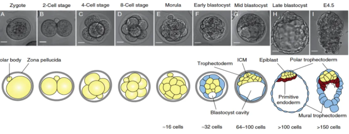
During early gestation, the zygote (Fig. 1A) cleavage produces the blastomeres, which organize to form the morula, a compact mass of approximately 16 cells (Fig 1E), and then the blastocyst (Fig. 1h), a structure formed by an inner cell mass (ICM), a fluid field cavity and an outer layer of cells named the trophoblast.(1)(3)(4) Throughout this process, cells become restricted in their developmental potential by cell lineage specification and spatial segregation. These events result in the establishment of the trophectoderm (TE), which will give rise to the placenta and extraembryonic tissues, and the ICM, which is composed by pluripotent stem cells that will eventually generate the definitive structures of the embryo (Fig.1G).(5)
As embryonic development progresses, a second major cell fate decision occurs in the pluripotent cells of the ICM, which segregate into two new tissues: the epiblast (EPI), which will give rise to the embryo proper; and the primitive endoderm (PrE), which contributes to extraembryonic membranes (Fig.1H) (6)(4)(7)
How these pluripotent stem cells decide their fate and enter differentiation is a question poorly understood, and several studies have proposed a variety of mechanisms to be involved in cell lineage specification, including cell position, polarization, molecular signaling and division plane when exposed in the same environment(2). Independently of the mechanisms that prove to be true, it is well established that they must, on one hand, provide stability in cell fate options to support self-renewal and maintenance of the uncommitted state, but must also, on the other hand, afford flexibility in cell-fate choice to allow cell-type diversification and differentiation in response to intrinsic cues or extrinsic signals. (7)
Figure 1 Schematic view of early mouse embryonic development.(A-E) The zygote
divides and forms the blastocyst, which is characterized by the presence of the ICM and the fluid cavity, both surround by TE cells (F-H). In the late blastocyst stage the embryo gives rise to the Epiblast (EPI) and primitive endoderm (PrE).
1.2 STEM CELLS
Stem Cells (SCs) are undifferentiated cells which are able to self-renew indefinitely and to differentiate into multiple cell lineages (Leitch et al. 2010; Wray et al. 2010; Ahn et al. 2008; Pera & Tam 2010). SCs can be found in embryonic tissues, as well as in the various adult tissues and organs and, throughout development and adulthood, they are responsible for the formation and maintenance of an organism.
SCs may be classified according to their origin or to their developmental potential. According to their in vivo origin, SCs are usually grouped into: embryonic stem cells (ESCs); fetal stem cells; and adult stem cells. In addition, there are also SCs generated in vitro by reprogramming of somatic cells, which are named “induced Pluripotent Stem” (iPSCs) (8) .According to their developmental potential, SCs are classified as: totipotent, pluripotent; multipotent ; oligopotent; and unipotent .(9)
Due to their properties, SCs are promising candidates for next generation therapeutics and constitute also a valuable tool to study cell fate specification, either during early embryonic development or during adulthood. As a consequence, these cells constitute a subject of thorough investigation, and in this thesis we have focused on mouse ESCs.
1.3 EMBRYONIC STEM CELLS
Embryonic stem cells (ESCs) are pluripotent stem cells (i.e. they can give rise to cells from the three germ layers), which can be derived in vitro from the ICM of mouse blastocysts (reviewed in Nichols & Smith 2011).
The first experiments that led to the isolation of these cells were motivated by previous studies using teratocarcinoma cells, which include undifferentiated embryonal carcinoma cells (ECCs) and allow tumor development when injected into host blastocysts(10)(11)(12). One important idea that emerged from embryonic teratocarcinoma studies was the notion that EPI derivation, during embryo development, is transient and may be susceptible for conversion to ECCs(13). This idea led to the attempt to directly isolate stem cells from mouse blastocysts, and in 1981 the first mouse ESCs were derived by Evans and Kaufman (14) and Martin (10). The derivation of ESCs from non-human primates occurred in 1995 (15) and from humans in 1998 (16)
Similarly to ECCs, ESCs are able to form teratocarcinomas upon transplantation into adult mice and also share similar morphology, growth behavior and gene expression (Nichols & Smith 2012; Chambers & Smith 2004b). In addition, ESCs are able to reintegrate in the embryo and contribute to chimera formation.
In order to derive ESCs in vitro, several culture conditions have been used. Initially, ESCs were derived in the presence of inactivated embryonic fibroblasts and fetal calf serum (14). Later, it was discovered that inactivated fibroblasts act by producing signals that can inhibit ESCs differentiation, a fact that was support by the efficient replacement of fibroblasts with medium prepared from Buffalo rat liver cells (BRL) (12).(17) Analysis of conditioned medium produced by feeder cells allowed the identification of leukaemia inhibitory factor (LIF) as the active component that allowed ESC self-renewal (Chambers & Smith 2004a), and this cytokine has been extensively used for ESCs maintenance (Lanner& Rossant 2010; Ying et al. 2008a). In 2003, Ying and co-workers reported that bone morphogenetic proteins (BMPs) are also able to sustain pluripotency when acting in combination with LIF (18). More recently, a combination of inhibitors of FGF/ERK and GSK3 has been reported to efficiently derive and maintain ESCs, without extrinsic signaling (19). These conditions suggest that pluripotency is an uninstructed ground state that is intrinsically self-maintaining if protected from inductive differentiation stimuli.
The different culture conditions that have been used for ESCs growth suggest that a balance of several signaling pathways has to be achieved so that self-renewal is sustained. In this sense, the various genetic and molecular signals that have been associated with pluripotency maintenance (see section 1.3) are responsible for gene activation or repression, which culminate in a cell fate decision to either self-renew or differentiate.(3)(2)
Although these different in vitro culture conditions represent a different environment from the in vivo scenario, they are able to provide a reasonable counterpart for in vivo cells and have been extensively used in several studies. Nonetheless, it is crucial to understand the extent of the similarities and differences between in vivo and in vitro conditions, so that an accurate understanding of the in vitro cell behavior and how it reflects in vivo events is achieved. To accomplish this aim, more detailed and comparative analysis of these cells should be performed in order to define more precisely the in vivo counterparts of ESCs and their role during embryonic development.
1.4:GENETIC AND MOLECULAR SIGNALS ASSOCIATED WITH PLURIPOTENCY
Pluripotency is an extraordinary property of ESCs that reflects the inner workings of a complex gene regulatory network, comprising both positive and negative regulators, and which have, at its core, the pluripotent genes Nanog, Oct4 and Sox2(20). Characterization of the transcriptional programs caused by these genes has revealed that they act by activating other pluripotency genes and repressing lineage-associated genes, but that they might also induce expression of differentiation signals (21).For instance, it has been shown that Oct4 and Sox2 also induce the expression of FGF4, which acts as an auto inductive differentiation signal. These findings suggest that pluripotency is a highly dynamic state, where the balance between Nanog, Oct4, Sox2 and also FGF/ERK signaling has to be carefully maintained. (22)(23)
In vitro, this dynamic equilibrium, between a stable self-renewing (ground state) and transient intermediate state prone to enter differentiation, may be sustained by the use of different culture conditions (which include different signaling molecules).(24) Therefore, in order to understand how this balance is achieved it is essential to elucidate how the different signaling molecules are integrated by the gene regulatory network. However, the complexity of these networks and signaling molecules is immense and is currently an extensively studied topic. In the next sections I will discuss the main transcriptional pathways responsible for stem cell maintenance and their interactions and known cross talks between each another.
BMP4 WNT
Integrin
JAK JAK
Ids, β-catenin, SMADs
SOX2–OCT4 Nanog STAT3
?
Trophectoderm ES Primitive endoderm gp130 MEK ERK LIFR LIF Frizzled GSK3 GAB1 SHP2 Cell membrane Nuclear membrane DIAPAUSE In general, a period of physiologically enforced dormancy between periods of activity. In the
pre-implantation-stage embryo, it is a transient/reversible state of reduced vitality during which the blastocyst floats in the uterus without implanting.
the undifferentiated state of mouse ESCs. Notably, BMP proteins are suggested to be targets of another ligand cascade that is initiated on binding of WNT to its receptor (see below)32.
WNT proteins. WNT proteins are secreted
glycopro-teins that have widespread roles in tissue differentia-tion and organogenesis33. In vivo, Wnt5a and Wnt11 are
expressed in mouse embryos that undergo the morula-to-blastocyst transition, but they are only weakly expressed or are not expressed at all in embryos that do so in vitro. This is consistent with the morbidity of embryos that are cultured in vitro. The upregulation of Wnt11 is temporally coordinated with the surge of maternal oestradiol at E4 — the time of implantation34.
The canonical WNT pathway is activated on binding of the WNT protein to the Frizzled receptor on the cell membrane (FIG. 2). Activation of the pathway leads to inhibition of glycogen-synthase kinase-3 (GSK3), subsequent nuclear accumulation of β-catenin and the expression of target genes. Sato and colleagues used a new and specific reversible inhibitor of GSK3, 6-bro-moindirubin-3´-oxime (BIO), to demonstrate that activation of the canonical WNT pathway maintains the undifferentiated phenotype in both mouse and human ESCs, and sustains expression of the pluripotent-state-specific transcription factors OCT4, REX1 (also known as zinc-finger protein-42; ZFP42) and Nanog27 in the
absence of supplemented LIF. The genetic manipulation of WNT signalling in ESCs — by either inactivating the adenomatous polyposis coli (APC) complex (a multiprotein complex tumour suppressor that mediates signal transduction from the WNT receptor on the cell surface to β-catenin in the nucleus) or by introducing a dominant-active form of β-catenin — results in the inhibition of neural differentiation in vitro32. This also
occurs after activation of the downstream targets of WNT signalling, such as cyclin-D1 REF. 35, MYC36 and BMP proteins32. Neural differentiation can be partially
restored by addition of the BMP4 antagonist Noggin32.
Emerging transcriptional regulators of ESCs. Most
recent studies have pointed to one signalling pathway through which nutrients and environmental cues — that is, amino acids37,38 and inositols39 — regulate
survival of early mouse embryos and ESCs. The mam-malian target of rapamycin (mTOR) is a serine/threo-nine kinase that was originally identified as a target of rapamycin in Saccharomyces cerevisiae and was then found to be highly conserved among eukaryotes40,41.
This signalling pathway ultimately regulates the inter-action between mTOR and two downstream substrates, eukaryotic translation-initiation factor 4E (eIF4E)-binding protein-1 and ribosomal-protein-S6 kinase42.
Inositols also participate in another pathway that is centered on the phosphatase and tensin homologue deleted on chromosome 10 (Pten) gene.
Inactivation of PTEN increases the proliferation of mouse primordial germ cells and enhances the deri-vation of EGCs43. In ESCs, it leads to increased entry
into S phase and increased cell survival44. PTEN is a
phosphatase that has a wide range of functions45 and
its lipid-phosphatase activity is associated with tumour suppression. The chief substrate of PTEN is phos-phatidylinositol-3,4,5-triphosphate (PtdIns(3,4,5)P3).
PTEN therefore terminates phosphatidylinositol 3-kinase (PI3K) signalling by dephosphorylation of the PtdIns(3,4,5)P3 second messenger43. The serine/
threonine kinase AKT is a downstream effector of PI3K that phosphorylates mTOR in vitro, indicating a direct linkage between mTOR and the PI3K–AKT pathway, which is negatively regulated by PTEN46.
Through a double-gene approach, in which both Pten and Akt1 were mutated, it was demonstrated that AKT1 upregulation is essential for the tumorigenic phenotype observed for Pten-null ESCs44.
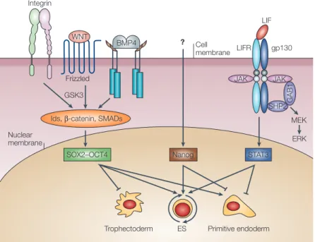
Figure 2 | Combinatorial signalling pathways involved in maintaining mouse ESC pluripotency. Cell-surface receptors initiate signals that are conveyed (thin black lines) to the
nucleus and affect key pluripotency transcription factors such as octamer-binding
transcription factor-4 (OCT4) and Nanog, and self-renewal transcription factors such as signal transducer and activator of transcription-3 (STAT3). So far, only the leukaemia inhibitory factor (LIF) receptor (LIFR)–STAT3 pathway has been defined in detail. The cytokine LIF functions by binding to LIFR at the cell surface, which causes it to heterodimerize with another
transmembrane protein, glycoprotein-130 (gp130). This is followed by the activation of kinases
that amplify and drive the signal to the nucleus, or that recruit other factors for docking to the activated LIFR–gp130 receptor. On binding LIF, the intracellular domains of the LIFR–gp130 heterodimer can recruit the non-receptor Janus tyrosine kinase (JAK) and the
antiphosphotyrosine immunoreactive kinase (TIK) and become phosphorylated. The
phosphorylated intracellular domains of the LIFR–gp130 heterodimer function as docking sites for proteins that contain Src-homology-2 (SH2) domains, which include the transcription factor STAT3. At the nuclear level, STAT3, OCT4 and Nanog cause changes in gene expression that result in (pointed arrow) or counteract (truncated line) phenotypic traits of embryonic stem cells (ESCs). OCT4 functions in connection with SRY-related high-mobility group (HMG)-box protein-2 (SOX2) and other cofactors to determine whether target genes are activated or repressed. Aspects of this figure are speculative as, in some cases (bone morphogenic protein-4; BMP4), the surface receptors are known but the transducers are not, and in others both have yet to be identified (OCT4, Nanog). The current lack of information is highlighted by a question mark. GAB1, GRB2-associated binding protein-1; GSK3, glycogen synthase kinase-3; Id, inhibitor of differentiation, JAK, janus kinase; MEK, mitogen-activated protein kinase (MAPK) and extracellular signal regulated kinase (ERK) protein kinase; SMAD, similar to mothers against decapentaplegic homologue; SHP2, SH2-domain-containing protein tyrosine phosphate-2; WNT, wingless type protein.
876 | NOVEMBER 2005 | VOLUME 6 www.nature.com/reviews/molcellbio
R E V I E W S
Figure 2 Combination of signalling pathway maintaining mouse ESCs pluripotency. LIF
binds to LIF receptor at cell surface and heterodimerize gp130 receptor. This is followed by the activation JACK- STAT3 pathway and also MAPK pathway pro-differentiation. BMP proteins bind with transmembrane receptor, which interact with different protein SMAD TF and induce Id genes. The canonical WNT pathway is activate by frizzled receptor on the cell membrane and lead the inhibition of GSK3 pathway. The interplay of this combination of pathway results in activation signals that are translocated to the nucleus and affect key pluripotent factors like Sox2, Oct4 and Nanog (Adapted from Boiane, 2005).
1.4.1 SIGNALLING MOLECULES
1.4.1.1 LIF
LIF belongs to the family of IL-6-type cytokines, which binds to a heterodimeric receptor complex that consists of two related subunit cytokine receptors: gp130 in association with the low affinity LIF receptor (LIFR), a ligand-specific subunit receptor. The binding of LIF to this receptor complex results in JAK (Janus associated tyrosine kinase) recruitment, which creates binding sites for proteins containing SH2 (Src homology 2) domains, such as the signal transducer and activator of transcription 3 (STAT3) and the extracellular regulated kinases ERK1 and ERK2. (24)
Upon phosphorylation, STAT3 proteins dimerize and translocate to the nucleus, where they activate target gene transcription that prevents stem cell differentiation, while inducing proliferation (25). The proliferative effects of STAT3 are counteracted by ERKs, which are components of a signal-transduction pathway that inhibits ES cell self-renewal and work as pro differentiation signals promoting the transition of a naïve to primed state (26).
Although mouse ESCs can be efficiently derived and maintained in the presence of LIF and serum, this last component is not ideal due to its variability and origin: not only it contains a non-defined variety of inductive stimuli that might also activate commitment and differentiation programs, but it can also be a source of contaminations due to its animal origins. This has led researchers to find alternatives where serum is not required and better growth conditions are defined, such as the use of BMPs (27)(28). Furthermore, the overexpression of the homeodomain protein Nanog has been reported to support LIF/STAT3-independent self-renewal, suggesting that other factors may play a key role in the pluripotency of mouse ESCs.(29)
1.4.1.2 BMP4
In serum free conditions, LIF alone is not sufficient to maintain pluripotency of murine ESCs, which enter neural differentiation, suggesting that other signals are necessary for suppressing this differentiation pathway. Indeed, studies by Ying and co-workers have reported that serum use can be bypassed through addition of BMPs to sustain the pluripotency (18). BMP proteins belong to the transforming grow factor β (TGF-β) family, and have evolutionarily conserved function.(30)
Upon binding of BMPs to the transmembrane type I e II receptors, oligomerization of serine/ threonine receptor kinases and phosphorylation of the cytoplasmic signaling molecules Smad2 and Smad3 occurs. This induces the expression of inhibitor or differentiation (Id) genes, via the Smad pathway, which promotes differentiation of ESCs to non-neural lineages. However, when BMPs are used in combination with LIF, proliferation of mouse ESCs is maintained even in the absence of feeder cells (18). Therefore, the two pathways LIF/STAT3 and BMP/Smad seem to act in a concerted way to maintain pluripotency. Nonetheless, it has also been reported that Nanog overexpression is able to replace BMP/serum stimulation, although it has a limited capacity to inhibit Smad differentiation pathway.(31)(32)
1.4.1.3 GSK3
GSK3 (Glycogen Synthase Kinase-3) is a ubiquitously expressed serine/threonine protein kinase, found in all eukaryotes. Identified originally as a regulator of glycogen metabolism, GSK3 acts as a downstream regulatory switch for numerous signaling pathways, including cellular responses to Wnt signaling. Sato and co-workers have demonstrated that the activation of this signaling pathway, through canonical Wnt signaling, is able to maintain both mouse and human ESCs in an undifferentiated state, independently of LIF/STAT3.(33) In brief, while Wnt ligands promote nuclear
accumulation of β-catenin, which associates with transcription factors that activate transcription, Gsk3 phosphorylates β-catenin, thereby negatively regulating Wnt signaling (34). This negative regulation is prevented by the use of inhibitors of GSK3, which promote Wnt stimulation and, consequently, ESCs self-renewal.
Recently, chemically defined serum free conditions were established, based on combinations of small molecules inhibitors of the mitogen-activated protein kinase (MAPK)/extracellular signal-related kinase (ERK1/2) and GSK3 (19) These conditions, known as 2i and 3i conditions, have been used to efficiently maintain mouse ESC in a undifferentiated state called “ground state” of ESC self renewal.
1.4.2 PLURIPOTENCY GENE REGULATORY NETWORK
At the core of the ESCs pluripotency gene regulatory network lies a unique set of transcriptional factors (TFs) – Oct4, Sox 2 and Nanog – that regulate cells “stemness” potential and prevent the onset of differentiation. These pluripotency factors are crucial for the maintenance of the equilibrium between self-renewal and differentiation and can be used as accurate pluripotency “readouts”.(35)
Oct4 belongs to the class of POU transcription factors and is expressed in
pre-implantation embryos, EPI, germ cells, ECCs and ESCs(36).In ESCs, Oct4 levels regulate pluripotency, with its inactivation causing differentiation into trophoectoderm (TE) cells, and its overexpression leading to mesoderm and endoderm differentiation. Although Oct4 levels have to be tightly regulated, its expression is not sufficient to sustain ESCs self-renewal upon withdrawal of LIF (37).
Sox2 is a member of the Sry-related HMG-box family that has been reported to interact
with Oct4, being required for ESCs pluripotency maintenance. It is expressed during early embryogenesis (morula and ICM of the blastocyst), EPI, germ cells, ESCs and neural SCs(38) Sox2 null embryos have been shown to result in early embryonic lethality. However, this gene does not seem to be a master pluripotency regulator, since it is not able to prevent ESCs differentiation upon removal of LIF.
Nanog is a transcription factor expressed in preimplantation embryos, germ cells,
ECCs and ESCs, which is able to support LIF/STAT3-independent self-renewal when overexpressed (39)(29).NANOG is a homeodomain protein of 280 amino acids that shares a high degree of homology and structural resemblance with NK proteins family (40)
The Nanog gene was initially identified in functional screening studies aimed at identifying factors that were capable of maintaining ESCs self-renew in a LIF-independent manner. Since then, overexpression of Nanog in ESCs has then been shown to allow pluripotency maintenance in the absence of LIF or BMP4, while Nanog deletion results in differentiation towards the primitive endoderm lineage. These findings place Nanog as a master transcriptional regulator of in vitro ESCs self-renewal, which, consequently, has an important role in cell fate decisions. (31)(41) In vivo, during early mouse embryonic development, the expression of Nanog is first observed in the interior cells of the compacted morulae until the blastocyst stage. Later in development, the expression of Nanog is restricted to the EPI stage. Therefore, Nanog plays also a critical role in in vivo regulation of cell fate and maintenance of the pluripotent EPI. Furthermore, Nanog has been shown to prevent differentiation into primitive endoderm, with a DNA motif recognizable by the NANOG protein found in the promoter region of Gata6, a gene known to play a critical role during primitive endoderm differentiation(42)(43). Actually, both Nanog and Gata6 have been reported to be heterogeneously expressed in early blastocysts, an observation that is also attained in ESCs. These genes identify, respectively, the EPI and the PrE, the two cell lineages that differentiate from the ICM.
Published data also shows that Nanog action requires maintenance of Oct4 and Sox2 expression. While OCT4 and SOX2 are homogenously expressed in undifferentiated ESCs, NANOG expression has been shown to fluctuate between states of high NANOG expression and low NANOG expression. It has been proposed that the heterogeneous expression of NANOG, observed both in vitro (in ESCs) as in vivo (in the ICM of mouse blastocysts), might confer different degrees of responsiveness to differentiation signals. More precisely, Nanog heterogeneity may be important to guarantee that a pool of stem cells is maintained, while, at the same time, each individual ESC is primed to differentiate and able to follow a differentiation path. This implies that Nanog may play a crucial role during cell fate decision in ESCs, and that its levels may be used to identify different ESCs sub-populations. In this thesis, this subject is addressed, by trying to identify the various types of NANOG expressing sub-populations using a Nanog reporter ESC line (Nd) (44)(31)(39) and several lineage-affiliated cell surface markers (described in more detail in the next sections).
1.4.2.1 Lineage specific markers
Due to their properties ESCs constitute an important tool to understand early embryonic development as well as to use in many biomedical applications, such as
drug screening, cellular therapies and tissue engineering. However, several problems still hinder the wide use of these cells. One of these main problems results from the lack of understanding of the molecular aspects underlying the dynamics of the ESC state. How is the observed heterogeneity in Nanog expression biologically relevant and how this correlates with distinct behavioral outputs upon differentiation cues are two interesting questions that still remain unanswered.(45)
In order to start dissecting these issues, a more detailed characterization of the different sub-populations present within an apparently homogeneous ESC population is essential. To do so, the use of specific cell surface markers is essential, so that precise identification, isolation and characterization of ESCs may be achieved. However, for mouse ESCs, most of the known markers are still not ideal and result in ambiguous identification of cell surface proteins. Nonetheless, recently Rungg-Gunn and co-workers have analyzed the cell-surface proteome of stem cell lines derived from early mouse embryos. Their work has enabled the identification of several protein markers that can be used to study the cell fate decisions that occur during early differentiation. The use of combinations of these markers with known pluripotency regulators, namely with Nanog, shall reveal some of the molecular issues underlying heterogeneity in ESCs.(46)
For mouse ESCs, several surface markers have already been described as fairly good markers, such as the stage specific embryonic antibody 1 (SSEA1), the platelet endothelial cell adhesion molecule (PECAM1, also known as CD31), the Ephrin type-B receptor 4 (Ephb4) and CD81(Cluster of Differentiation 81).
SSEA-1 (Stage specific Embryonic Antigen 1) is a carbohydrate molecule required for
cell adhesion that is involved in controlling cell surface interactions during development. This molecule is expressed, on the surface of murine embryonal carcinoma and embryonic stem cells, early mouse embryos at pre implantation stage, murine and human germ cells. However, upon differentiation, human cells have been shown to have increased expression of SSEA-1, in contrast to what is observed in mouse cells, where the expression of SSEA-1 decreases. Overall, SSEA-1 is regarded as an excellent cell surface marker to monitor early stages of mouse embryogenesis and ESCs differentiation (45).
Pecam1 (Platelet/endothelial cell adhesion molecule 1, also known as CD31) is a
plasma membrane-spanning molecule that contains six Ig-like extracellular domains and is associated with undifferentiated and differentiated cells (47). During development, Pecam1 expression is first detected on blastocysts, the main source of
ESCs. Some studies have shown that this marker plays an important role in vasculogenesis and angiogenesis, in cell adhesion and signal transduction within the vascular compartment.
Ephb4 is a classe of ephrin tyrosine kinase receptor, whose ligands, the ephrins, are
membrane-anchored proteins, usually grouped in two subclasses: ephrins A and ephrins B. These Ephrin receptor –ligant complexes play important roles in various processes during embryonic development, including modulation of the targeting behavior of migratory neurons, vascular cell assembly, and angiogenesis regulation. The receptor tyrosine kinase EphB4 are widely expressed in fetal and adult tissues and your expression is turned on in response to cell proliferation.(48)
CD81 (TAPA-1) is a widely expressed cell-surface protein involved in several biologic
responses. This molecule is associate with adhesion, morphology, activation, proliferation, and differentiation into T and B cells. During development, this molecule is expressed in all stages of the preimplantation embryo.(49)
For lineages related to early differentiation, the TS cell marker CD40 and the EPI cell marker Notch3 have been described.
CD40 (TNFR5) is a cell surface receptor that belongs to the tumor necrosis factor-R
(TNF-R) family. This receptor was first identified and functionally characterized in the adult immunological compartment and has been shown to be expressed at the earliest stages of blood specification, both in vitro and in vivo. Overall, CD40 is a co-stimulatory molecule associated with angiogenesis and inflammation.
Notch3 is a transmembrane protein receptor that belongs to the canonical Notch
pathway. Notch pathway is a highly conserved signaling mechanism that participates in a variety of cellular processes: cell fate specification, differentiation, proliferation, apoptosis, adhesion, epithelial-mesenchymal transition, migration, and angiogenesis. Notch signals are able to link fate decisions of one cell to those of cell to those of its neighbours (50 ) and have been shown to be essential for the transition from the primitive neural stem cell to the definitive neural stem cell stage, controlling cellular fate choices. In ESCs, Notch receptors and ligands has been shown to regulate the transition from a naïve to a primed EPI state.
The antibodies described above are promising candidates to label different subpopulation of cells and that therefore have the potential to be used for understanding cell fate decisions during development.
GOAL
Given that Low-Nanog is a transient state into differentiation, where ESCs express
different priming genes, the main goal of this project was to identify and characterize molecularly co-existing ESCs subpopulations containing low Nanog or high Nanog levels. This was performed by taking advantage of a Nanog reporter ESC line and using a panel of specific antibodies that have been reported to label ESCs subsets. In addition, I aimed to find a combination of antibodies that correlates with transient pluripotent states and functional heterogeneity of ESCs.
Chapter 2:
2.1 MATERIALS AND REAGENTS 2.1.1 REAGENTS
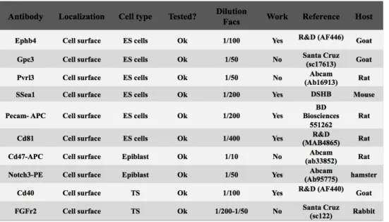
All reagents, solutions, media and antibodies used in this study are described in Appendix I(section 6.1. 1)
2.1.2 Cell Lines
Two mouse ESC lines were used: E14tg2a and Nd (51). Nd cell line is a novel mouse ESC line containing a transgenic Bacterial Artifical Chromossome (BAC) with a short-lived fluorescent protein VNP under the control of Nanog regulation. This strategy has been development at the Developmental Biology Unit to study the role of NANOG dynamic expression in pluripotency maintenance.
2.2 METHODS
2.2.1 ESC CULTURE
All steps involved in the manipulation of ESCs were performed in a sterile laminar flow hood class II, type A/B3.
2.2.1.1 Expansion of ESCs
ESCs were thawed in pre-heated Glasgow Modified Eagles Medium 1x (GMEM) and plated on 0.1% (v/v) gelatin-coated dishes in supplemented GMEM (with 2ng/ml LIF). Medium was changed 6-12 hours later to eliminate DMSO residues. ESCs were grown at 37ºC in a 5% (v/v) CO2 incubator on gelatin-coated dishes in supplemented GMEM.
The morphology and health of the cells were assessed daily by direct visualization on a bright field microscope and cells were passaged every other day, at a constant plating density of 3x10⁴ cells/cm2. For each passage, cells were washed twice with PBS and
dissociated with 0,025% trypsin for 2-3min at 37ºC. Cells were immediately resuspended in GMEM (in order to neutralize trypsin), centrifuged at 1200 rpm for 4min and resuspended again in GMEM. Cells were counted using trypan blue dye exclusion method and the required amount of cells was plated on gelatin-coated dishes in supplemented GMEM (Serum/LIF conditions). To prepare ESC stocks, 3x10⁶ cells
were frozen in GMEM 1x with 10% Dimethyl sulphoxide (DMSO) and stored in liquid N2. Every time cells were frozen, a sample was collected to test for Mycoplasma contamination. Alternatively, ESCs were grown in 2i medium, supplemented with LIF, a serum-free based medium that contains inhibitors of the ERK pathway and of GSK3ß (2i/LIF conditions), or in ESGRO medium supplemented with BMP4 and LIF (BMP4/LIF conditions).
2.2.1.2 Differentiation of ESCs
To analyse ESCs ability to enter differentiation, ESCs were plated at 3x10⁴ cells/cm2 in
GMEM 1x without LIF supplementation, on gelatin-coated plates, for 48 hours (Serum conditions).
2.2.2 PROTEIN EXPRESSION ANALYSIS
2.2.2.1 Fluorescence Activated Cell Sorting 2.2.2.1.1 Nanog:VNP analysis
For live cells FACS analysis of Nanog:VNP, cells were dissociated and resuspended in PBS. The expression of nanog:VNP was measured on a FACS Calibur cytometer (Becton Dickinson), using Cell Quest software. Live cells were gated based on forward scatter and side scatter and by propidium iodide dye exclusion. For each sample, 10000 gated events were acquired and the obtained data was analyzed using the FlowJO software.
2.2.2.1.2 Cell surface protein expression analysis
For live cells FACS analysis of several cell surface proteins (see Table 2), cells were dissociated and distributed into eppendorf tubes at a concentration of 0.5x10⁶ cells/tube
in the respective growth media (Serum, Serum/LIF, BMP4/LIF or 2i/LIF). For each antibody analysed, samples with no antibodies (negative control) or with no primary antibody (2nd antibody control) were used as controls. Cells were washed twice with
PBS and centrifuged 5 min at 4000 rpm. The supernatant was removed and cells were ressuspend in 300µl of blocking solution (10% of FBS in PBS) to block non-specific ligations. After 1hour incubation at room temperature, cells were centrifuged again (5min, 4000rpm) and the supernatant was carefully removed. Cells were ressuspended in 100ul of PBS (controls) or appropriate primary antibody and incubated in the dark for 1 hour at room temperature. Cells were washed twice with PBS. Cells labeled with conjugated antibodies were transferred into 0.1% BSA pre-coated FACS tubed and were kept at 4ºC in the dark until data acquisition. Cells labeled with non- conjugated antibodies, were ressuspended in 100µl of PBS (negative control) or appropriate secondary antibody and were again incubated in the dark for 1 hour at room temperature. Cells were washed twice with PBS, transferred into 0.1% BSA pre-coated FACS tubed and were kept at 4ºC in the dark. Data acquisition was performed on a FACS Calibur cytometer (Becton Dickinson), using Cell Quest software. Live cells were
gated based on forward scatter and side scatter. For each sample, 10000 gated events were acquired and the obtained data was analyzed using the FlowJO software.
2.2.3 DNA ANALYSIS
2.2.3.1 PCR for Mycoplasma detection
To check for the absence of mycoplasma contamination in ESC cultures, samples were routinely collected using the following procedure. 106 cells were centrifuged at 2000rpm for 5min, resuspended in wash buffer and centrifuged again in the same conditions. The pellet was then resuspended in a 1:1 mix of solution A and solution B and incubated for 1h at 60ºC. The suspension was denatured, to inactivate proteinase K, by incubation at 90ºC for 10min.
The PCR for mycoplasma detection was performed using rTaq Polymerase, amplifying a conserved region in the 16S RNA gene. The amplification was performed with an initial step of denaturation at 95ºC for 5min, followed by 30 cycles of denaturation at 95ºC for 30sec, annealing at 58ºC for 1.5min and extension at 72ºC for 1.5min, and a final step of extension at 72ºC for 10min. The reactions were prepared for a final volume of 25µL: 3µL of sample, 1x buffer, 0.2mM dCTP, 0.2mM dGTP, 0.2mM dATP, 0.2mM dTTP, 25pmol of each primer and 2.5U of rTaq Polymerase. The PCR products were analyzed by agarose gel electrophoresis. The quality of the DNA preparation was confirmed by performing a PCR to detect GAPDH, a housekeeping gene that functions as an internal control. Also, a plasmid that carries an insertion that corresponds to the amplified fragment was used as a positive mycoplasma control.
Chapter 3:
3.1 CARACTERIZATION OF EMBRYONIC STEM CELL CULTURES
ESCs may be maintained in different cell culture media, such as Serum/LIF, BMP4/LIF or 2i/LIF. Additionally, they can be induced to differentiate by removal of LIF (Serum conditions). In order to characterize cells growth under these different conditions, E14TG2Atg2a and Nd ESCs self-renewal potential was examined by analysing cells morphology, viability and growth rates (section 3.1.1). Furthermore, Nanog:VNP expression was analysed by FACS (section 3.1.2) and the detection of the expression of several ESCs and early differentiation markers was optimized using flow cytometry(FC) analysis (section 3.1.3).
3.1.1 SELF-RENEWAL POTENCIAL
In order to assess the self-renewal potential of ESCs cultured in different cell culture media and through different passages, three different parameters were analysed: cells morphology; viability; and growth rates, measured as fold increase.
The morphology and general health of the cells were evaluated on an invert microscope by direct observation. In figure 3, the typical morphology of Nd ESCs grown in Serum (Fig.3A), Serum/LIF (Fig.3B), BMP4/LIF (Fig.3C) and 2i/LIF (Fig.3D) can be observed, with significant differences being detected. Similar data was obtained for both ESC lines studied. In all media with LIF, we can observe cells organized in undifferentiated clusters, which are more compact in 2i/LIF than in Serum/LIF or BMP4/LIF conditions. In contrast, in Serum medium cells are more adherent to the substrate and clusters are less compact, suggesting a higher differentiation degree.
Figure 3: Nd ESCs morphology in different cell culture media.A) Serum medium for 48
hours. These conditions allow ESCs to commit into differentiation. B) Serum/LIF medium. C) BMP4/LIF medium. D) 2i/LIF medium.
For each ESC passage the viability and the growth rate (measured as fold increase) was calculated. Cell’s viability was evaluated using Trypan Blue dye exclusion. The obtained data regarding cells viability is shown in Figure 4 for both E14TG2Atg2a and Nd ESC lines. As it can be observed, cells viability is high (always above 95%) and is very similar between the different cell lines (Fig. 4A). Furthermore, no significant
differences are observed through serial passaging, confirming the general “well-being” of the cultures (Fig. 4B).
Figure 4 Self-renewal capacity of Nd and E14TG2Atg2a cell lines – Viability data. A)
Average viability for all cell passages in culture (n=13). B) Viability data for each cell passage. Grey and black bars/lines depict, respectively, E14TG2Atg2a and Nd ESCs data. No statistically significant differences were observed between ESC lines (unpaired t-test).
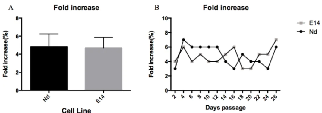
Regarding the fold increase (calculated as the ratio between the final number of cells at the end of each passage and the initial cell number that was plated), the obtained data may be observed in Figure 5. Both fold increase and viability are very similar for both cell lines and throughout serial passaging, confirming self-renewal potential maintenance.
Figure 5: Self-renewal capacity of Nd and E14TG2Atg2a cell lines – Fold Increase data. A)
Average fold increase for all cell passages in culture (n=13). B) Fold increase data for each cell passage. Grey and black bars/lines depict, respectively, E14TG2ATG2A and Nd ESCs data. No statistically significant differences were observed between ESC lines (unpaired t-test)
3.1.2 NANOG:VNP EXPRESSION
NANOG expression has been shown to be heterogeneous in ESCs, particularly when cells are grown in Serum/LIF conditions. This results in the co-existence of different sub-populations of cells expressing various NANOG levels. It has also been shown that when ESCs are grown in 2i/LIF media, heterogeneity is reduced (although not eliminated).
In order to characterize NANOG expression when ESCs are grown in different culture conditions, Nd ESCs were were grown for 48 hour in Serum/LIF, BMP4/LIF, 2i/LIF and
Serum (without LIF) and the expression of Nanog:VNP was measured by FACS in live cells. In addition to the different morphologies, ESCs also show different Nanog:VNP expressions when exposed to different conditions (Fig.6). FC analysis reveals that in Serum conditions Nd cells have lower Nanog:VNP expression (20.4±7.6%) with a characteristic distribution (Fig. 6A,E), which correlates to the loss of NANOG expression associated with cells committing to differentiation. In contrast, in all pluripotency conditions, higher levels of Nanog:VNP expression were observed, which are associated with pluripotency maintenance. In Serum/LIF 56.5±4.5% of the cells show Nanog:VNP expression (Fig. 6B,F), and this value increases in BMP4/LIF (72.76±4.6%) (Fig. 6C,G) and 2i/LIF (85.0±3.5%) (Fig. 6D,H).
Figure 6Expression of Nanog:VNP in Nd and E14TG2Atg2a ESC, grown in different media.The picture shows the distributions of the cells grown in different culture conditions (A-D)
and the respective averages of Nanog:VNP expression (E-F) (n=8) for Nd and E14TG2Atg2a ESCs grown in Serum (A and E), Serum/LIF (B and F), BMP4/LIF (C and G) and 2i/LIF (D and H) media. Error bars represent the standard deviation.
These results are in agreement with previously published data [REF] and with unpublished data obtained in the lab. In the presence of Serum, the addition of LIF is sufficient to sustain pluripotency. In the absence of Serum, the presence of BMP4 or FGF/ERK inhibitors is enough to maintain pluripotency and even increase the levels of NANOG expression. BMP4 acts by suppressing differentiation through expression of inhibitor of differentiation genes (Id genes) while the inhibitors present in the 2i media act by the simultaneous inhibition of Gsk-3 and MEK pathways. The addition of LIF to supplement both BMP4 and 2i media is important to prevent differentiation into specific lineages as well as for cell survival.(19)
The obtained distributions when ESCs are grown in different culture media, as well as the observation that, upon medium change, Nanog:VNP expression changes rapidly
(within 24-48h), suggests that NANOG expression is dynamic and dependent on environmental cues. This data also suggest that heterogeneous expression of Nanog is an intrinsic property of pluripotent cells and that it might have a functional role during pluripotency cell fate decisions.
3.1.3 OPTIMIZATION OF THE DETECTION OF SURFACE PROTEINS
In order to characterize different sub-populations of cells present in ESCs cultures, the expression of several surface proteins was analysed by FACS. Although this technology is highly sensitive and broadly used to assess protein expression, optimization assays are usually required before analysis.
In this work, 10 antibodies that have been described to be expressed by ESCs, EPI or TS cells were tested for ESCs grown in different culture media. See Appendix I(section 6.1.2). From these, 6 yielded reasonable results and are described below. The remaining 4, failed to provide proper protein detection even after changes in experimental conditions, namely antibody concentration, blocking solution and incubation time(48)
3.1.3.1 ESCs markers: Ephb4, CD81, Ssea1 and Pecam1.
In order to characterize the expression of pluripotency markers in ESCs cultures, Ephb4, CD81, Ssea1 and Pecam1 antibodies were tested in ESCs grown in different cell culture conditions.
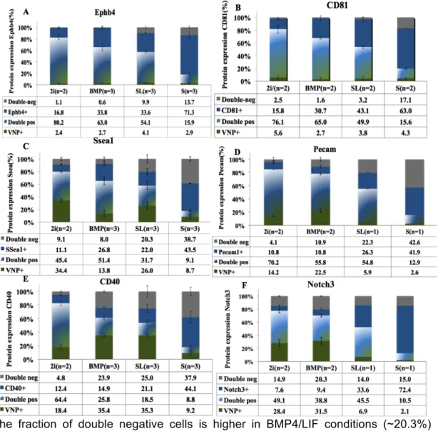
Ephb4 In ESCs, EphB4 has been shown to modulate the response to mesoderm
induction signals. The data obtained in this thesis for EphB4 (Fig. 7A) does not show statistically significant differences among different ESCs culture conditions (~88-97% of positive cells), with p-values higher than 0.05 (Appendix I – Table 3). This suggests that this marker is broadly expressed in ESCs and not dependent on cell exposure to various pluripotency conditions. Furthermore, only a slight reduction is observed in the expression of this marker when cells are exposed to differentiation (82-85% of expressing cells), suggesting that it is stably expressed.
CD81 Similarly to EphB4, our results (Fig. 7B) show that this protein is expressed in all
pluripotent culture conditions, with no statistical significant differences (Table S4). When ESCs were grown in serum alone for 48h, a small, but statistically significant decrease in the expression of CD81 was observed (p=0,036
Appendix I Table 3),
suggesting that this marker is slightly more specific of the pluripotent state than EphB4.SSEA1 In ESCs, this marker has been shown to be correlated with pluripotency and to
using SSEA1 antibody (Fig.7C) shows that this marker has a specific cell surface expression,with statistically significant higher levels observed in BMP/LIF conditions (~75-81% positive cells) compare to Serum/LIF (~61-63%) and 2i/LIF (~56-67%) conditions. Although the observed decrease in Serum/LIF is consistent with the lower Nanog levels, the decrease observed in 2i/LIF was not expected. Indeed, in 2i/LIF conditions, cells are usually associated with higher pluripotency degrees and higher Nanog levels. One possible explanation for the observed decrease in SSEA1 expression may be presence of GSK3 inhibitors in 2i/LIF, which are able to activate the WNT canonical pathway that regulates various cell mechanisms, which might interfere with this markers expression. In Serum alone, upon exposure to differentiation condition, a decrease in the expression of this marker was observed (~52-53% of positive cells), an expected finding since SSEA1 is a known pluripotency marker commonly used for ESCs characterization.
Pecam1 Similarly to SSEA1, this marker has also been shown to be correlated with
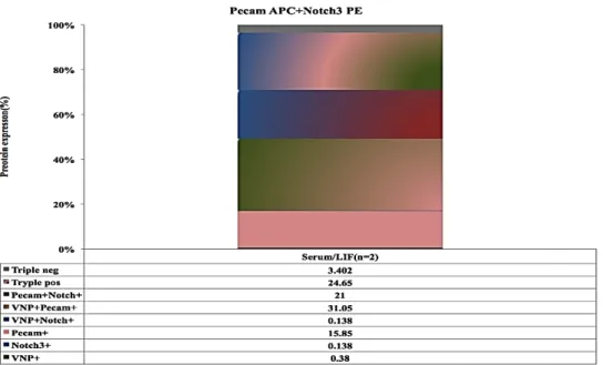
pluripotency and to ESCs ability to incorporate into the EPI of chimeric embryos. The obtained results (Fig. 7D) show that this marker has specific cell surface expression, with higher levels being observed in 2i/LIF (~74-83% of positive cells) and Serum/LIF (~67-81% of positive cells) conditions. In BMP/LIF conditions, a slight but not statistically significant reduction in the expression of this marker is observed (~57-67% of positive cells). As expected, lower expression is always observed upon exposure to differentiation conditions (~55% of positive cells). To note, only 1 or 2 biological replicates were analysed using this marker and a considerable variability is observed between the two cell lines analysed. Nonetheless, the obtained data was interesting enough to proceed with the analysis (see section 3.2.1).
3.1.3.2 TS cells marker: CD40
In order to characterize the presence/absence of trophoblast cells in ESCs cultures, CD40 antibody was tested in different cell culture conditions.
The obtained data for CD40 (Fig. 7E) shows that this marker has cell-surface specific expression, with significantly higher levels being observed in 2i/LIF culture conditions (~76-77% of positive cells) compared to BMP4/LIF and Serum/LIF conditions (~32-40% of positive cells). These observations, namely the increase in CD40 marker expression in 2i/LIF, suggest that, in these conditions, ESCs are more prone to differentiate into trophoblast. This is in agreement with recently published data showing that 2i/LIF supports a totipotent state similar to the in vivo morula or early blastocyst embryonic stages.
In Serum alone, i.e., upon exposure to differentiation conditions, approximately 53% of cells express CD40. This value, although not as high as the one observed in 2i/LIF conditions, is statistically significantly higher than the expression levels observed in BMP4/LIF and Serum/LIF conditions, in agreement with the advanced differentiation state of these cells.
3.1.3.2 Epiblast cells marker: Notch3
In order to characterize the presence/absence of early EPI cells in ESCs cultures, Notch3 antibody was tested in different cell culture conditions.
During this work, two Notch3 antibodies labelled with different fluorochromes (PE and Alexa647) were tested. In Figure 7F, the results obtained for Notch3:Alexa647 in 4 different condition are shown. The obtained date shows a statistically significant higher expression in Serum/LIF (~79-85% of positive cells), when compared with 2i/LIF (~52-53% of positive cells) and BMP/LIF condition.(~43-45% of positive cells). In Serum alone, i.e., upon exposure to differentiation conditions, an increase to approximately 84% of cells expressing Notch3 is observed. This increase is expected, since in Serum alone cells are exposed to differentiation cues and start transiting from an ESCs to an EPI state.
Figure 7 Expression of several surface proteins in Nd and E14TG2Atg2a cells grown in different culture media. Nd (blue) and E14tg2a (grey) ESCs were grown for 48h in 2i/LIF (2i),
BMP4/LIF (BMP), Serum/LIF (SL) or Serum alone (S) and the expression of and four ESCs markers (EphB4 (A), CD81 (B), Ssea1 C) and Pecam1 (D)), one TS cells marker (CD40 (E)) and one EPI cells marker (Notch3 (F)) was analysed by FACS.Number of replicates and mean protein expression is depicted for each condition in the x-axis. p-values for each comparison are shown as supplementary data (Table 4).
Overall, the results described above are in agreement with previously published data regarding the expression of the analysed markers in ESCs and, therefore, we proceeded to the analysis of their co-expression with the Nanog:VNP reporter in Nd ESCs (section 3.2).
3.2 CARACTERIZATION OF EMBRYONIC STEM CELLS SUB-POPULATION
Within an apparently homogeneous ESC population, pluripotent cells in various states co-exist, which parallel the in vivo developmental program, from the pre-implantation to the post-implantation EPI. These different states have been associated with ESCs heterogeneity observed for several pluripotency markers, and have been shown to be interconvertible within the dynamic pluripotency window (53). However, what drives these cells to a specific direction and how does heterogeneity define different subpopulations in ESCs culture are still questions poorly understood.
The use of cell surface markers to define specific genetic signatures linked with pluripotency-associated heterogeneity constitutes a potential tool to identify different ESCs sub-populations. In this work we employed a gated strategy for FC data analysis (See Appendix I -6.31 section) to uncover different cell profiles based on Nanog:VNP expression and on the cell surface markers expression that were described in section 3.1.3. Nd cells grown in different cell culture conditions were classified into four different categories: Nanog:VNP positive cells (VNP+); cell surface marker positive cells (Single+); Nanog:VNP and cell surface marker positive cells (Double-Pos); and Nanog:VNP and cell surface marker negative cells (Double-neg). The obtained data is described in the next sections.
3.2.1 ESCs markers: EphB4, CD81, Ssea1 and Pecam1
In order to identify different ESCs sub-populations, the expression of the the Nanog:VNP reporter and of the ESCs markers EphB4, CD81, Ssea1 and Pecam1 was analysed.
The data obtained using the cell surface marker EphB4 is shown in Figure 8A and is hereafter described in detail: