The gene expression profiles of induced pluripotent stem cells (iPSCs)
generated by a non-integrating method are more similar to embryonic stem
cells than those of iPSCs generated by an integrating method
Yajun Liu, De Cheng, Zhenzhen Li, Xing Gao and Huayan Wang
College of Veterinary Medicine, Shaanxi Center for Stem Cell Engineering and Technology,
Northwest A&F University, Yangling, Shaanxi, China.
Abstract
Induced pluripotent stem cells (iPSCs) obtained by the ectopic expression of defined transcription factors have tre-mendous promise and therapeutic potential for regenerative medicine. Many studies have highlighted important dif-ferences between iPSCs and embryonic stem cells (ESCs). In this work, we used meta-analysis to compare the global transcriptional profiles of human iPSCs from various cellular origins and induced by different methods. The in-duction strategy affected the quality of iPSCs in terms of transcriptional signatures. The iPSCs generated by non-integrating methods were closer to ESCs in terms of transcriptional distance than iPSCs generated by integrat-ing methods. Several pathways that could be potentially useful for studyintegrat-ing the molecular mechanisms underlyintegrat-ing transcription factor-mediated reprogramming leading to pluripotency were also identified. These pathways were mostly associated with the maintenance of ESC pluripotency and cancer regulation. Numerous genes that are up-regulated during the induction of reprogramming also have an important role in the success of human preimplan-tation embryonic development. Our results indicate that hiPSCs maintain their pluripotency through mechanisms similar to those of hESCs.
Key words:DNA microarray analysis, embryonic stem cells, gene expression profiling, induced pluripotent stem cells.
Received: February 9, 2012; Accepted: May 14, 2012.
Introduction
Induced pluripotent stem cells (iPSCs) are derived from somatic cells by transfecting two pluripotent tran-scription factors, Oct4 (O) and Sox2 (S), and two proto-oncogenes, c-Myc (M) and Klf4 (K). These four transcrip-tion factors globally reset the epigenetic and transcriptranscrip-tional state of fibroblasts into that of pluripotent cells (Takahashi
et al., 2007). This technology provides alternative pluri-potent cells that closely resemble blastocyst-derived em-bryonic stem cells (ESCs), which are considered the gold standard for stem cells (Takahashiet al., 2007; Kanget al., 2010). The replacement of ESCs with iPSCs in the field of regenerative medicine is based on the assumption that iPSCs are as potent as ESCs in their ability to differentiate and in their safety for clinical applications (Boueet al., 2010). Mouse iPSCs have the same functional characteris-tics as mouse ESCs, as shown by their capacity to generate mice in tetraploid complementation experiments (Bolandet al., 2009; Kanget al., 2009; Zhaoet al., 2009). In contrast, this convincing pluripotency test is difficult to execute in human iPSCs (hiPSCs). Genome-wide profiling analysis of
gene expression (Ghosh et al., 2010), DNA methylation patterns (Doi et al., 2009) and differentiation properties have detected incomplete reprogramming in hiPSCs. These findings suggest that there are substantial differences be-tween hESCs and hiPSCs.
The advantages and disadvantages of the delivery method for each factor have been discussed elsewhere (Achiwaet al., 2005; Gonzalezet al., 2011). Since the first report on iPSCs produced by retroviral delivery of four fac-tors (OSKM), a substantial number of alternative ap-proaches have been developed to induce pluripotency. In this report, we describe a meta-analysis of gene expression information from multiple independent but related studies (summarized in Table 1). For this, we compared the tran-scription signatures of hiPSCs generated by different meth-ods and transcriptional factors, with hESCs serving as the gold standard. We also determined the detailed molecular events involved in human cell reprogramming by compar-ing the transcriptomes of hiPSCs and fibroblasts.
Materials and Methods
Source of gene expression data
All of the microarray information and individual cell intensity (CEL) files in the HG-U133Plus2 microarray
Genetics and Molecular Biology, 35, 3, 693-700 (2012)
Copyright © 2012, Sociedade Brasileira de Genética. Printed in Brazil www.sbg.org.br
Send correspondence to Huayan Wang. Department of Animal Bio-technology, College of Veterinary Medicine, Northwest A&F
Univer-sity, 712100 Yangling, Shaanxi, China. E-mail:
Liu
et
al.
iPSCs-ESCsindicates the number of differentially expressed genes between iPSCs and ESCs, andiPSCs-fibroblastsindicates the number of differentially expressed genes between iPSCs and fibroblasts. K: Klf4, L: Lin28, M: c-Myc, N: Nanog, O: Oct4, S: Sox2.
Donor cells iPSCs Method of induction
Reprogramming factors
ESCs Sex GEO number iPSCs-ESCs iPSCs-fibroblasts Reference
NPC iPS 462813 Episome OS HUES6 F GSE18618 9507 7688 Marchettoet al.(2009)
MSC fibroblasts MSC-derived iPSC line Retroviral OSKM BG01 ESCs M 1424 15106 Marchettoet al.(2009)
Foreskin iPS cells Episome OSNL H13B ESC M GSE15148 8823 9689 Yuet al.(2009)
Foreskin JT-iPSC Episome OSNL H13 M GSE20014 1843 10673 Jiaet al.(2010)
Foreskin iPSC Minicircle DNA OSNL H13 M 7668 12092 Jiaet al.(2010)
Fibroblast hiPSC Retroviral OSKMN Hsf1 p51 F GSE22392 10048 9598 Chinet al.(2010)
Fibroblast hiPS Inducible system OSK hES_BG01 M GSE22499 6209 15684 Guentheret al.(2010)
Fibroblast hiPSC Proteins OSKMN H9 hESCs F GSE16093 1610 9104 Kimet al.(2009)
dH1f fibroblast dH1f-iPS3 Retroviral OSKM H1-OGN hES cells M GSE9832 8658 8614 Parket al.(2008)
dH1cf16 dH1cf16-iPS5 Retroviral OSKM H1-OGN hES cells M 11159 7793 Parket al.(2008)
BJ fibroblast BJ_iPS Retroviral OSKM H1_ES-1 M 14523 15424 Parket al.(2008)
dH1F fibroblast dH1F_iPS3 Retroviral OSKM H1_ES-1 M GSE23583 8658 15243 Warrenet al.(2010)
dH1F fibroblast dH1F_RiPS mRNA OSKM H1_ES-1 M 9125 11490 Warrenet al.(2010)
BJ1 fibroblast BJ1- iPSC 2 Retroviral OSKM BG01 ESCs M 2188 12499 Warrenet al.(2010)
BJ fibroblast BJ_RiPS mRNA OSKM H1_ES-1 M 10173 11072 Warrenet al.(2010)
MRC5 fibroblast MRC5_RiPS mRNA OSKM H1_ES-1 M 10445 12092 Warrenet al.(2010)
BJ1 fibroblast BJ1-iPS1 Retroviral OSKM H1-OGN hES cells M GSE24182 2920 15424 Loeweret al.(2010)
CB_CD133+ CBiPS_4F Retroviral OSKM ES2_1 F GSE16694 15414 16810 Giorgettiet al.(2009)
CB_CD133+ CBiPS_2F Retroviral OS ES2_1 F 14523 8614 Parket al.(2008)
platform (Affymetrix) were obtained online at the Gene Expression Omnibus (GEO), a public repository for a wide range of high-throughput experimental data. The donor cells and different hiPSC lines are summarized in Table 1.
Microarray analysis
We imported datasets from GEO into GeneSpring GX 11.0 using a guided workflow step to identify potential targets that were both statistically and biologically mean-ingful. Probe sets with gene-level normalized intensities greater than log (base 2) of 5.0 in a least one sample were excluded from ANOVA. The data were then filtered based on their flag values (P – present and A – absent) to remove probe sets for which the signal intensities for all the treat-ment groups were in the lowest 20 percentile of all intensity values. ANOVA in conjunction with the Benjamini-Hochberg FDR multiple test correction was used to identify genes that were differentially expressed between different groups. The level of significance was set at p < 0.05.
Gene ontology (GO) annotation and pathway analysis
The functions of up- or down-regulated genes in iPSCsvs.somatic cells were investigated by using the
Da-tabase for Annotation, Visualization and Integrated Dis-covery (DAVID) v 6.7 (Huanget al., 2009) based on gene ontology (GO) (Ashburneret al., 2000) annotations. In ad-dition, groups of genes associated with specific pathways (based on the Kyoto Encyclopedia of Genes and Genomes – KEGG) were analyzed together to assess pathway regula-tion during reprogramming.
Network analysis
We investigated the possible functional associations between the top 484 noticeably significant unregulated genes in iPSCs compared with fibroblasts using the STRING database (STRING score of at least 0.5) (von Meringet al., 2007). Gene networks for which there was high confidence as interacting partners were visualized us-ing MEDUSA (Hooper and Bork, 2005).
Results
Comparative global transcriptomic analysis of iPSCs and ESCs
Figure 1 provides a flowchart of the global trans-criptomic analysis. Reprogramming methods can be di-vided into two classes,i.e., those that are integration-free
Gene expression profiles of iPSCs and ESCs 695
Figure 1- A schematic overview of the approach used in this study. The microarray data for ESCs, iPSCs and their donor cells were obtained from the
(including synthetic modified mRNA, episomes, proteins and minicircles) and those involving the integration of ex-ogenous transcription factors (lentiviral and retroviral me-thods and inducible reprogramming systems). Most (75%) of the iPSCs analyzed in this study used fibroblasts as the donor cell type. ANOVA was used to determine the degree of reprogramming within hiPSCs derived using different methods of induction and transcription factors, and to ex-amine the “distance”, i.e., number of differentially ex-pressed genes (based on cut-off criteria of p < 0.05 and a fold-change = 2), among hESCs, hiPSCs and their corre-sponding donor cells (Figure 1). To eliminate the influence of micro-environmental factors associated with different laboratories and the genetic background of donor cells, the differentially expressed genes were identified by compar-ing iPSCs and ESCs derived from the same laboratory and donor animals of the same sex (Table 1). Table S1 (Supple-mentary material) provides a detailed list of the genes that were differentially expressed between iPSCs and ESCs.
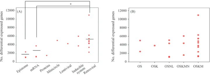
We also analyzed the relationship between the “dis-tance” of iPSCsvs.ESCs and the method used to deliver the transcription factor(s). iPSCs generated by integrating viral vectors (moloney-based retrovirus and HIV-based lenti-virus) were not as close to ESC lines as iPSCs generated by non-integrating methods (episomes, synthetic modified mRNA, proteins and minicircle DNA) (Figure 2A). The type of transcription factor used had little impact on the gene expression signature of iPSCs (Figure 2B). No over-lapping genes were differentially expressed between hESCs and hiPSCs derived from various reprogramming experiments,i.e., there were no consistent differences in the global gene expression between human ESCs and iPSCs. These findings supported the idea that reprogram-ming progressed through a series of stochastic events to produce pluripotency.
Functional analysis of significantly altered genes between iPSCs and donor cells
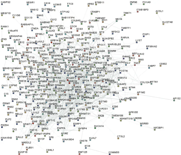
The detailed molecular events involved in reprogram-ming to produce iPSCs remain largely unknown. To ad-dress this issue, we undertook an in-depth analysis of the biological functions of differentially expressed genes in all 20 iPSC linesvs. donor fibroblasts; the selection criteria were again p < 0.05 (Student’st-test) and at least a two-fold difference in gene expression. Table 1 summarizes the number of differentially expressed genes between the iPSC lines and the original cell lines. Of these, 312 genes up-regulated in each iPSC line were compared with fibroblasts (Table S2). We defined the 312 up-regulated probes as es-sential for maintaining the pluripotency of hiPSCs (EMP genes). The STRING database was used to visualize all known functional interactions between EMP genes in iPSC lines using the default cutoff suggested by STRING. One hundred and fifty-nine genes in this set (32%) interacted with each other (Figure 3). The functional network of genes with higher expression levels in iPSCs showed a central, highly interconnected area in which common pluripotency regulators such as Pou5f1, Nanog, Lin28, Dnmt3 and Dppa4 were identified. This finding indicated that hiPSCs and hESCs shared a similar core network to maintain pluripotency. The absence of Sox2 in this analysis reflects the fact that Marchettoet al.(2009) used mouse neural stem cells (NSCs), which have a high endogenous expression of Sox2, as the donor cell lines to induce reprogramming. Hence, Sox2 was not included in the 312 genes unregulated in iPSCs. This protein interaction network for pluripotency provides a model for exploring neo-factors that may en-hance the induction of reprogramming.
We took advantage of a recently published micro-array dataset (Xieet al., 2010) to study the dynamic chan-ges in EMP genes during mammalian preimplantation
bryonic development (Table S3). One hundred and twenty EMP genes, including Pou5f1 Dppa4 and Lin28, were up regulated during the transitional phase from the four-cell stage to the eight-cell stage of human early embryonic de-velopment, known as the human zygotic genome activation period (Hoffertet al., 1997) (Figure 4). This pluripotent network, which is essential for maintaining the self-renewal of iPSCs, also plays a pivotal role in establishing embryos in vivo. The 101 EMP genes that were down-regulated during the process could contribute to the differ-entiation of stem cellsin vivoandin vitro.
The functions associated with genes that were signifi-cantly altered in reprogramming were examined by analyz-ing the over-represented annotations and pathways usanalyz-ing DAVID, with a cut-off criterion of p < 0.01. The over-represented GO terms focused on “regulation of transcrip-tion” and “regulation of cell proliferatranscrip-tion” (Table S4). The results of this analysis supported the idea that an increase in
proliferation rate was necessary for fully cellular repro-gramming (Smithet al., 2010).
We also analyzed whether significant pathways in iPSCs were enriched in significantly altered genes. The re-sults showed that hiPSCs were responsive to the TGF-b
signaling pathway that regulates the maintenance of pluri-potency, self-renewal and proliferation of hESCs (Table S4). These results demonstrated that hiPSCs repro-grammed from somatic or embryonic cells relied on similar signaling pathways to control their pluripotency.
Discussion
The results described herein show that the overall transcriptional profiles of different human iPSC lines shared a common “signature” with hESCs, although there were certain differences. Notably, the transcriptomes of hiPSCs produced by a delivery method that avoided
geno-Gene expression profiles of iPSCs and ESCs 697
mic integration shared a greater gene expression signature with hESCs than did iPSCs produced by a virus-based method. Gene-delivery methods can affect the quality of the resulting iPSCs by influencing the amount, balance, continuity and silencing of transgene expression. Potent oncogenes such asmycapparently have little effect on the transcriptional signature of iPSCs. Our findings provide a basis for selecting the most suitable method for clinical or basic applications and a better understanding of the repro-gramming process.
This study also improves our understanding of the mechanisms of cellular reprogramming. The trans-criptional network maintains the self-renewal and pluri-potency of iPSCs established primarily during preimplan-tation at the stage of zygote genome activation. Detailed analysis showed that increased proliferation and the up-regulation of genes that drive the cell cycle are necessary events for fibroblast reprogramming. Recent reports have shown that hiPSCs are more tumorigenic than hESCs based on a comparison of protein-coding point mutations (Goreet al., 2011), copy number variations (Husseinet al., 2011) and DNA methylation (Listeret al., 2011). Together, these results stress the link between pluripotency and tumorige-nicity. Given that self-renewal is a hallmark of ESCs and cancer cells, the ability to induce tumors during cellular re-programming implies that there are potential risks involved in the use of iPSCs for regenerative therapy.
In addition, non-coding RNA, including microRNA (miRNA) and large intergenic non-coding (lincRNA), which may represent a distinct layer to fine-tune the trans-criptional network of stem cells, has a role in modulating the induction of reprogramming (Judsonet al., 2009; Loe-weret al., 2010). Significantly, recent work has shown that a single miRNA cluster rapidly reprogrammed mouse and human fibroblasts into iPSCs and totally avoided the use of transcription factors (Anokye-Danso Fet al., 2011). The mechanism underlying reprogramming by miRNA differs from that of transcription factor-induced reprogramming in that there is no requirement for protein translation; the for-mer method also targets hundreds of ESC-related mRNAs directly.
In conclusion, we have examined the gene expression profiles of iPSCs obtained by different methods and from donor cell of different of origins. iPSCs produced by non-integrative methods are more closely resembled the fully reprogrammed pluripotent state than did iPSCs obtained by using integrative delivery systems, although the efficiency and kinetics were lower. Some of the results described here may reflect the markedly different circumstances in which they were generated,e.g., the culture conditions, the pas-sage number at which the cells were used and the age of the donor cells. Another limitation in our analysis was that only the initial state (donor cell) and end state (pluripotent cell) of reprogramming were examined.
Further research on each aspect of reprogramming,
e.g., the initial transcriptional response to the induction of reprogramming, the epigenetic roadblocks, the partially pluripotent state and the late events leading to pluripotency, is required in order to understand how reprogramming leads to pluripotency. A comprehensive understanding of the events involved in reprogramming a set of iPSCs can only be reached by examining the changes in the corre-sponding transcriptome (protein coding RNA, microRNA and lincRNA expression), epigenome (genome imprint, X chromosome activation, histone modifications and DNA methylation), metabolome and proteome.
Acknowledgments
This work was supported by the Chinese National Natural Science Foundation (#30871786) and the Major Project of Chinese National Programs for Fundamental Re-search and Development (“973” plan, 2009CB941002).
References
Achiwa Y, Hasegawa K and Udagawa Y (2005) Molecular mech-anism of ursolic acid induced apoptosis in poorly differenti-ated endometrial cancer HEC108 cells. Oncol Rep 14:507-512.
Anokye-Danso F, Trivedi CM, Juhr D, Gupta M, Cui Z, Tian Y, Zhang Y, Yang W, Gruber PJ, Epstein JA, et al.(2011) Highly efficient miRNA-mediated reprogramming of
Figure 4- The gene expression tendency of 312 transcripts (EMP genes)
mouse and human somatic cells to pluripotency. Cell Stem Cell 8:376-388.
Ashburner M, Ball CA, Blake JA, Botstein D, Butler H, Cherry JM, Davis AP, Dolinski K, Dwight SS, Eppig JT, et al.
(2000) Gene ontology: Tool for the unification of biology. The Gene Ontology Consortium. Nat Genet 25:25-29. Boland MJ, Hazen JL, Nazor KL, Rodriguez AR, Gifford W,
Martin G, Kupriyanov S and Baldwin KK (2009) Adult mice generated from induced pluripotent stem cells. Nature 461:91-94.
Boue S, Paramonov I, Barrero MJ and Belmonte JCI (2010) Anal-ysis of human and mouse reprogramming of somatic cells to induced pluripotent stem cells. What is in the plate? PLoS One 5:e12664.
Chin MH, Pellegrini M, Plath K and Lowry WE (2010) Molecular analyses of human induced pluripotent stem cells and em-bryonic stem cells. Cell Stem Cell 7:263-269.
Doi A, Park IH, Wen B, Murakami P, Aryee MJ, Irizarry R, Herb B, Ladd-Acosta C, Rho J, Loewer S,et al.(2009) Differen-tial methylation of tissue- and cancer-specific CpG island shores distinguishes human induced pluripotent stem cells, embryonic stem cells and fibroblasts. Nat Genet 41:1350-1353.
Ghosh Z, Wilson KD, Wu Y, Hu SJ, Quertermous T and Wu JC (2010) Persistent donor cell gene expression among human induced pluripotent stem cells contributes to differences with human embryonic stem cells. PLoS One 5:e8975. Gonzalez F, Boue S and Izpisua Belmonte JC (2011) Methods for
making induced pluripotent stem cells: Reprogramming a la carte. Nat Rev Genet 12:231-242.
Gore A, Li Z, Fung HL, Young JE, Agarwal S, Antosiewicz-Bourget J, Canto I, Giorgetti A, Israel MA, Kiskinis E,et al.
(2011) Somatic coding mutations in human induced pluri-potent stem cells. Nature 471:63-67.
Giorgetti A, Montserrat N, Aasen T, Gonzalez F, Rodriguez-Piza I, Vassena R, Raya A, Boue S, Barrero MJ, Corbella BA,et al.(2009) Generation of induced pluripotent stem cells from human cord blood using OCT4 and SOX2. Cell Stem Cell 5:353-357.
Guenther MG, Frampton GM, Soldner F, Hockemeyer D, Mitali-pova M, Jaenisch R and Young RA (2010) Chromatin struc-ture and gene expression programs of human embryonic and induced pluripotent stem cells. Cell Stem Cell 7:249-257. Hoffert KA, Anderson GB, Wildt DE and Roth TL (1997)
Transi-tion from maternal to embryonic control of development in IVM/IVF domestic cat embryos. Mol Reprod Dev 48:208-215.
Hooper SD and Bork P (2005) Medusa: A simple tool for interac-tion graph analysis. Bioinformatics 21:4432-4433. Huang DW, Sherman BT and Lempicki RA (2009) Systematic
and integrative analysis of large gene lists using DAVID bioinformatics resources. Nat Protoc 4:44-57.
Hussein SM, Batada NN, Vuoristo S, Ching RW, Autio R, Narva E, Ng S, Sourour M, Hamalainen R, Olsson C,et al.(2011) Copy number variation and selection during reprogramming to pluripotency. Nature 471:58-62.
Jia F, Wilson KD, Sun N, Gupta DM, Huang M, Li Z, Panetta NJ, Chen ZY, Robbins RC, Kay MA,et al.(2010) A nonviral minicircle vector for deriving human iPS cells. Nat Methods 7:197-199.
Judson RL, Babiarz JE, Venere M and Blelloch R (2009) Embry-onic stem cell-specific microRNAs promote induced pluri-potency. Nat Biotechnol 27:459-461.
Kang L, Wang J, Zhang Y, Kou Z and Gao S (2009) iPS cells can support full-term development of tetraploid blastocyst-com-plemented embryos. Cell Stem Cell 5:135-138.
Kang L, Kou ZH, Zhang Y and Gao SR (2010) Induced pluri-potent stem cells (iPSCs) - A new era of reprogramming. J Genet Genomics 37:415-421.
Kim D, Kim CH, Moon JI, Chung YG, Chang MY, Han BS, Ko S, Yang E, Cha KY, Lanza R,et al.(2009) Generation of hu-man induced pluripotent stem cells by direct delivery of re-programming proteins. Cell Stem Cell 4:472-476.
Lister R, Pelizzola M, Kida YS, Hawkins RD, Nery JR, Hon G, Antosiewicz-Bourget J, O’Malley R, Castanon R, Klugman S,et al.(2011) Hotspots of aberrant epigenomic reprogram-ming in human induced pluripotent stem cells. Nature 471:68-73.
Loewer S, Cabili MN, Guttman M, Loh YH, Thomas K, Park IH, Garber M, Curran M, Onder T, Agarwal S,et al.(2010) Large intergenic non-coding RNA-RoR modulates repro-gramming of human induced pluripotent stem cells. Nat Genet 42:1113-1117.
Marchetto MC, Yeo GW, Kainohana O, Marsala M, Gage FH and Muotri AR (2009) Transcriptional signature and memory re-tention of human-induced pluripotent stem cells. PLoS One 4:e7076.
Maherali N, Ahfeldt T, Rigamonti A, Utikal J, Cowan C and Hochedlinger K (2008) A high-efficiency system for the generation and study of human induced pluripotent stem cells. Cell Stem Cell 3:340-345.
Park IH, Zhao R, West JA, Yabuuchi A, Huo H, Ince TA, Lerou PH, Lensch MW and Daley GQ (2008) Reprogramming of human somatic cells to pluripotency with defined factors. Nature 451:141-146.
Smith ZD, Nachman I, Regev A and Meissner A (2010) Dynamic single-cell imaging of direct reprogramming reveals an early specifying event. Nat Biotechnol 28:521-526.
Takahashi K, Tanabe K, Ohnuki M, Narita M, Ichisaka T, To-moda K and Yamanaka S (2007) Induction of pluripotent stem cells from adult human fibroblasts by defined factors. Cell 131:861-872.
von Mering C, Jensen LJ, Kuhn M, Chaffron S, Doerks T, Kruger B, Snel B and Bork P (2007) STRING 7 - Recent develop-ments in the integration and prediction of protein interac-tions. Nucleic Acids Res 35:D358-362.
Warren L, Manos PD, Ahfeldt T, Loh YH, Li H, Lau F, Ebina W, Mandal PK, Smith ZD, Meissner A,et al.(2010) Highly ef-ficient reprogramming to pluripotency and directed differ-entiation of human cells with synthetic modified mRNA. Cell Stem Cell 7:618-630.
Xie D, Chen CC, Ptaszek LM, Xiao S, Cao X, Fang F, Ng HH, Lewin HA, Cowan C and Zhong S (2010) Rewirable gene regulatory networks in the preimplantation embryonic de-velopment of three mammalian species. Genome Res 20:804-815.
Yu JY, Hu KJ, Smuga-Otto K, Tian SL, Stewart R, Slukvin II and Thomson JA (2009) Human induced pluripotent stem cells free of vector and transgene sequences. Science 324:797-801.
Zhao XY, Li W, Lv Z, Liu L, Tong M, Hai T, Hao J, Guo CL, Ma QW, Wang L,et al.(2009) iPS cells produce viable mice through tetraploid complementation. Nature 461:U86-U88.
Internet Resources
Gene Expression Omnibus (GEO), http://www.ncbi.nlm.nih.gov/geo/.
Supplementary Material
The following online material is available for this ar-ticle:
Table S1 - Genes differentially expressed between iPSC lines and their original donor cells.
Table S2 – A detailed list of genes dynamically ex-pressed during early embryonic development.
Table S3 - The over-represented classification of GO annotations for differentially expressed genes in iPSCs and ESCs compared with donor cells.
Table S4 - The over-represented classification of pathways for 1942 differentially expressed genes in iPSCs compared with donor cells.
This material is available as part of the online article from http://www.scielo.br/gmb.
Associate Editor: Carlos F.M. Menck